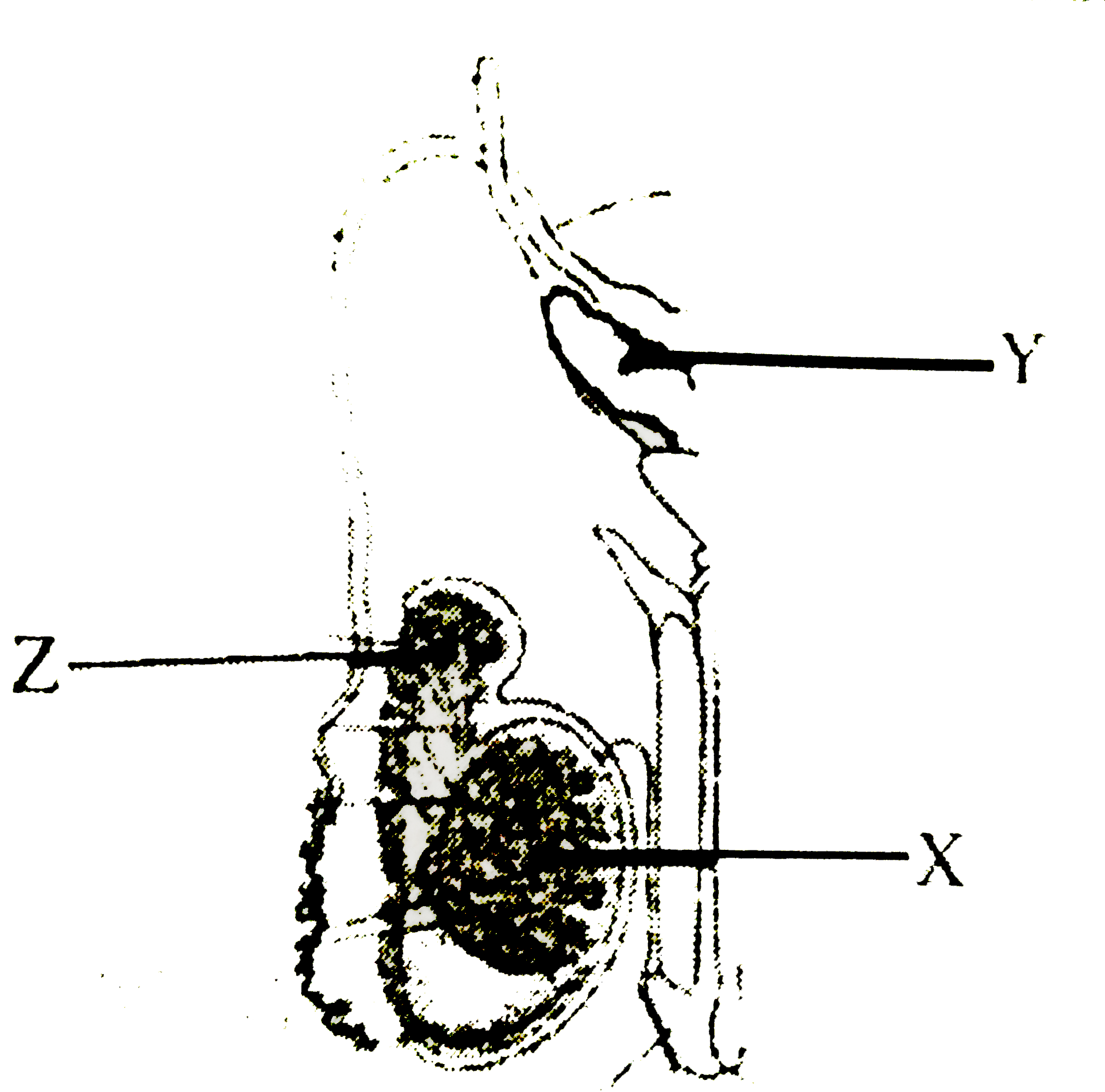
The above diagrams shows human male reproductive system (one side only )
(a) Identify 'X' and write its location in the body .
(b) Name the accessory gland 'Y' and its reaction.
(c) Name and state the function of 'Z'.
(a) X→Testis
The seminiferous tubules of testis open into the vasa efferentia through rete testis.
(b) Y→ Seminal vesicle
The male accessory glands include paired seminal vesicles. Secretion of this gland constitute the seminal plasma which is rich in fructose, calcium and certain enzymes.
(c) Z→ Epididymis
The epididymis leads to vas deferens that ascends to the abdomen and loops over the urinary bladder. It receives a duct from seminal vesicle and opens into urethra as the ejaculatory duct.
Topper's Solved these Questions
- BOOK - XII BOARD PREVIOUS YEAR PAPER ENGLISHCHAPTER - XII BOARDSEXERCISE - Set II9videos
XII BOARDS
- BOOK - XII BOARD PREVIOUS YEAR PAPER ENGLISHCHAPTER - XII BOARDSEXERCISE - Set III9videos
XII BOARDS
- BOOK - XII BOARD PREVIOUS YEAR PAPER ENGLISHCHAPTER - XII BOARDSEXERCISE - Delhi Board : Set II Section - C3videos
XII BOARDS
- BOOK - XII BOARD PREVIOUS YEAR PAPER ENGLISHCHAPTER - SAMPLE PAPER 2019EXERCISE - Section E Part B3videos
SAMPLE PAPER 2019
Similar Questions
The diagram given below represents two reproductive cells A and B. Study the same and then answer the questions that follow:
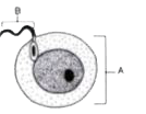
(i) Identify the reproductive cells A and B.
(ii) Name the specific part of the reproductive system where the above cells are produced
(iii) Where in the female reproductive system do these cells unite ?
(iv) Name the main hormones secreted by the (1) ovary (2) testes.
(v) Name an accessory gland found in the male reproductive system and state the function of its secretion.View Solution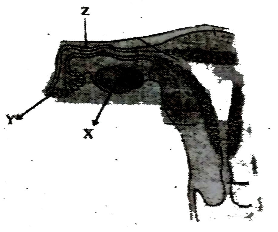
The diagram above shows a part of the human female reproductive system.
(a) Name the gamete cells that would be present in 'X' if taken from a newborn baby.
(b) Name 'Y' and write its function.
(c) Name 'Z' and write the events that take · place here.View SolutionGiven below is the diagram of the male reproductive system and its associated parts in the human body. Study the diagram and answer the questions that follow:
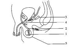
State the functionn of the part marked 2and 3.View SolutionGiven below is the diagram of the male reproductive system and its associated parts in the human body. Study the diagram and answer the questions that follow:
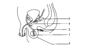
Draw a neat labelled diagram of a human sperm.View SolutionFollowing is the diagram of the male reproductive system of frog. Select the correct set of names labelled A, B, C and D.
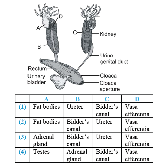 View Solution
Compound X and aluminium are used to join railway tracks. (a) Identify the compound X (b) Name the reaction (c) Write down its reaction.
View SolutionThe diagram given below represents an endocrine gland in the human body. Study the diagram and answer the following questions :
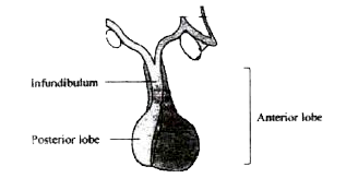
(i) Identify the endocrine gland. Where is it located?
(ii) Why is the above gland referred to as the 'Master gland' ?
(iii) Name the hormone which in deficiency causes Diabetes Insipidus. How does this disorder differ from Diabetes Mellitus ?
(iv) Explain the term 'Hormone'.
What is the role of Tropic hormones in the human body?
(v) Which lobe of the above gland secretes:
1. Oxytocin
2. ACTH
3. Growth hormone.View SolutionGiven below is the diagram of the male reproductive system and its associated parts in the human body. Study the diagram and answer the questions that follow:
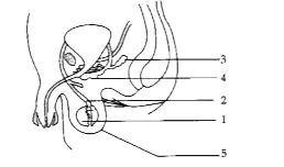
Mention the hormone secreted by the part labelled 1.View SolutionGiven below is the diagram of the male reproductive system and its associated parts in the human body. Study the diagram and answer the questions that follow:
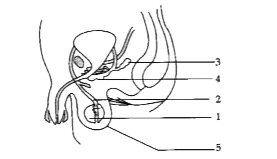
What is the significance of the part labelled 5.View SolutionGiven below is the outline of the male reproductive system :
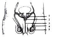
(i) Name the parts labelled 1 to 5.
(ii) State the functions of the parts labelled 1 and 4.
(iii) Name the cells of part 5 that produce testosterone.
(iv) Why is the structure 5 present outside the body in the scrotal sacs ?
(v) What is semen ?
(vi) Give the exact location of prostate gland.View Solution

