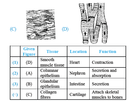
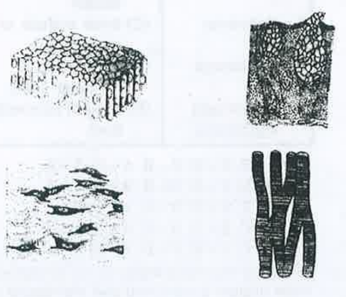
The four figures (A, B, C and D) given below represent four different types of animal tissues. Which one of these is correctly identified in the given options along with its correct location and function ?

Tissue Location FunctionB-Glandular epithelium Intestine Secretion
Tissue Location FunctionC-Collagen fibres Cartilage Attach bone to bone
Tissue Location FunctionD-Smooth muscle tissue Heart Heart contraction
Tissue Location FunctionA-Columnar epithelium Nephron Secretion and absorption
The correct Answer is:A
Step by step video, text & image solution for The four figures (A, B, C and D) given below represent four different types of animal tissues. Which one of these is correctly identified in the given options along with its correct location and function ? by Biology experts to help you in doubts & scoring excellent marks in Class 11 exams.
Updated on:21/07/2023Topper's Solved these Questions
- STRUCTURAL ORGANISATION IN ANIMALSBOOK - NCERT FINGERTPS ENGLISHCHAPTER - STRUCTURAL ORGANISATION IN ANIMALSEXERCISE - HOTS
5 Videos
- STRUCTURAL ORGANISATION IN ANIMALSBOOK - NCERT FINGERTPS ENGLISHCHAPTER - STRUCTURAL ORGANISATION IN ANIMALSEXERCISE - EXEMPLAR
9 Videos
- RESPIRATION IN PLANTSBOOK - NCERT FINGERTPS ENGLISHCHAPTER - RESPIRATION IN PLANTSEXERCISE - Respiration In Plants
130 Videos
- TEST PAPERBOOK - NCERT FINGERTPS ENGLISHCHAPTER - TEST PAPEREXERCISE - Test Paper 3
50 Videos
Similar Questions
The four sketches (A, B, C and D) given below, represent four different types of animal tissues. Which one of these is correctly identified in the options given, along with its correct location and function ?

View SolutionThe four sketches A, B,C and D given below
represent four different types of animal tissues .
which one of these is correctly identified
in the options given along with its correct location and function ?
View Solution
Knowledge Check
Identify A,B,C and D in the given flow chart and slect the correct option.
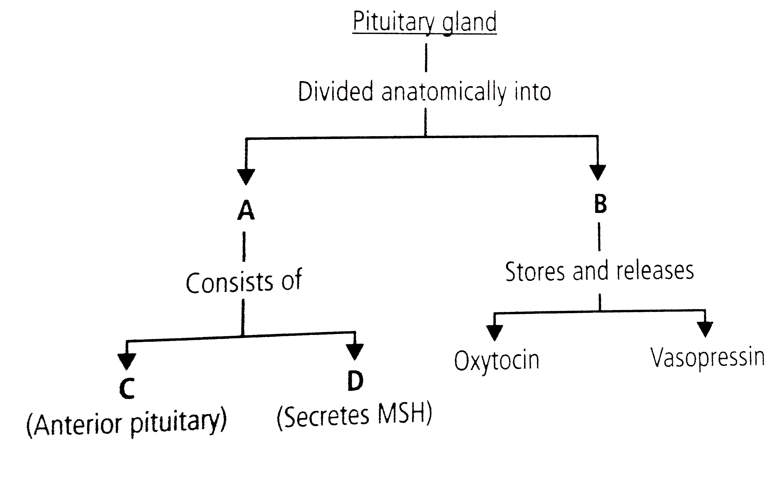 AABCDNeurohypophysisAdenohypophysisParsdistalisPars intermediaBABCDAdeno-hypophysisNeurohypophysisPars intermediaPars distailsCABCDAdenohypophysisNeuro-hypophysisParsdistailsPars intermediaDABCDNeuro-hypophysisAdeno-hypophysisPars intermediaPars distails
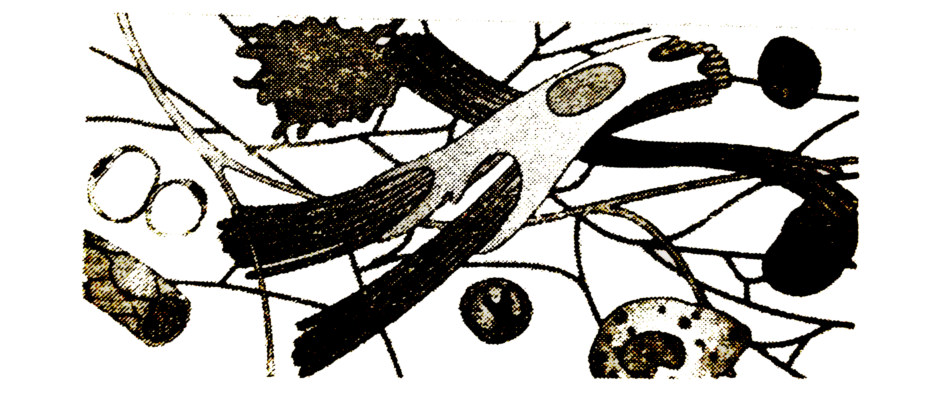
Identify the figure of animals tissue given below, along with it's correct location :-
View SolutionIdentify A and B in the given figure and select the correct option.
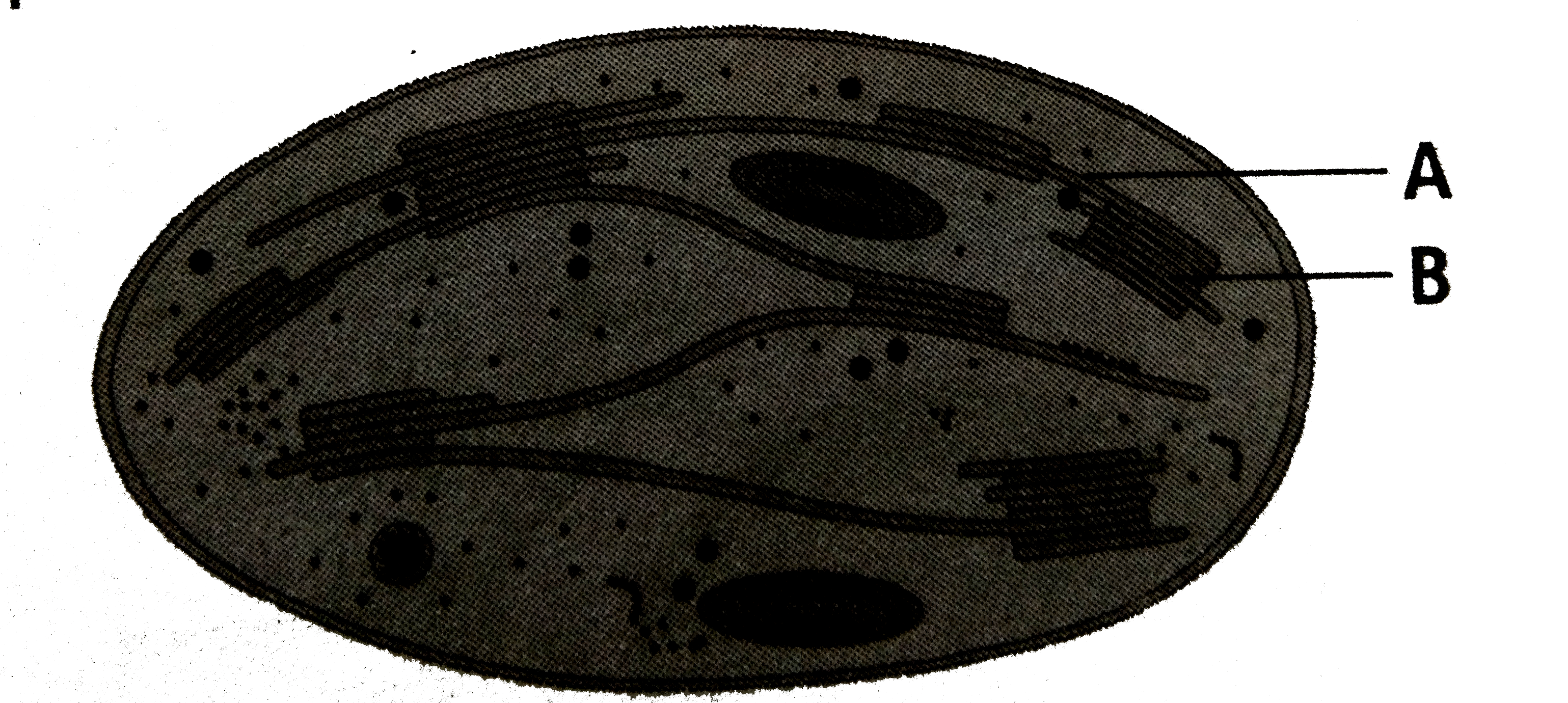
View Solution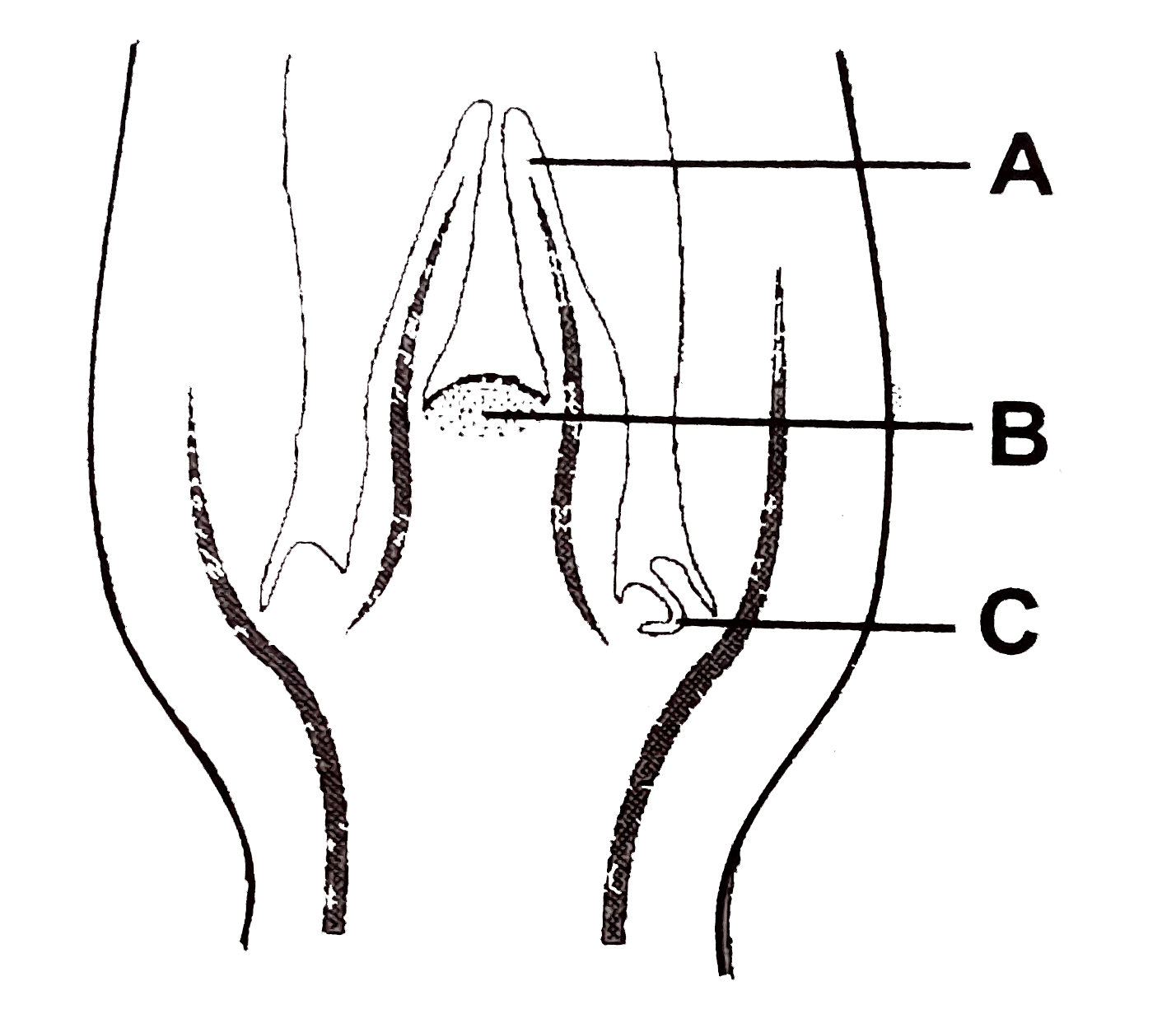
Identify the given figure and select the correct option for A, B and C
View SolutionIdentify the figures A, B ,C and D given below and select the correct option.
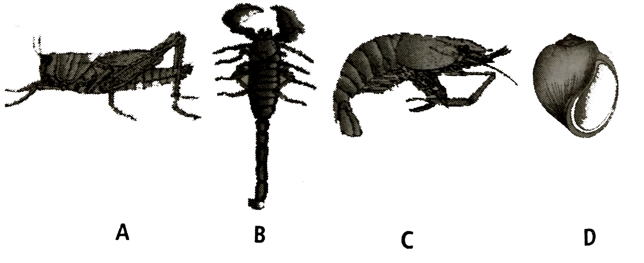
View SolutionIdentify the given figures A, B and C and select the correct option.
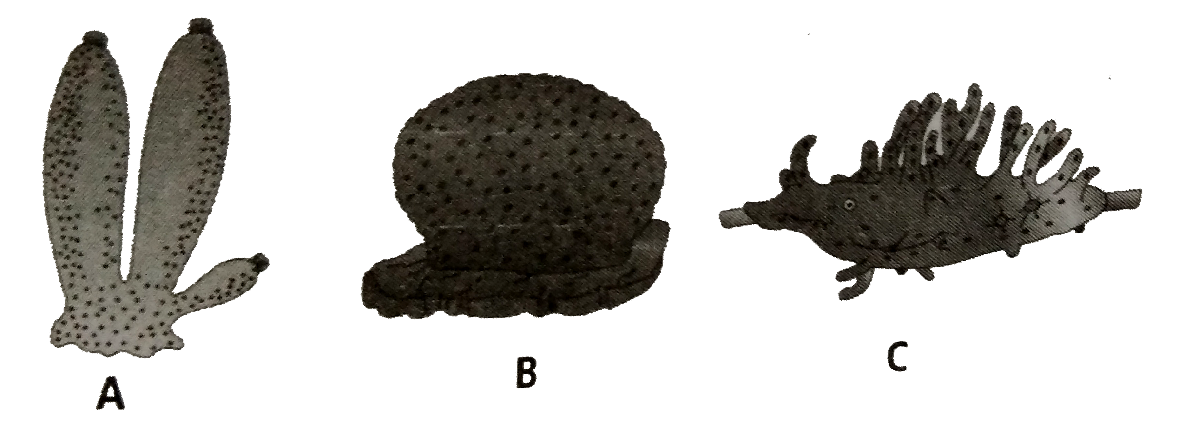
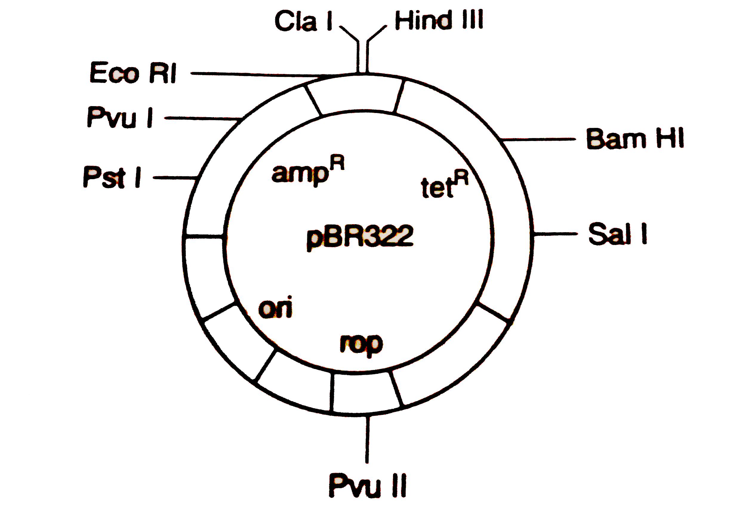
View SolutionThe given figure is the diagrammatic representation of the E. coli vector pBR322. Which one of the given options correctly identifies its certain component (s) ?
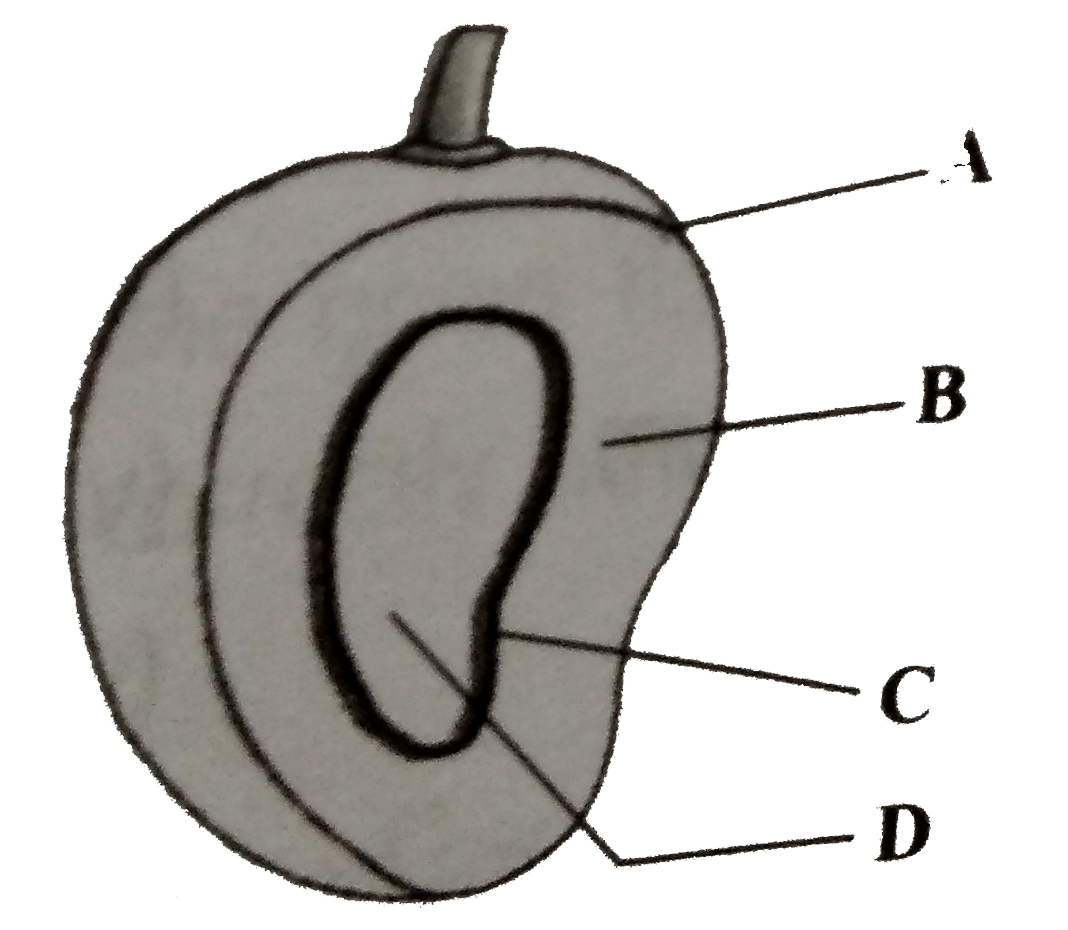
View SolutionGiven figure represents a drupe of mango. Select the option that correctly identifies A, B, C and D
View Solution
