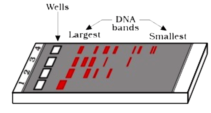
Study the diagram given below and answer the questions that follow:a Why have DNA fragments in band ‘D’ moved farther away in comparison to those in band ‘C’? b Identify the anode end in the diagram. c How are these DNA fragments visualized?
a DNA fragments in band ‘D’ are smaller in size than fragments in band ‘C’. Therefore they moved faster and farther away. b The anode end is ‘B’. c The separated DNA fragments can be visualised by staining the DNA with ethidium bromide followed by exposure to UV radiation.
Related Solutions
Diagram shows a typical agarose gel showing migration of DNA fragments . a. Which of the bands has the largest and smallest DNA fragments ? b. How can you make fragments of DNA for electrophoresis ? c. Explain separation of DNA fragments using electrophoresis. d. Point out a method to visualize the separated DNA fragments after electrophoresis .
View SolutionAfter electrophoresis the separated DNA fragment can be vishualised ethidium bromide gel exposed UV light . These DNA fragments appear as _______ coloured bands :
View SolutionAfter electrophoresis the separated DNA fragment can be visualized ethidium bromide gel exposed UV light . These DNA fragments appear as _______ coloured bands :
View SolutionStudy the linking of DNA fragments shown below: Name 'a' DNA and 'b" DNA
View SolutionGiven below are four statements pertaining to separation of DNA fragments using gel electrophoresis. Identify the incorrect statements. (a) DNA is negatively charged molecule and so it is loaded on gel towards the Anode terminal (b) DNA fragments travel along the surface of the gel whose concentration does not affect movement of DNA. (c) Smaller the size of DNA fragment larger is the distance it travels through it. (d) Pure DNA can be visualized directly by exposing UV radiation. Choose correct answer from the options given below
View SolutionGiven diagram showing a typical agarose gel electrophoresis showing migration of DNA fragments . The undigested DNA fragment will be present in the lane

View SolutionGiven diagram showing a typical agarose gel electrophoresis showing migration of DNA fragments . The undigested DNA fragment will be present in the lane

View SolutionWhy do DNA fragments move towards the anode during gel electrophoresis ?
View SolutionWhy do DNA fragments move towards the anode during gel electrophoresis ?
View SolutionDNA fragments move towards anode during Denaturation.
View SolutionThe fragmented DNA can be visualized by staining DNA with
View SolutionA: During gel electrophoresis, the DNA fragments move towards the anode.
R: DNA fragments are negatively charged molecules .
View SolutionA: During gel electrophoresis, the DNA fragments move towards the anode.
R: DNA fragments are negatively charged molecules .
View SolutionA: During gel electrophoresis, the DNA fragments move towards the anode. R: DNA fragments are negatively charged molecules .
View SolutionA: During gel electrophoresis, the DNA fragments move towards the anode.
R: DNA fragments are negatively charged molecules .
View SolutionIdentify a , band c in the following diagram .
View SolutionStudy the given figure carefully and select the incorrect statements regarding this: A. It represent a typical agarose gel electrophoresis in which lane in contains undigested DNA B. Smallest DNA bands are formed at A and largest DNA bands are formed at B. C. The separated DNA fragments can be visualized alter staining in the visible light D The separated DNA bands arc cut out from the agarose gel and extracted from the gel piece. This step's known as elution.
View SolutionImagine a gel through which DNA fragments have moved in response to an applied electrical current.The band on this gel that is farthest from the top(that is,from the place where the DNA fragments were added to the "well")represents the
View SolutionImagine a gel through which DNA fragments have moved in response to an applied electrical current. The band on this gel this is farthest from the top (that is, from the place where the DNA fragments were added to the "well") represents the
View SolutionImagine a gel through which DNA fragments have moved in response to an applied electrical current. The band on this gel this is farthest from the top (that is, from the place where the DNA fragments were added to the "well") represents the
View Solution