(a) List two reasons for the appearance of variations among the progeny formed by sexual reproduction.
(b) (1) Name the part marked 'A' in the diagram alongside.
(ii) How does 'A' reaches part 'B' ?
(iii) State the importance of the part 'C'.
(iv) What happens to the part marked 'D' after fertilisation is over ?
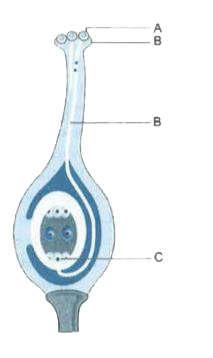
(a) Variation is seen among progeny formed by sexual reproduction because of:
(i) Involvement of two different individuals.
(ii) Creation of new combination of variants.
(b) A-pollen or pollen grain. (i) It reaches the stigma (B) by agents of pollination.
(iii) C (pollen tube) helps male gamete to reach the egg (ovule).
(iv) After fertilsation it converts into embryo.
Topper's Solved these Questions
- HOW DO ORGANISMS REPRODUCE ?BOOK - VK GLOBAL PUBLICATIONCHAPTER - HOW DO ORGANISMS REPRODUCE ?EXERCISE - LONG ANSWER QUESTIONS
20 Videos
- HOW DO ORGANISMS REPRODUCE ?BOOK - VK GLOBAL PUBLICATIONCHAPTER - HOW DO ORGANISMS REPRODUCE ?EXERCISE - HOTS ( Higher Order Thinking Skills )
8 Videos
- HOW DO ORGANISMS REPRODUCE ?BOOK - VK GLOBAL PUBLICATIONCHAPTER - HOW DO ORGANISMS REPRODUCE ?EXERCISE - SHORT ANSWER QUESTIONS -I
36 Videos
- HEREDITY AND EVOLUTION BOOK - VK GLOBAL PUBLICATIONCHAPTER - HEREDITY AND EVOLUTION EXERCISE - HOTS ( Higher Order Thinking Skills )
12 Videos
- LIFE PROCESSES BOOK - VK GLOBAL PUBLICATIONCHAPTER - LIFE PROCESSES EXERCISE - HOTS (HIGHER ORDER THINKING SKILLS)
22 Videos
Similar Questions
Study the diagram given below
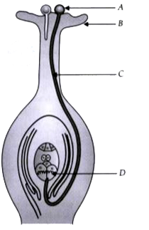
What happens to the part marked 'D' after fertilization is over?
View SolutionIn the diagram drawn alongside
(i) Label the parts a, b and c.
(ii) In which part does the light reaction occur?
(iii) Name the part which the dark phase of photosynthesis occur in.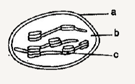
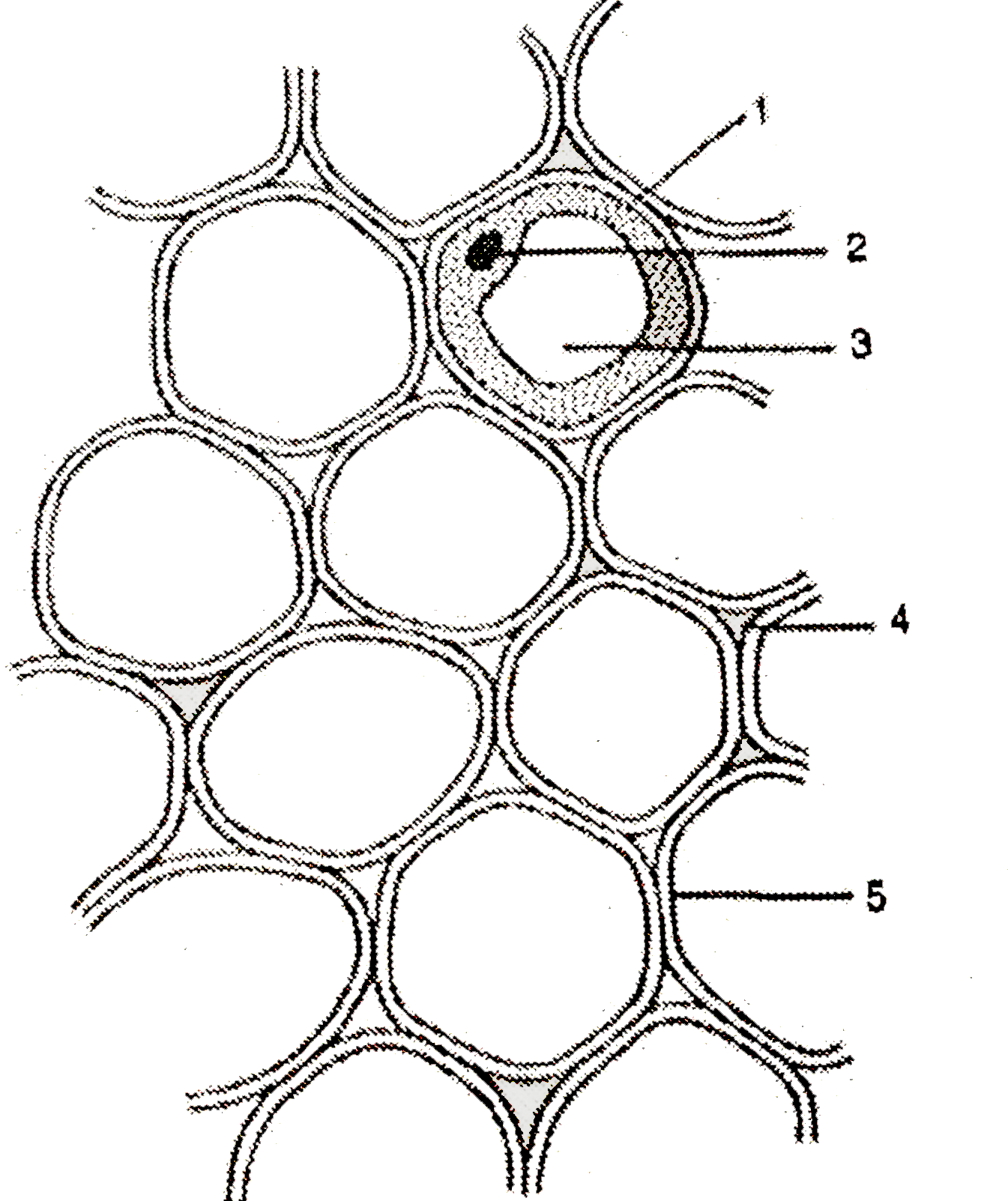
View Solution(a) Identify the plant tissue given in figure alongside.
(b) Label the parts marked 1 to 5.
(c) Mention the function of this tissue.
View Solution(a) To which group this organism belongs ?
(b) Lebel the parts marked 1 to 7
(c) What is the function of part marked 7 ?

View SolutionThe following diagram represents the human brain as seen in an external view. Study the same and then answer the questions that follow :
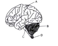
(i) Name the part labelled A, B, C and D.
(ii) Mention the difference in the arrangement of the nerve cells in the parts marked 'A' and 'D'
(iii) What is the main function of the parts marked 'C and 'D'?
(iv) Name the sheet of nerve fibres that connect the two halves of the part labelled.
(v) Name the basic unit of brain.
View SolutionStudy the diagram given alongside showing a plant cell kept in a strong sugar solution.
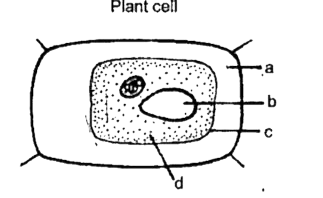
Name the parts a, b, c, and d.
View Solution(a) What is pollination ? State its significance
(b) How does fertilisation occurs in flowers ? Name the parts of the flower that develop into (i) seed and (ii) fruit after fertilisation.
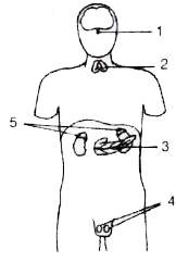
View SolutionThe given diagram is an outline of human body showing the posttion of certain organs. In this diagram
(I) Name the parts labelled 1-5.
(ii) Name the hormone released from 1 that influences the working of part 2.
(iil) How do part labelled as 3 differs from parts labelled as 1 and 2 in general? Name the endocrine cells present in part 3.
(iv) What will happen if the part labelled as 4 is removed ?
(11) Name the hormones released by part 5.
(VI) Why the part labelled as 1 called master gland? Which part of the forebrain controls the gland labelled 1 ?
(vii) Name the gland that secretes 'emergency hormone.
View Solution(a) Draw a diagram showing germination of pollen on stigma of a flower and mark on it the following organs/parts:
(i) Pollen Grain
(ii) Pollen tube
(iii) Stigma
(iv) Female germ cell
(b) State the significance of pollen tube.
(c ) Name the parts of flower that develop after fertilisation into
(i) Seed (ii) Fruit
View SolutionStudy the diagram given below and then answer the questions that follow :

(i) Name the part labelled A. Name any two hormones produced by the part labelled A.
(ii) What happens to the part labelled B if :
(1) fertilisation takes place?
(2) fertilisation does not take place ?
(iii) Where does fertilisation occur ?
(iv) Draw a neat diagram of the human sperm as seen under high magnification and label the following parts: (a) Acrosome (b) Mitochondria.
View Solution
