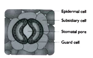
A student is observing temporary mount of leaf peel under the microscope. Draw labelled diagram of the structure of the stomata as seen under the microscope.Solution in Kannada
a] 
b]
Role of stomata in photosynthesis- Mainly for exchange of gases. Plants closes this pore during night time when it does not get sunlight.
It helps to expel the excess water out from the leaves in the form of water vapour.
Topper's Solved these Questions
- LIFE PROCESSESBOOK - ZEN PUBLICATIONCHAPTER - LIFE PROCESSESEXERCISE - ZEE ADDITIONAL QUESTIONS SECTIONS (SHORT ANSWER [SA] TYPE 2 QUESTIONS)
47 Videos
- LIFE PROCESSESBOOK - ZEN PUBLICATIONCHAPTER - LIFE PROCESSESEXERCISE - ZEE ADDITIONAL QUESTIONS SECTIONS (LONG ANSWER [LA] TYPE QUESTIONS)
32 Videos
- LIFE PROCESSESBOOK - ZEN PUBLICATIONCHAPTER - LIFE PROCESSESEXERCISE - ZEE ADDITIONAL QUESTIONS SECTIONS (VERY SHORT ANSWER [VSA] TYPE QUESTIONS)
42 Videos
- HOW DO ORGANISMS REPRODUCE?BOOK - ZEN PUBLICATIONCHAPTER - HOW DO ORGANISMS REPRODUCE?EXERCISE - ZEN ADDITIONAL QUESTIONS SECTION (SHORT ANSWER [SA] TYPE 2 QUESTIONS)
36 Videos
- OUR ENVIRONMENT BOOK - ZEN PUBLICATIONCHAPTER - OUR ENVIRONMENT EXERCISE - ZEN ADDITIONAL QUESTIONS SECTION ( LONG ANSWER (LA) QUESTIONS )
4 Videos
Similar Questions
How does Paramecium obtain its food ?
View SolutionExplain the mechanism by which humans regulate their body temperature.
View SolutionWhat are life processes?
View SolutionExplain fission with examples. Differentiate between binary and multiple fission. Draw a well labelled diagram of binary fission in amoeba.
View SolutionIndicate the tlow of energy In an ecosystem. ny s t unidirectional? Justify.
OR
AS energy moves progressively through various trophic levels of food chain it is no longer available to the previous level . Give reasons .
View SolutionTo prepare stained temporary mounts of (a) onion peel, and (b) human cheek cells and to record observations and draw their labelled diagram. The cels are first focussed in microscope under
View SolutionThe temporary mount of the leaf epidermal peel which looked pinkish red under the microscope was:
View SolutionA well stained leaf peel mount when observed under the high power of a microscope shows nuclei in:
View SolutionRecord your observations when a stained a mounted leaf peel is viewed by you under high power (45 xx ) microscope.
View SolutionIdentify the observed various parts of temporary mount of well stained leaf peel, when focused under the high power of a microscope.
View SolutionList the steps of preparation of temporary mount of a leaf peel to observe stomata.
View SolutionA student is observing the temporary mount of a leaf peel under a microscope. Draw
labelled diageram of the structure (of stomata) as seen under the microscope.
View Solutionक्लोरोप्लास्ट की संरचना का चित्र बनाओं जैसा कि इलेक्ट्रॉन सूक्ष्मदर्शी में दिखाया गया है और इसका कार्य दीजिए।
View SolutionDraw a labelled diagram of the cross section of chloroplastid as seen under microscope.
View SolutionName the green dot like structures in some cell observed by a student when a leaf peel was viewed under a microscope, what is this green colour due to ?
View SolutionWhy was the temporary mount of the leaf epidermal peel pinkish-red under the microscope?
View Solution
