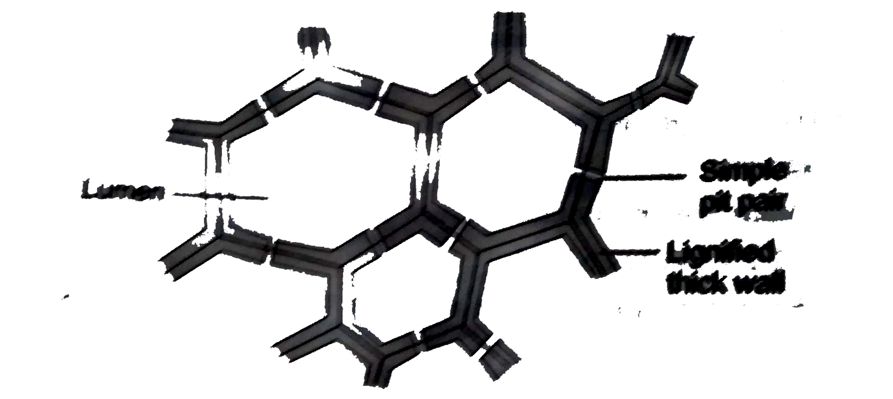
(a) Tarun was asked to draw a diagram of sclerenchyma as seen in longitudinal section and label it . His figure is given below. Point out the mistake in the figure.
(b) Are the cells of sclerenchyma live or dead ?
(a) The figure is transverse section instead of longitudinal section.
(b) The cells of sclerenchyma are dead cells.
Topper's Solved these Questions
- TISSUESBOOK - PRADEEPCHAPTER - TISSUESEXERCISE - Long Answer Questions
15 Videos
- TISSUESBOOK - PRADEEPCHAPTER - TISSUESEXERCISE - Additional Questions (very short answer questions)
46 Videos
- TISSUESBOOK - PRADEEPCHAPTER - TISSUESEXERCISE - NCERT Exampler Problems
33 Videos
- THE FUNDAMENTAL UNIT OF LIFE BOOK - PRADEEPCHAPTER - THE FUNDAMENTAL UNIT OF LIFE EXERCISE - ANALYTICAL
11 Videos
Similar Questions
Knowledge Check
Mohit was asked to draw a diagram of sclerenchyma as seen in longitudinal section and label it. His diagram is as shown. Find out the mistake he has made in the diagram.
 AThe pit pair is bordered not simple.BThe cell walls are not lignified in sclerenchyma.CThe nucleus and cytoplasm are not shown.DIt is a transverse section and not the longitudinal section.
For the circuit shown in figure given below, the equivalent resistance points A and B is
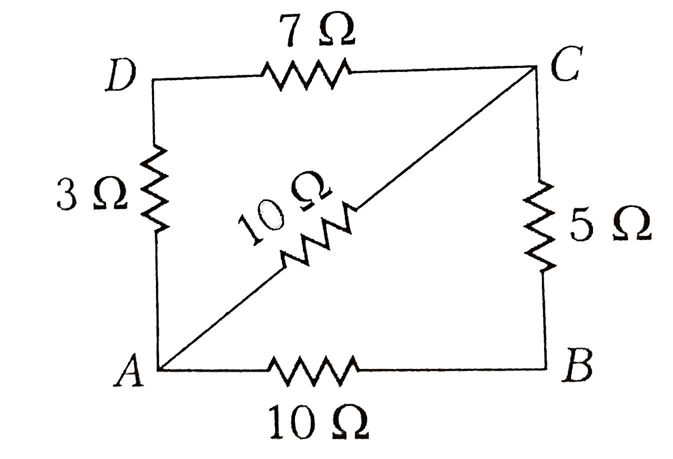 A10ΩB5ΩC103ΩD2Ω
Study the given diagram and answer the questions given below:
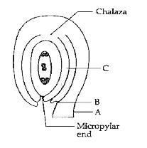
Tegmen develops from the part labelled C in the figure is calledAInner integumentBOuter IntegumentCFunicleDChalazal pole
Examine the figure given below and select the correct option for labelled parts, a,b,c,d
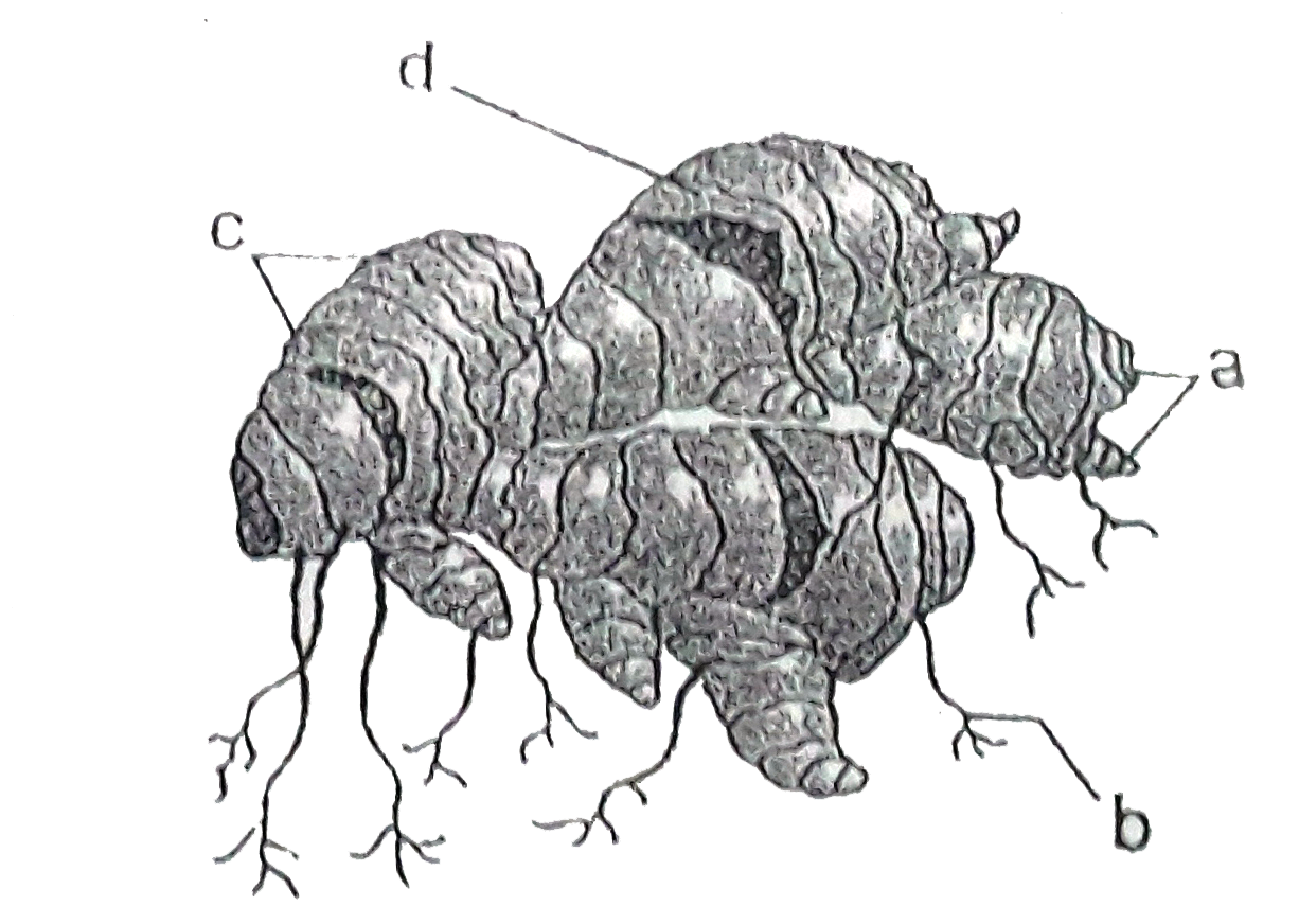
View Solution(a) A student observed a nerve cell and drawn the following figure. He did a mistake in labelling. Find out the mistake
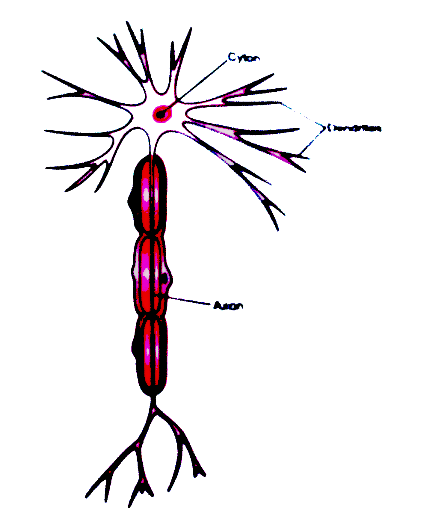
(b) Where would you find nerve cells ? What is the function of nerve cell ?
View SolutionThere are two large identical open tanks as shown in figure. In tanks 1 there is a small hole of cross sectional area A at its base. Tank II has a similar hole, to which a pipe of length h has been connected as shown. The internal cross sectional area of the pipe can be considered to be equal
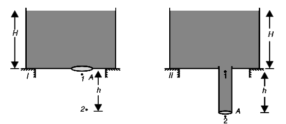
to A. Point 1 marked in both figures, is a point just below the opening in the tank and point 2 marked in both figures, is a point h below point 1 (In fig II, point 2 is just outside the opening in the pipe)
(a) Find the speed of flow at point 2 in both figures.
(b) Find the ratio of speed of flow at point 1 is first the ratio of speed of flow at point 1 is first figure to that in second figure.
(c) Find the difference in pressure at point 1 in both figures.
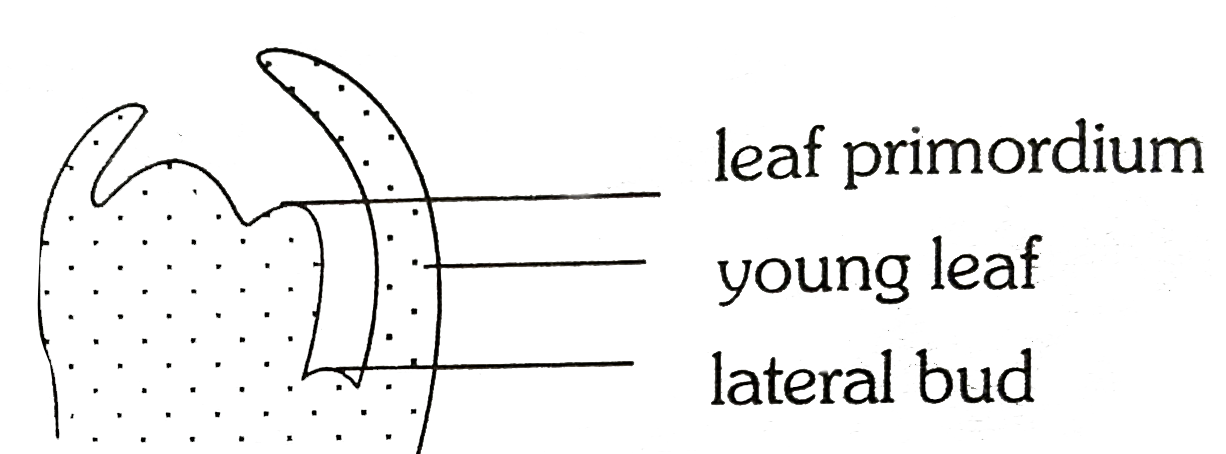
View SolutionThe following diagram opposite illustrates a longitudinal section through a shoot apex. Which of the figures gives below shows the correct appearance of this shoot apex at the formation of the next leaf primordium
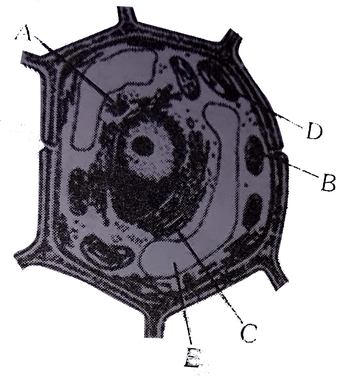
View SolutionThe diagram of the ultranstructure of a plant cell is given below. Identify the functions of the organelles labelled. A,B,C,D,E in the diagram
View Solution

