Describe in detail structure of Eye, various parts and functions of it.
Text Solution

Paired eyes are located in sockets of the skull called orbits.
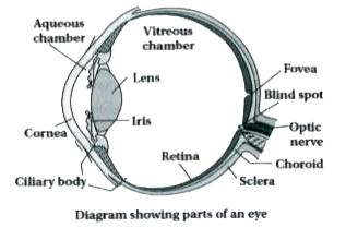
Parts of an eye : Each eye ball is nearly a spherical in structure.
Its diameter is approximately 2.5 cm and weighs around 6 to 8 gm.
The wall of the eyeball is composed of three layers. (see figure)
(a) Sclera (b) Choroid (c) Retina
Sclera : It is fibrous layer, white layer and have transparent membrane post. 5/6 part is white membrane formed of collagen fibres where as anterior 1/6 part formed of fibrous tissue devoid of blood vessel is called cornea.
Conjunctiva : Conjunctiva is thin, transparent, made up of stratified epithelium. It externally surrounds transparent membrane and open part of white layer. It is thinnest epithelium of animal body.
(b) Choroid : It is middle layer, contains blood vessels and looks bluish in colour.
The choroid layer is thin over the posterior two-thirds of the eye ball, but it becomes thick in the anterior part to form the ciliary body.
Ciliary body continues forward to form a pigmented and opaque structure called Iris. Iris is the visible coloured portion of the eye.
The eye ball contains a transparent crystalline Lens which is held in place by ligaments attached to the ciliary body.
In front of the lens, the aperture surrounded by the iris is called the pupil. The diameter of the pupil is regulated by the muscle fibres of iris.
(c ) Retina : It is the inner most layer and it contains three layers of neural cells from inside to outside - Ganglionated Cells, Bipolar Cells and Photoreceptor Cells.
Photoreceptor cells are of two types rod cells and cone cells.
These cells contain the light-sensitive proteins called the photopigments.
The daylight photopic vision and colour vision are functions of cones and the twilight scotopic vision is the function of the rods.
The rods cell contain purplish-red protein called Rhodopsin, which contains derivative of Vitamin A.
In the human eye, there are three types of cones which possess their own photopigments that respond to red, green and blue lights.
The sensations of different colours are produced by various combinations of these cones and their photopigments.
When these cones are stimulated equally, a sensation of white light is produced.
The optic nerves leave the eye and the retinal blood vessels enter in it at a point medial to and slightly above the posterior pole of the eye ball.
Photoreceptor cells are not present in that region and hence it is called the blind spot.
At the posterior pole of the eye lateral to the blind spot, there is a yellowish pigmented spot called macula lutea with a central pit called the fovea.
The fovea is a thinned-out portion of the retina where only the cones are densely packed. It is the point where the resolution is the greatest.
The space between the cornea and lens is called the aqueous chamber and contains a thin watery fluid called aqueous humor.
The space between the lens and the retina is called the vitreous chamber and is filled with a transparent gel called vitreous humor.
|
Updated on:21/07/2023
Topper's Solved these Questions
- BOOK - KUMAR PRAKASHANCHAPTER - NEURAL CONTROL AND COORDINATIONEXERCISE - SECTION - B (DIFFERENCE/SCIENTIFIC REASONS)11videos
NEURAL CONTROL AND COORDINATION
- BOOK - KUMAR PRAKASHANCHAPTER - NEURAL CONTROL AND COORDINATIONEXERCISE - SECTION - C (DEFINITION/EXPLANATION - TERMS/IMPORTANCE)21videos
NEURAL CONTROL AND COORDINATION
- BOOK - KUMAR PRAKASHANCHAPTER - MORPHOLOGY OF FLOWERING PLANTSEXERCISE - questions from module -(Important MCQ for NEET)4videos
MORPHOLOGY OF FLOWERING PLANTS
- BOOK - KUMAR PRAKASHANCHAPTER - PHOTOSYNTHESIS IN HIGHER PLANTSEXERCISE - FILL IN BLANKS10videos
PHOTOSYNTHESIS IN HIGHER PLANTS
Similar Questions
Describe the structure , types and functions of plastids.
View SolutionDescribe structure and functions of bone.
View SolutionDescribe the functions of various parts of brain.
View SolutionDescribe the structure and functions of epithelium.
View SolutionDescribe the structure and functions of bone.
View SolutionDraw a neat-labelled diagram of a typical bacterial cell to show the detailed structure and describe the functions of important organelles.
View SolutionDescribe the structure and function of pancreas.
View SolutionDescribe the structure and functions of mitochondria.
View SolutionDescribe the structure and functions of nucleus.
View SolutionDescribe the structure and functions of nucleus.
View Solution

