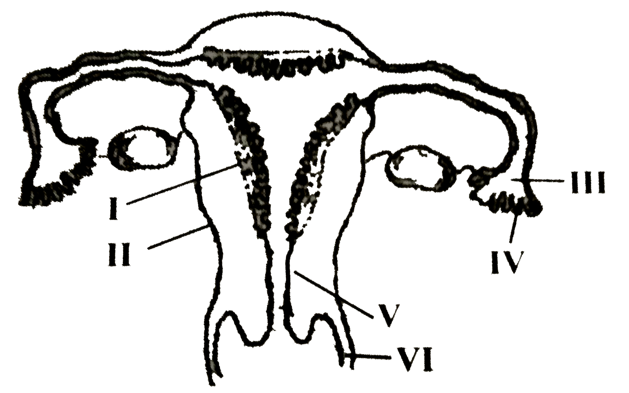
Draw a neat labelled, diagrammatic sectional view of female reproductive system.
Step-by-Step Text Solution
1. Start with the Basic Structure: Begin by drawing a vertical section of the female reproductive system. This should include the ovaries, fallopian tubes, uterus, cervix, vagina, and external genitalia.
2. Draw the Ovaries: Place the ovaries at the top of your diagram. They should be represented as two small oval shapes, one on each side. Label them as "Ovary".
3. Add the Fallopian Tubes: Draw the fallopian tubes extending from each ovary towards the uterus. The tubes should have three distinct regions:
- Infundibulum: The funnel-shaped end near the ovary.
- Ampulla: The middle section of the tube.
- Isthmus: The narrow section that connects to the uterus.
Label these parts accordingly.
4. Illustrate the Uterus: Below the fallopian tubes, draw the uterus as a pear-shaped structure. Indicate the three layers of the uterus:
- Perimetrium: The outer layer.
- Myometrium: The thick middle muscular layer.
- Endometrium: The inner lining where implantation occurs.
Label these layers and also mark the "Uterine Cavity".
5. Draw the Cervix: At the lower end of the uterus, draw the cervix, which connects the uterus to the vagina. Label it as "Cervix" and indicate the "Cervical Canal" inside it.
6. Illustrate the Vagina: Below the cervix, draw the vagina as a muscular tube leading to the external genitalia. Label it as "Vagina".
7. Add External Genitalia: At the bottom of your diagram, depict the external genitalia, which includes:
- Mons Pubis: The fatty tissue covering the pubic bone.
- Labia Majora: The larger outer folds.
- Labia Minora: The smaller inner folds.
- Hymen: A thin membrane partially covering the vaginal opening.
- Clitoris: A small protrusion located at the top of the labia minora.
Label all these structures clearly.
8. Final Touches: Ensure that all parts are neatly drawn and clearly labeled. Use arrows or lines to connect labels to the respective parts of the diagram for clarity.
- Infundibulum: The funnel-shaped end near the ovary.
- Ampulla: The middle section of the tube.
- Isthmus: The narrow section that connects to the uterus.
Label these parts accordingly.
- Perimetrium: The outer layer.
- Myometrium: The thick middle muscular layer.
- Endometrium: The inner lining where implantation occurs.
Label these layers and also mark the "Uterine Cavity".
- Mons Pubis: The fatty tissue covering the pubic bone.
- Labia Majora: The larger outer folds.
- Labia Minora: The smaller inner folds.
- Hymen: A thin membrane partially covering the vaginal opening.
- Clitoris: A small protrusion located at the top of the labia minora.
Label all these structures clearly.
Topper's Solved these Questions
- HUMAN REPRODUCTIONBOOK - MODERN PUBLICATIONCHAPTER - HUMAN REPRODUCTIONEXERCISE - REVISION EXERCISES (LONG ANSWER TYPE QUESTION (B) QUESTION FROM CBSE EXAMINATION)
25 Videos
- HUMAN REPRODUCTIONBOOK - MODERN PUBLICATIONCHAPTER - HUMAN REPRODUCTIONEXERCISE - COMPETITION FILE (OBJECTIVE TYPE QUESTIONS (A) MULTIPLE CHOICE QUESTIONS)
183 Videos
- HUMAN REPRODUCTIONBOOK - MODERN PUBLICATIONCHAPTER - HUMAN REPRODUCTIONEXERCISE - REVISION EXERCISES (SHORT ANSWER TYPE QUESTION (B )QUESTIONS FROM CBSE EXAMINATIONS)
19 Videos
- HUMAN HEALTH AND DISEASESBOOK - MODERN PUBLICATIONCHAPTER - HUMAN HEALTH AND DISEASESEXERCISE - Chapter Practice Test
2 Videos
- MICROBES IN HUMAN WELFAREBOOK - MODERN PUBLICATIONCHAPTER - MICROBES IN HUMAN WELFAREEXERCISE - CHAPTER PRACTICE TEST (Long Answer Type Question )(Five marks each)(Section -E)
8 Videos
Similar Questions
Draw a well labelled diagram of sectional view of human female reproductive system.
View Solution(a) Draw a diagrammatic sectional view of the female reproductive system of human and label the parts
(i) where the secondary oocytes develop
(ii) which helps in collection of ovum after ovulation
(iii) where fertilization occurs
(iv) where implantation of embryo occurs.
(b) Explain the role of pituitary and the ovarian hormones in menstrual cycle in human females.
View Solution
Knowledge Check
The given figure depicts a diagrammatic sectional view of the human female reproductive system. Which set of three parts out of I−Vi have been correctly identified?
 A(II) endometrium, (III) infundibulum, (IV) fimbriaeB(III) infundibulum, (IV) fimbriae, (V) cervixC(IV) oviducal funnel, (V)uterus,(VI)cervixD(I) perimetrium, (II) myometrium, (III) Fallopian tube
Draw a neat labelled diagram of nucleus.
View Solutiona) Draw a labelled diagmmatic view of human male reproductive system.
b) Differentiate between:
i) Vas deferens and vasa efferentia
ii) Spermatogenesis and spermeogenesis
View SolutionDraw a neat labelled diagram of an animal cell.
View SolutionDraw a well labelled diagrammatic sectional view of mammary gland. Describe its structure. (
View SolutionDraw a labeled diagram of male reproductive system.
View SolutionWith the help of a neat and labelled diagram, describe the human male reproductive system.
View SolutionDraw neat labelled diagram of a cross section of maize root.
View Solution
