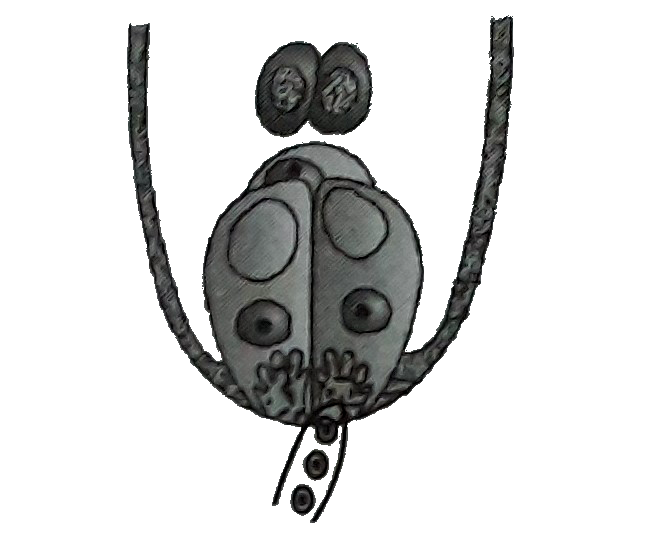
Draw diagram of embryo sac and label the following:
(i) Egg cell (ii) Polar Nuclei (iii) Synergids
Step-by-Step Solution:
1. Draw the Outline of the Embryo Sac: Start by sketching a long oval shape to represent the embryo sac. This will serve as the container for the various components you need to label.
2. Identify the Micropilar and Chalazal Ends: Label one end of the oval as the "Micropilar End" and the opposite end as the "Chalazal End". The micropilar end is where the egg cell and synergids are located, while the chalazal end contains the antipodal cells.
3. Draw the Egg Cell: At the micropilar end, draw a small circle to represent the egg cell. Label this circle as "Egg Cell".
4. Draw the Synergids: Next to the egg cell, draw two small oval shapes (or circles) on either side of the egg cell to represent the synergids. Label these as "Synergids".
5. Draw the Central Cell: In the center of the embryo sac, draw a larger oval shape to represent the central cell. This is where the polar nuclei are located.
6. Draw the Polar Nuclei: Inside the central cell, draw two small circles close to each other to represent the polar nuclei. Label these circles as "Polar Nuclei".
7. Draw Antipodal Cells: At the chalazal end of the embryo sac, draw three small circles to represent the three antipodal cells. Label these as "Antipodal Cells".
8. Final Labeling: Ensure all parts are clearly labeled: Egg Cell, Synergids, Polar Nuclei, and Antipodal Cells.
Diagram Summary:
- Micropilar End: Egg Cell and Synergids
- Central Cell: Polar Nuclei
- Chalazal End: Antipodal Cells
- Micropilar End: Egg Cell and Synergids
- Central Cell: Polar Nuclei
- Chalazal End: Antipodal Cells
Topper's Solved these Questions
- SEXUAL REPRODUCTION IN FLOWERING PLANTSBOOK - MODERN PUBLICATIONCHAPTER - SEXUAL REPRODUCTION IN FLOWERING PLANTSEXERCISE - Revison Exercises (VI. Long Answer Type Questions)
25 Videos
- SEXUAL REPRODUCTION IN FLOWERING PLANTSBOOK - MODERN PUBLICATIONCHAPTER - SEXUAL REPRODUCTION IN FLOWERING PLANTSEXERCISE - Competition File (Objective Type Questions (MCQ))
89 Videos
- SEXUAL REPRODUCTION IN FLOWERING PLANTSBOOK - MODERN PUBLICATIONCHAPTER - SEXUAL REPRODUCTION IN FLOWERING PLANTSEXERCISE - Revison Exercises (B. (Questions From Cbse Board Examinations)
19 Videos
- REPRODUCTIVE HEALTH BOOK - MODERN PUBLICATIONCHAPTER - REPRODUCTIVE HEALTH EXERCISE - CHAPTER PRACTICE TEST (SECTION D (LONG ANSWER QUESTION))
2 Videos
- STRATEGIES FOR ENHANCEMENT IN FOOD PRODUCTIONBOOK - MODERN PUBLICATIONCHAPTER - STRATEGIES FOR ENHANCEMENT IN FOOD PRODUCTIONEXERCISE - CHAPTER PRACTICE TEST
15 Videos
Similar Questions
(a) Draw an L.S. of pistil showing pollen tube entering into the embryo sac. Label the following :
(i) Nucellus (ii) Antipodals
(iii) Synergids (iv) Micropyle
(b) Write the functions of the following :
(i) Synergids (ii) Micropyle
View SolutionDraw a diagram of human excretory system and label the following :
(i) Urinary bladder
(ii) Left kidney
(iii) Left ureter
View Solution(a) Draw a diagram of a mature embryo sac of an angiosperm and label the following parts in it :
(i) Filiform apparatus (ii) Synergids
(iii) Central cell (iv) Egg cell
(v) Polar nuclei (vi) Antipodals
(b) Write the fate of egg cell and polar nuclei after fertilization.
View SolutionDraw diagram of a germinating seed and label the following parts of it:
(i) Part that gives rise to shoot system
(ii) Part that gives rise to root system.
(iii) Part that contains stored food.
View SolutionDraw a diagram of internal structure of human heart and label the following:
(i) Mitral valve (ti) Eustachius valve (iii) Chordae tendinae (iv) Pulmonary arch.
View SolutionDraw a well labelled diagram of embryo sac.
View SolutionIn the figure given below label the following parts-male gametes, egg cell, polar nuclei, synergid and pollen tube

View SolutionDraw the labelled diagram of the following : (i) gram seed (ii) V.S. of maize seed.
View SolutionDraw a diagram of an animal cell annd label the various parts.
View SolutionEgg cell, Polar nuclei,Pollen tube, Antipodals
View Solution
