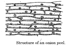
Draw the diagram of an onion peel as shown by the compound microscope.Solution in Punjabi
Topper's Solved these Questions
- BOOK - NAND LAL PUBLICATIONCHAPTER - THE FUNDAMENTAL UNIT OF LIFEEXERCISE - EXERCISE11videos
THE FUNDAMENTAL UNIT OF LIFE
- BOOK - NAND LAL PUBLICATIONCHAPTER - NATURAL RESOURCESEXERCISE - ADDITIONAL QUESTIONS18videos
NATURAL RESOURCES
- BOOK - NAND LAL PUBLICATIONCHAPTER - TISSUESEXERCISE - ADDITIONAL QUESTIONS6videos
TISSUES
Similar Questions
Make a comparison and write down ways in which plant cells are different from animal cells.
01:50View SolutionWhat two things are essential for existence of life on the earth?
01:52View SolutionAre plants and animal cells similar?
View SolutionTo prepare stained temporary mounts of (a) onion peel, and (b) human cheek cells and to record observations and draw their labelled diagram. The cels are first focussed in microscope under
03:39View SolutionWhat type of cells will you observe in an onion peel examined under a microscope ?
View SolutionWhat type of cells with you observe in an onion peel examined under a microscope ?
03:06View SolutionDraw a ray diagram for the formation of image by a compound microscope.
View SolutionOnion peel cells are
View SolutionA student is observing the temporary mount of a leaf peel under a microscope. Draw labelled diagram of the structure (of stomata) as seen under the microscope.
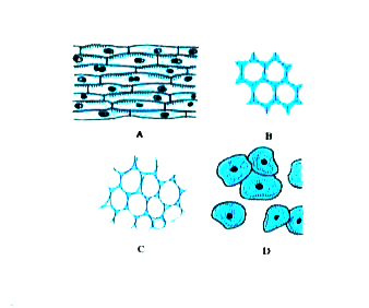
View SolutionWhich one of the following diagram depicts the structure of onion peel?

The correct answer is ...02:40View SolutionAmit is observing temporary mount of an onion peel stained with safranin under a microscope. The colour of the cell wall appears...
03:30View SolutionThe cellular component NOT seen while observing the slide of an onion peel under a compound microscope is...
03:12View SolutionDraw the ray diagram of image formation in case of compound microscope
03:36View SolutionWhich one of the following statements is correct about the onion peel cells and cheek cells that we observed under microscope?
03:53View SolutionDraw a ray diagram to show the formation of final image by a compound microscope. Find an expression for magnification of an image formed by a compound microscope.
View SolutionDraw the ray diagram for the formation of image by a compound microscope.
View Solution


