Draw the structure of a neuron and label it.
To draw the structure of a neuron and label its parts, follow these steps:
1. Draw the Cell Body (Soma):
- Start by drawing a large circle or oval shape in the center of your paper. This represents the cell body of the neuron, which contains the nucleus and organelles.
2. Add the Nucleus:
- Inside the cell body, draw a smaller circle to represent the nucleus. Label this as "Nucleus".
3. Draw Dendrites:
- From the cell body, draw several branching lines extending outward. These are the dendrites, which receive impulses from other neurons. Label these as "Dendrites".
4. Draw the Axon:
- From one side of the cell body, draw a long, thin line extending outward. This is the axon, which transmits impulses away from the cell body. Label this as "Axon".
5. Add the Myelin Sheath:
- Along the axon, draw a series of segmented lines or ovals to represent the myelin sheath, which insulates the axon and speeds up impulse transmission. Label this as "Myelin Sheath".
6. Draw the Nodes of Ranvier:
- Between the segments of the myelin sheath, leave small gaps. These gaps are called the Nodes of Ranvier. Label these as "Node of Ranvier".
7. Draw Axon Terminals:
- At the end of the axon, draw small branches or knobs. These are the axon terminals, which release neurotransmitters to communicate with the next neuron. Label this as "Axon Terminals".
8. Add Schwann Cells:
- Surrounding the myelin sheath, you can draw small cells or ovals to represent Schwann cells, which are responsible for the formation of the myelin sheath. Label these as "Schwann Cells".
Final Labeling:
- Ensure all parts are clearly labeled: Dendrites, Nucleus, Cell Body, Axon, Myelin Sheath, Nodes of Ranvier, Axon Terminals, and Schwann Cells.
- Start by drawing a large circle or oval shape in the center of your paper. This represents the cell body of the neuron, which contains the nucleus and organelles.
- Inside the cell body, draw a smaller circle to represent the nucleus. Label this as "Nucleus".
- From the cell body, draw several branching lines extending outward. These are the dendrites, which receive impulses from other neurons. Label these as "Dendrites".
- From one side of the cell body, draw a long, thin line extending outward. This is the axon, which transmits impulses away from the cell body. Label this as "Axon".
- Along the axon, draw a series of segmented lines or ovals to represent the myelin sheath, which insulates the axon and speeds up impulse transmission. Label this as "Myelin Sheath".
- Between the segments of the myelin sheath, leave small gaps. These gaps are called the Nodes of Ranvier. Label these as "Node of Ranvier".
- At the end of the axon, draw small branches or knobs. These are the axon terminals, which release neurotransmitters to communicate with the next neuron. Label this as "Axon Terminals".
- Surrounding the myelin sheath, you can draw small cells or ovals to represent Schwann cells, which are responsible for the formation of the myelin sheath. Label these as "Schwann Cells".
- Ensure all parts are clearly labeled: Dendrites, Nucleus, Cell Body, Axon, Myelin Sheath, Nodes of Ranvier, Axon Terminals, and Schwann Cells.
Topper's Solved these Questions
- TISSUES BOOK - CENGAGE BIOLOGYCHAPTER - TISSUES EXERCISE - OLYMPIAD AND NTSE LEVEL EXERCISES
9 Videos
- TISSUES BOOK - CENGAGE BIOLOGYCHAPTER - TISSUES EXERCISE - CHALLENGING EXERCISE
2 Videos
- TISSUES BOOK - CENGAGE BIOLOGYCHAPTER - TISSUES EXERCISE - MANDATORY EXERCISE (EXERCISE SET II) MCQs
61 Videos
- IMPROVEMENT IN FOOD RESOURCES : PLANTSBOOK - CENGAGE BIOLOGYCHAPTER - IMPROVEMENT IN FOOD RESOURCES : PLANTSEXERCISE - CHALLENGING EXERCISE
4 Videos
- WORLD OF MICROBES BOOK - CENGAGE BIOLOGYCHAPTER - WORLD OF MICROBES EXERCISE - CHALLENGING EXERCISE
3 Videos
Similar Questions
Knowledge Check
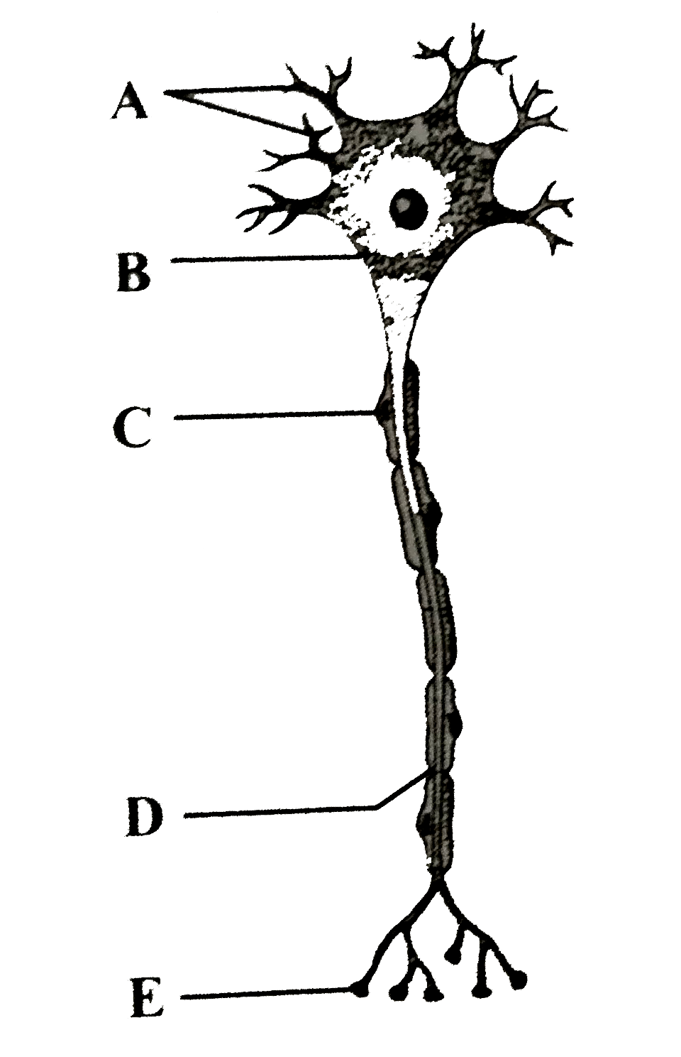
The given figure shows the structure of a neuron.Select the option that correctly identifies the parts labelled as A to E.
 AABCDENerve fibresCytonSchwann cellNode of RanvierSynaptic knobBABCDEDendritesCytonSchwann cellNode of RanvierSynaptic knobCABCDEDendritesNerve cellSchwann cellSynaptic knobNode of RanvierDABCDEAxonsCytonNerve cellNode of RanvierSynaptic knob
Draw the structure of heart and label it.
View Solution(a) Draw the structure of Neuron and explain its function.
(b) How does Phototropism occur in Plants?
View SolutionDraw the sketch of a neuron and label all the components.
View SolutionDraw a diagram to show the structure of a neuron with myelinated axon and label any six parts in it .
View SolutionDraw a suitable diagram for the structure of the atmosphere and label it and describe it.
View SolutionStructure Of Neurons
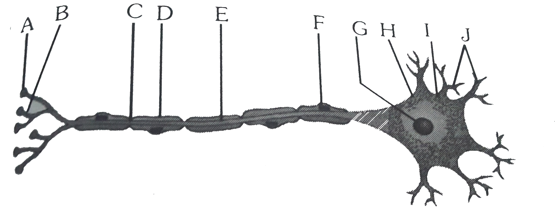
View SolutionGiven below is a structure of a neuron label the parts A to J

View Solution
