Give a labelled figure showing the structure of an animal cell or of a cell as shown by a an electron microscope.
Step-by-Step Solution for the Structure of an Animal Cell
1. Understanding the Animal Cell:
- An animal cell is a eukaryotic cell that lacks a cell wall and is surrounded by a cell membrane. It contains various organelles that perform specific functions.
2. Using an Electron Microscope:
- To observe the detailed structure of an animal cell, an electron microscope is used. This type of microscope provides high-resolution images of the cell's organelles.
3. Drawing the Structure:
- Begin by sketching the outline of the animal cell. The shape is usually irregular and roundish.
- Draw the outermost layer as the Cell Membrane.
4. Labeling the Organelles:
- Cytoplasm: Fill the inside of the cell with a gel-like substance, which is the cytoplasm where the organelles are suspended.
- Nucleus: Draw a large circle within the cell to represent the nucleus, which contains the genetic material (chromosomes).
- Nucleolus: Inside the nucleus, draw a smaller circle to represent the nucleolus.
- Mitochondria: Draw several oval shapes with inner folds to represent mitochondria, known as the powerhouse of the cell.
- Golgi Apparatus: Sketch a series of stacked, flattened membranes to represent the Golgi apparatus.
- Lysosomes: Draw small circular shapes to represent lysosomes, which are involved in cellular digestion.
- Vacuoles: Optionally, you can draw small vacuoles, which may be present in some animal cells.
- Nuclear Membrane: Draw a double membrane around the nucleus to indicate the nuclear membrane.
- Chromosomes: Inside the nucleus, draw squiggly lines to represent chromosomes.
5. Finalizing the Figure:
- Ensure all organelles are clearly labeled with arrows pointing to them. Use clear and legible text for the labels.
- An animal cell is a eukaryotic cell that lacks a cell wall and is surrounded by a cell membrane. It contains various organelles that perform specific functions.
- To observe the detailed structure of an animal cell, an electron microscope is used. This type of microscope provides high-resolution images of the cell's organelles.
- Begin by sketching the outline of the animal cell. The shape is usually irregular and roundish.
- Draw the outermost layer as the Cell Membrane.
- Cytoplasm: Fill the inside of the cell with a gel-like substance, which is the cytoplasm where the organelles are suspended.
- Nucleus: Draw a large circle within the cell to represent the nucleus, which contains the genetic material (chromosomes).
- Nucleolus: Inside the nucleus, draw a smaller circle to represent the nucleolus.
- Mitochondria: Draw several oval shapes with inner folds to represent mitochondria, known as the powerhouse of the cell.
- Golgi Apparatus: Sketch a series of stacked, flattened membranes to represent the Golgi apparatus.
- Lysosomes: Draw small circular shapes to represent lysosomes, which are involved in cellular digestion.
- Vacuoles: Optionally, you can draw small vacuoles, which may be present in some animal cells.
- Nuclear Membrane: Draw a double membrane around the nucleus to indicate the nuclear membrane.
- Chromosomes: Inside the nucleus, draw squiggly lines to represent chromosomes.
- Ensure all organelles are clearly labeled with arrows pointing to them. Use clear and legible text for the labels.
Topper's Solved these Questions
- CELL : THE UNIT OF LIFE BOOK - PRADEEPCHAPTER - CELL : THE UNIT OF LIFE EXERCISE - ANALYTICAL QUESTIONS WITH ANSWERS
20 Videos
- CELL : THE UNIT OF LIFE BOOK - PRADEEPCHAPTER - CELL : THE UNIT OF LIFE EXERCISE - PRACTICE QUESTIONS(MULTIPLE CHOICE QUESTIONS )
156 Videos
- CELL : THE UNIT OF LIFE BOOK - PRADEEPCHAPTER - CELL : THE UNIT OF LIFE EXERCISE - ADDITIONAL QUESTIONS (SHORT ANSWER QUESTIONS)
111 Videos
- BREATHING AND EXCHANGE OF GASES BOOK - PRADEEPCHAPTER - BREATHING AND EXCHANGE OF GASES EXERCISE - NOTABLE QUESTIONS
1 Videos
- CELL CYCLE AND CELL DIVISION BOOK - PRADEEPCHAPTER - CELL CYCLE AND CELL DIVISION EXERCISE - Assertion-Reason type questions
18 Videos
Similar Questions
Knowledge Check
Cell Cycle of an ordinary animal cell -
A2n Mitosis −−−−−−−→n Fertilization −−−−−−−−−→2n Meiosis −−−−−−−−→2nBn Meiosis −−−−−−−→2n Fertilization −−−−−−−−−→2n Mitosis −−−−−−−→nC2n Meiosis −−−−−−−−→n Fertilization −−−−−−−−−→2n Mitosis −−−−−−−−−→2nD2n Fertilization −−−−−−−−−−−→(n) Mitosis −−−−−−→2n Meiosis −−−−−−−−→n
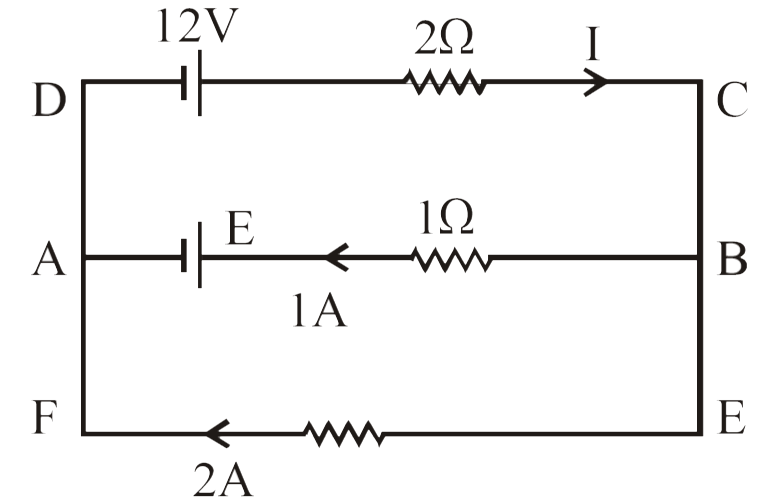
Find emf E of the cell as shown in figure.

View SolutionDraw a labelled diagram of the microscopic structure of a human sperm.
View SolutionName two structures found only in animal cells and not in plant cells.
View SolutionName two structures found only in animal cells and not in plant cells.
View SolutionMost cells are microscopic.
View SolutionWhy animal cells do not show plasmolysis?
View SolutionWith the help of well labelled diagrams describe the structure of a typically bacterial cell.
View Solution
