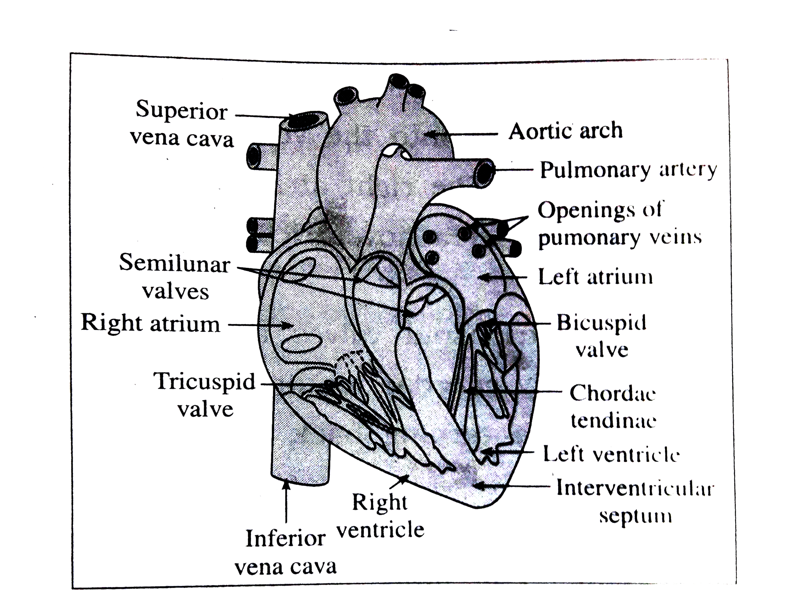
Sketch and label internal view of heart.
Text Solution

The heart shows four chambers with two atria and two ventricles.
I. Atria :
I. Right atrium :
(1) Interatrial septum separates two atria which are thin walled receiving chambers on the upper side.
(2) The right atrium receives deoxygenated blood from upper part of body through superior vena cava and from the lower part of the body by inferior vena cava. In the right atrium opens the coronary sinus which brings blood from the heart muscles.
(3) Eustachian valve guards the opening of inferior vena cava while opening of coronary sinus is guarded by Theebesian valve.
(4) On the right of interatrial septum is seen an oval depression called the fossa ovalis.
2. Left atrium :
The oxygenated blood from the lungs is brought into left atrium through four openings of pulmonary veins.
II. Ventricles :
(1) Two thick walled ventricles are separated from each other by interventricular septum. They are distributing chambers.
(2) The inner surface of the ventricle is thrown into a series of folds called columnae carnae or trabeculae carnae.
(3) Each atrium opens into the ventricle of its side through atrioventricular aperture. The right atrioventricular is guarded by tricuspid vlve. While the left atrioventricular valve by biscuspid valve or mitral valve.
(4) Bicuspid and tricuspid valves are attached to papillary muscles of ventricles by chordae tendinae. The chordae tendinae prevent the valves from turning back into the atria during the contraction of ventricles.
(5) From the right ventricle arises pulmonary trunk which carries deoxygenated blood to lungs for oxygenation. From the left ventricle arise systemic aorta which distributes oxygenated blood to all parts of the body.
(6) Pulmonary aorta and systemic aorta have three semilunar valves at the base which prevent backward flow of blood during ventricular diastole.
|
Topper's Solved these Questions
- LONG ANSWER QUESTIONSBOOK - NAVNEET PUBLICATION - MAHARASHTRA BOARDCHAPTER - LONG ANSWER QUESTIONSEXERCISE - Excretion and Osmoregulation
2 Videos
- LONG ANSWER QUESTIONSBOOK - NAVNEET PUBLICATION - MAHARASHTRA BOARDCHAPTER - LONG ANSWER QUESTIONSEXERCISE - Control and Coodination
13 Videos
- LONG ANSWER QUESTIONSBOOK - NAVNEET PUBLICATION - MAHARASHTRA BOARDCHAPTER - LONG ANSWER QUESTIONSEXERCISE - Reproduction in Plants
9 Videos
- GIVE REASONS BOOK - NAVNEET PUBLICATION - MAHARASHTRA BOARDCHAPTER - GIVE REASONS EXERCISE - Organisms and Environment - II
2 Videos
- MATCH THE COLUMNS/MATCH THE PAIRS/COMPLETE THE TABLEBOOK - NAVNEET PUBLICATION - MAHARASHTRA BOARDCHAPTER - MATCH THE COLUMNS/MATCH THE PAIRS/COMPLETE THE TABLEEXERCISE - Chapter 19 : Organisms and Environment - II
3 Videos
Similar Questions
Sketch and label ventral view of human heart .
View SolutionSketch and label the lateral view of human brain.
View SolutionWhat is reflex arc? Draw a labelled diagram to show reflex arc on touching a very hot object.
View SolutionSketch and label T.S of vein
View SolutionSketch and label lytic cycle.
View SolutionSketch and label Malpighian body.
View SolutionSketch and label Malpighian body.
View SolutionSTRUCTURE OF NEPHRON
View SolutionDraw a labelled diagram of human heart.
View SolutionSketch and label the structure of chromosome.
View Solution
