The coats of eyeball are :
(1) sclera (2) Retina (3) Conjunctiva (4)Otolith membrane
1, 2 and 3 are correct
1 and 2 are correct
2 and 4 are correct
1 and 3 are correct
The correct Answer is:B
To solve the question regarding the coats of the eyeball, we will analyze each of the options provided:
1. Sclera: The sclera is the white outer layer of the eyeball. It is a tough and protective covering that maintains the shape of the eye and provides a surface for the attachment of the eye muscles. Thus, the sclera is indeed one of the coats of the eyeball.
2. Retina: The retina is a thin layer of tissue located at the back of the eye. It plays a crucial role in vision as it receives light that the lens has focused, converts it into neural signals, and sends these signals to the brain for visual recognition. Therefore, the retina is also a coat of the eyeball.
3. Conjunctiva: The conjunctiva is a thin, transparent membrane that covers the front of the eye and lines the inside of the eyelids. While it does provide protection and lubrication to the eye, it is not classified as a coat of the eyeball itself.
4. Otolith membrane: The otolith membrane is part of the inner ear and is involved in the sense of balance and equilibrium. It is not related to the structure of the eyeball and thus is not a coat of the eyeball.
Based on this analysis, the correct coats of the eyeball are:
- Sclera
- Retina
Therefore, the answer to the question is that the coats of the eyeball are (1) Sclera and (2) Retina.
Final Answer:
The coats of the eyeball are: (1) Sclera and (2) Retina.
- Sclera
- Retina
The coats of the eyeball are: (1) Sclera and (2) Retina.
Topper's Solved these Questions
- NEURAL CONTROL AND COORDINATIONBOOK - MODERN PUBLICATIONCHAPTER - NEURAL CONTROL AND COORDINATIONEXERCISE - COMPETITION FILE (OBJECTIVE TYPE QUESTIONS)(B. CBSE - PMT (MAIN EXAMINATION)QUESTIONS: )
6 Videos
- NEURAL CONTROL AND COORDINATIONBOOK - MODERN PUBLICATIONCHAPTER - NEURAL CONTROL AND COORDINATIONEXERCISE - COMPETITION FILE (OBJECTIVE TYPE QUESTIONS)(C. MATCHING TYPE QUESTIONS: )
4 Videos
- NEURAL CONTROL AND COORDINATIONBOOK - MODERN PUBLICATIONCHAPTER - NEURAL CONTROL AND COORDINATIONEXERCISE - REVISION EXERCISES (Long Answer Questions)
3 Videos
- MORPHOLOGY OF FLOWERING PLANTS BOOK - MODERN PUBLICATIONCHAPTER - MORPHOLOGY OF FLOWERING PLANTS EXERCISE - CASE BASED SHORT ANSWER TYPE QUESTIONS
2 Videos
- PHOTOSYNTHESIS IN HIGHER PLANTSBOOK - MODERN PUBLICATIONCHAPTER - PHOTOSYNTHESIS IN HIGHER PLANTSEXERCISE - CHAPTER PRACTICE TEST
9 Videos
Similar Questions
Let A={1,2,3,4},R={(2,2),(3,3),(4,4),(1,2)} be a relation on A. Then R is 1) reflexive 2) symmetric 3) transitive 4) Both 1 & 2
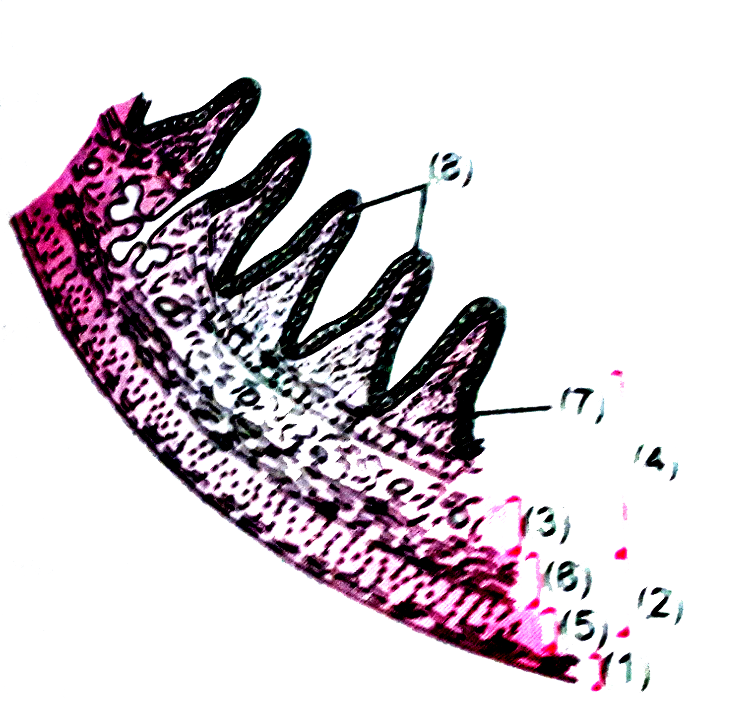
View SolutionFigure depicts microsopic structure of the intestinal wall of alimentary canal. Study the figure and answer the following question?
(i) The wall of alimentary canal consists of 4 main concentric coats labelled as 1,2,3 and 4. Name these coats.
(ii) The coat marked 2 has two layers marked as 5 and 6. Between these two layers is a network of nerve cells and para-sympathetic nerve fibres called..... It controls the .......
(iii) The coat marked 3 is highly vasucular connective tissue having another network of nerve cells and sympathetic nerve fibres called...... . it controls the secretion of ........
(iv) Name the parts labelled as 7 and 8

View Solution
Knowledge Check
Bipolar neurons occur in:
(1) Retina of eye (2) Olfactory epithelium (3) Inner ear (4) BrainA1, 2 and 3 are correctB1 and 2 are correctC2 and 4 are correctD1 and 3 are correctCochlea is divided into three chambers or spaces
1. Scala vestibuli 2. Scala media 3. Scala tympani
Basilar membrane and Reissner's membrane are respectively found betweenA(i) and (iii) and (i) & (ii)B(i), (ii) and (iii)C(ii) & (iii) and (i) & (iii)D(ii) and (iii) and (i) & (ii)Given the area of the retina
1. Macula 2. Fovea centralis 3. Optic disc 4. Periphery of the retina
Choose the arrangement that lists the areas according to the density of cones, starting with the area that has highest density of conesA2, 1, 4, 3B2, 4, 1, 3C1, 2, 3, 4D1, 3, 2, 4
The two squre faces of a rectangular dielectric slab (dielectric constant4.0) of dimensions 20cm xx20cm xx 1.0mm are matel -coated. Find the capacitance between the coated surfaces.
View SolutionWhat is related to Humility in the same way as Desire is related to
1)Cap
2)Tie
3)Coat
4)Cowan
5)None of these
View SolutionA cylindrical vessel, without lid, has to be tin-coated on its both sides. If the radius of the base is 70cm and its height is 1.4 m, calculate the cost of tin-coating at the rate of Rs. 3.50 per 1000cm2.
View SolutionA circular disc of which 2/3 part is coated with yellow and 1/3 part is with blue . It is rotated about its central axis with high velocity. Then it will be seen as
View SolutionA circular disc of which 2/3 part is coated with yellow and 1/3 part is with blue it is rotated about its central axis with high velocity then it will be seen as
View Solution
