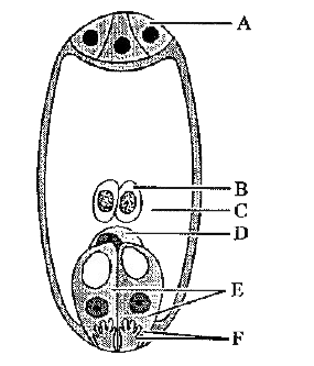
The diagram given below represents different cells of a typical angiospermic embryo sac labelled from A to F. Identify these cells and select the correct option.
A
A-Antipodals, B-Polar nuclei, C-Central cell, D-Egg, E-Synergids, F-Filiform apparatus
B
A-Antipodals, B-Central cell, C-Polar nuclei, D-Egg, E-Synergids, F-Filiform apparatus
C
A-Filiform apparatus, B-Polar nuclei, C-Central cell, D-Synergids, E-Egg, F-Antipodals
D
A-Filiform apparatus, B-Central cell, C-Polar nuclei, D-Antipodals, E-Egg: F-Synergids
Text Solution
The correct Answer is:A
Three cells grouped together at the micropylar end constitute the egg apparatus which, in turn, consists of two Synergids (labelled as E) and one egg cell (labelled as D). The special cellular thickenings at the micropylar tip of synergids is the filiform apparatus (labelled as F). Antipodals (labelled as A) are the three cells which are at the chalazal end. The large central cell (labelled as C) has two polar nuclei (labelled as B).
|
Similar Questions
Knowledge Check
What could be the number of cells in a typical angiosperm embryo sac?
A5B6C7D8The different cells of embryo sac are
ACentral cell and antipodal cellsBAntipodal cells and egg apparatusCCentral cell and egg apparatusDEgg apparatus, central cell and antipodal cells.Given figure represents the ultrastructure of a typical cyanobacterial cell. Identify the different parts and select the correct option for A and B
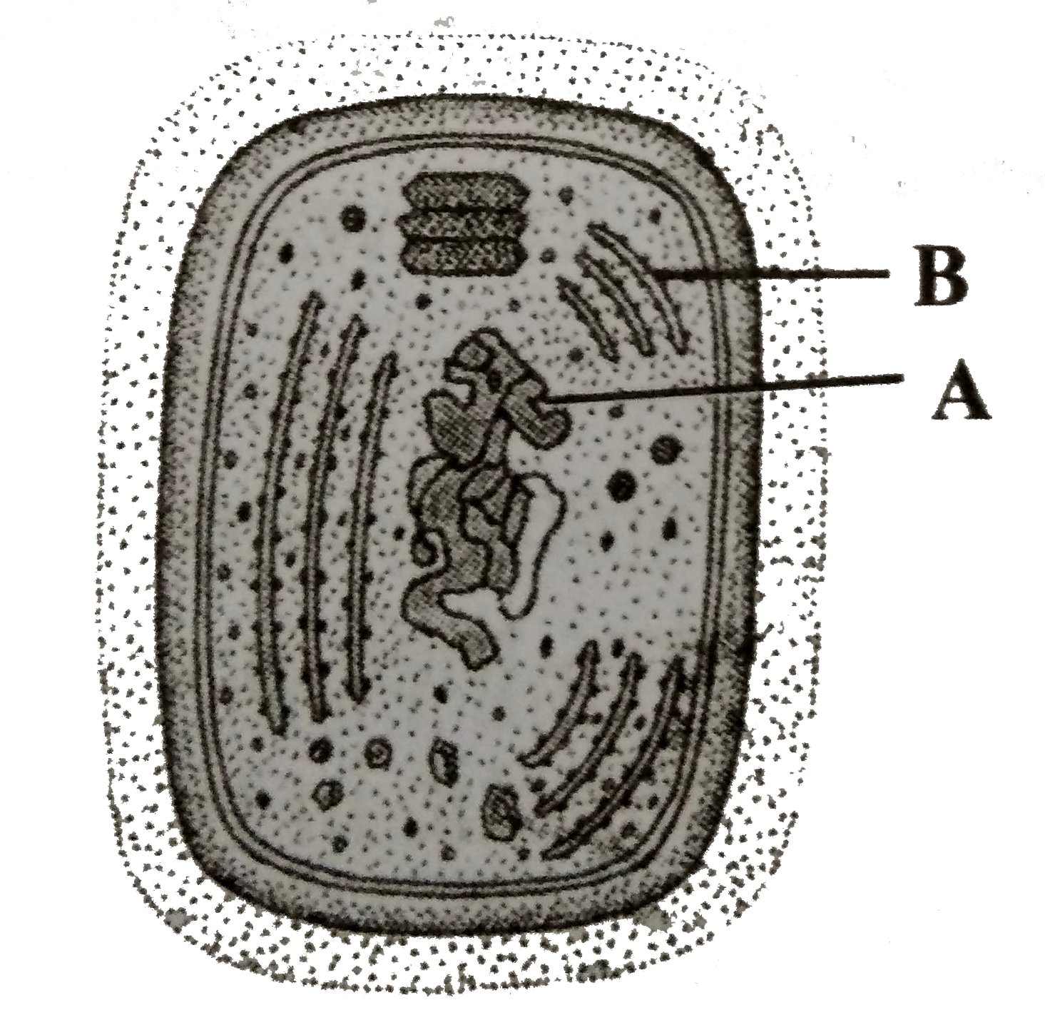 AAB(a)Naked DNAThylakoidBAB(b)ThylakoidNaked DNACAB(c)DNA+HistonesThylakoidDAB(d)DNA+Histone80S ribosomes
The different cells of embryo sac are
View SolutionIdentify parts labeled A and B in the given diagram and selecte the correct option.
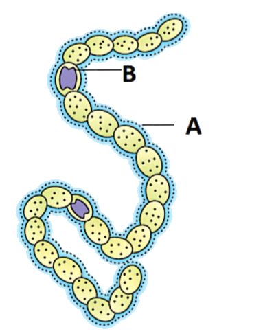
View SolutionIdentify parts labeled A and B in the given diagram and selecte the correct option.

View SolutionIdentify parts labelled A, B, and C in the given diagram and select the correct option.
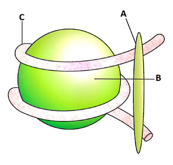
View SolutionIdentify parts labelled A, B, and C in the given diagram and select the correct option.

View Solution

