The given figure are related to castor seeds, identify A to D respectively
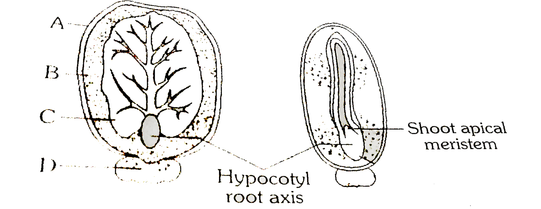
Seed coat, endosperm cotyledon and caruncle
Seed coat, cotyledon, endosperm and caruncle
Seed coat, endosperm, caruncle and cotyledon
Endosperm, seed coat, cotyledon and caruncle
The correct Answer is:A
Step by step video, text & image solution for The given figure are related to castor seeds, identify A to D respectively by Biology experts to help you in doubts & scoring excellent marks in Class 12 exams.
Updated on:21/07/2023Topper's Solved these Questions
- REPRODUCTION IN ORGANISMSBOOK - ERRORLESS CHAPTER - REPRODUCTION IN ORGANISMSEXERCISE - Reproduction In Organisms
116 Videos
- SMOKING,ALCOHOLISM,DRUG ADDITION, MENTAL HEALTH AND COMMUNITY HEALTHBOOK - ERRORLESS CHAPTER - SMOKING,ALCOHOLISM,DRUG ADDITION, MENTAL HEALTH AND COMMUNITY HEALTHEXERCISE - Smoking,Alcoholism,Drug Addition, Mental Health And Community Health
88 Videos
Similar Questions
Knowledge Check
The given figure refers to a T.S of anther. Identify A to E respectively
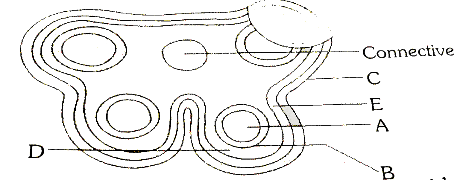 ASporogenous tissue, tapetum, middle layer, epidermis, endotheciumBSporogenous tissue, epidermis, middle layer, tapetum, endotheciumCSporogenous tissue, epidermis, tapetum, middle layer, endotheciumDSporogenous tissue, tapetum, epidermis, middle layer, endothecium
Seed of Castor is
ANonedospermic albuminousBEndospermic albuminousCEndospermic exalbuminousDNonendospermic albuminousSee the figure given below and identify A to D respectively
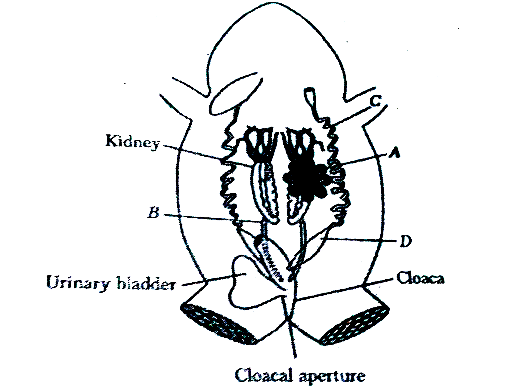
 AA- seminal vesicle, B- Testis, C- Prostate gland, D- Accessory glandBA- Testis, B- Seminal vesicle, C- Prostate gland, D- Accessory glandCA- Seminal vesicle, B- Testis, C- Accessory gland, D- Prostate glandDA- Testis, B-Seminal vesicle, C- Accessory gland, D- Prostate gland
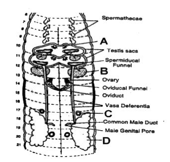
Identify A to D in the figure -
View SolutionThe given figure indicates the stages of seed gremination
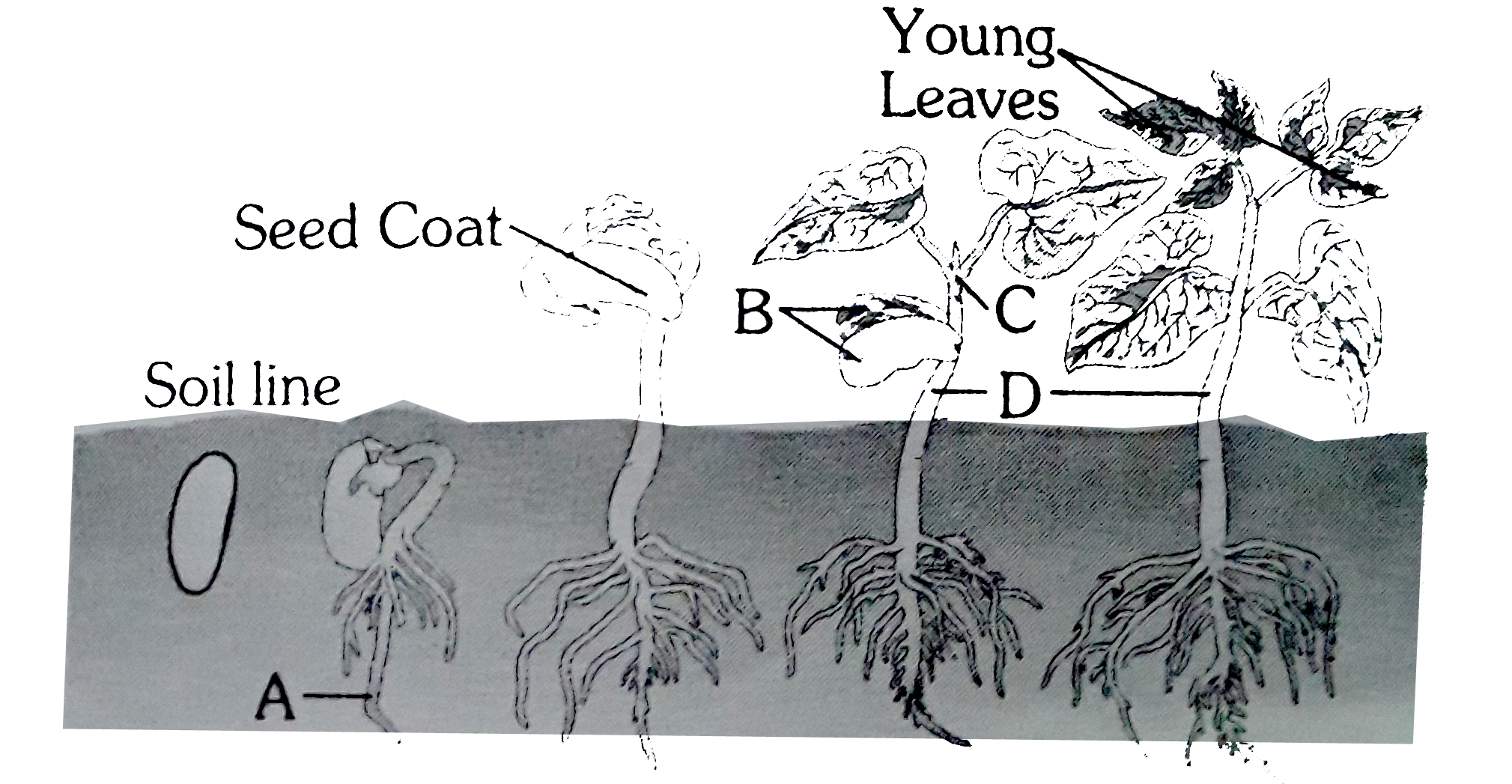
Identify A,B,C and D respectivelty
View SolutionThe given figure is related to diagrammatic representation of internal organs of frog. Identify A to E
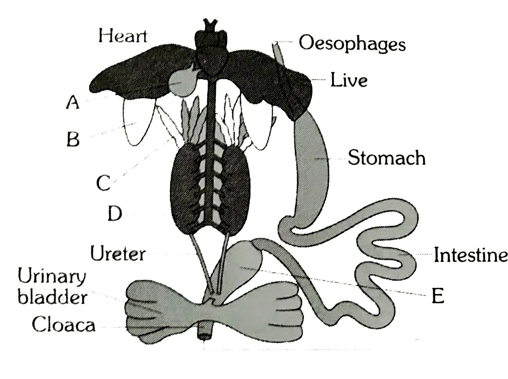
View SolutionThe given figure represents some stages in microgametogenesis identify A, B and C respectively

View SolutionThe given figure is related with myosin monomer (meromyosis). Identify the parts labelled from A to C and select the correct option .
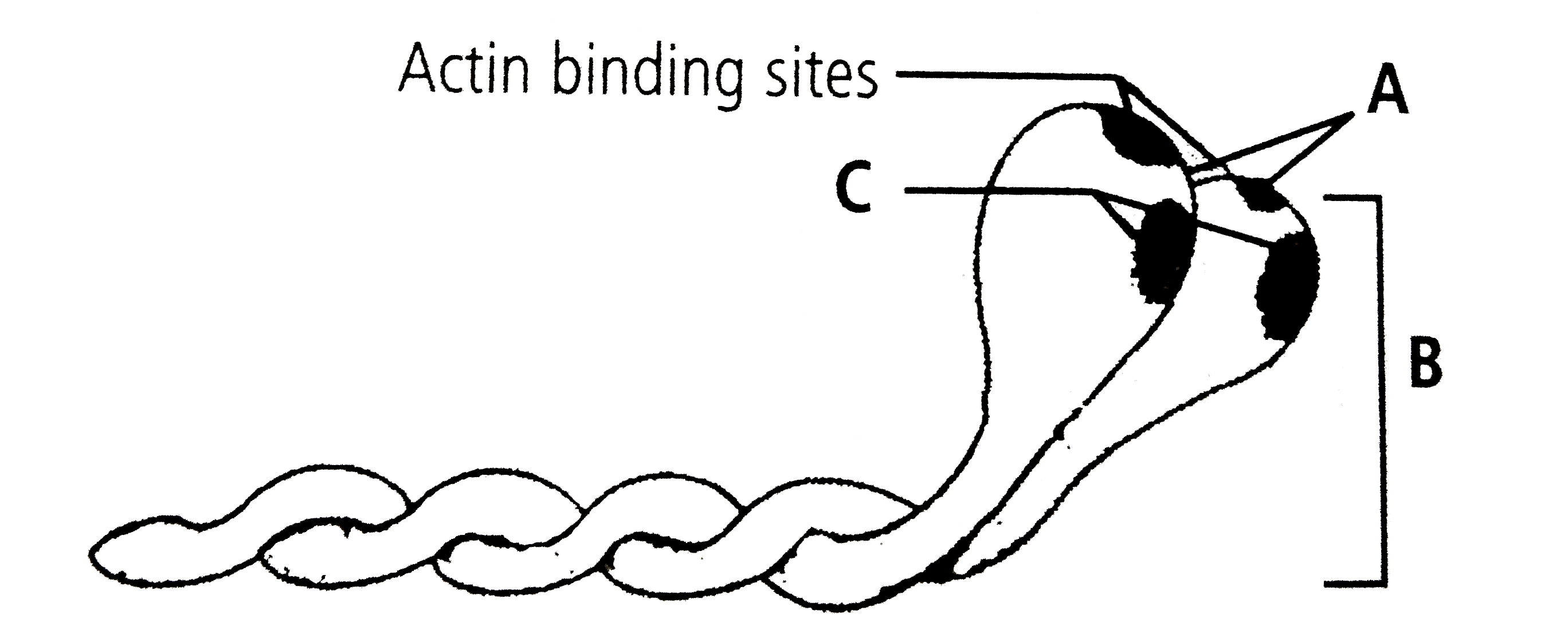
View Solution


