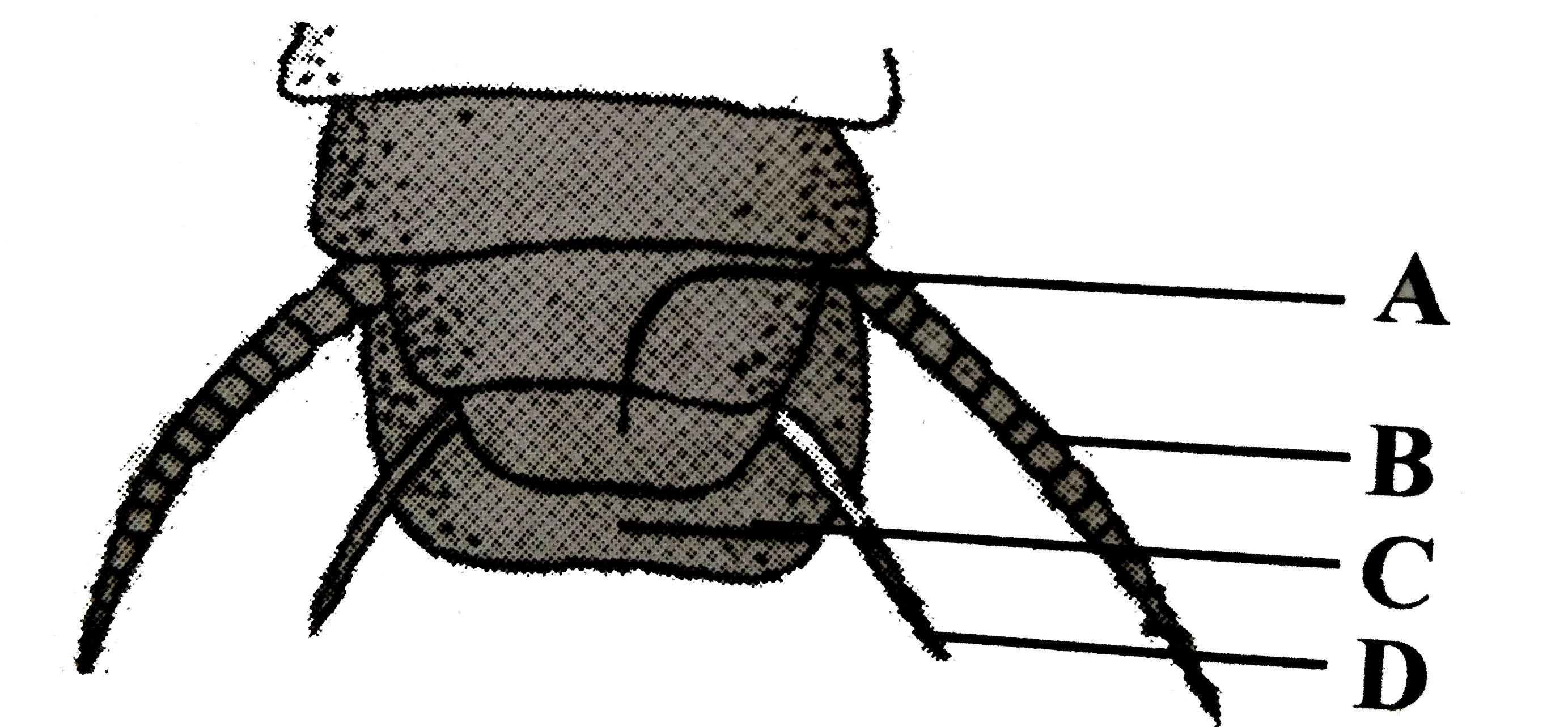
The given figure represents a mammalian spermatozoon.

(a) Name the parts labelled A-F.
(b) Explain briefly the functions of the parts A, B and D.
( c) What events occur in the egg immediately following the entry of the spermatozoon?
(d) What is place of fertilisation in female reproductive tract?
(e) At which stage does implantation take place?
(a) A-Acrosome, B-Nucleus, C-Distal centriole, D-Mitochondrial sheath, E-Axial Filament, F-Tail.
(b) (i) Function of part A (Acrosome): Acrosome has tissue dissolving enzymes or sperm lysins and help spermatozoon to penetrate throuth egg membrane. These are:
(1) Hyaluronidase-for dissolution of cementing substance amongst corona cells. (2) Corona penetrating enzyme-for passage of sperm through corona (3) Acrosin or Zona Lysin-for passage through zona pellucida.
(ii) Function of part B (Nucleus): It contributes haploid set of chromosomes to the ovum, so that the zygote formed restores diploid chromosomal complement.
(iii) Function of part D (Mitochondrial sheath): It provides energy to the sperm for the movement inside female genital tract to reach the ovum.
( c) (i) Cortical reaction results in formation of fertilisation membrane.
(ii) Completion of second maturation division results in release of second polar body and formation of secondary oocyte or ovum.
(d) Ampulla-Isthmus junction.
(e) Blastocyst stage.
Topper's Solved these Questions
- BOOK - ICSECHAPTER - HUMAN REPRODUCTIONEXERCISE - PRACTICE QUESTIONS (VERY SHORT ANSWER TYPE QUESTIONS)28videos
HUMAN REPRODUCTION
- BOOK - ICSECHAPTER - HUMAN REPRODUCTIONEXERCISE - PRACTICE QUESTIONS (SHORT ANSWER TYPE-I QUESTIONS)17videos
HUMAN REPRODUCTION
- BOOK - ICSECHAPTER - HUMAN HEALTH AND DISEASE EXERCISE - COMPETITION CORNER (OBJECTIVE TYPE QUESTIONS) (Assertion and Reason Type Questions (For AIIMS aspirants))28videos
HUMAN HEALTH AND DISEASE
- BOOK - ICSECHAPTER - ISC BOARD PAPER (2019 -BIOLOGY)EXERCISE - PART-II ( SECTION-C)12videos
ISC BOARD PAPER (2019 -BIOLOGY)
Similar Questions
Knowledge Check
- Question 1 - Select One
The given figure represents the histology of of a striated muscle. Identify the parts labelled as A, B, C and D, and select the correct option.
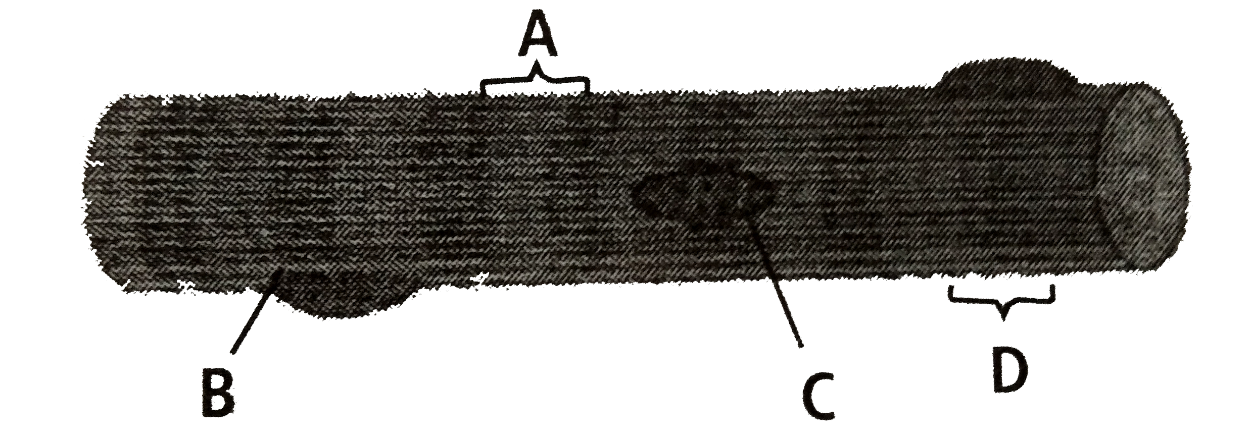 A(A)(B)(C)(D)(Sarcoplasm)(Sarcolemma)(Dark band)(Light band)B(A)(B)(C)(D)(Dark band)(Myofibril)(Nucleus)(Light band)C(A)(B)(C)(D)(Light band)(Myofibril)(Nucleus)(Dark band)D(A)(B)(C)(D)(Nucleus)(Dark band)(Light band)(Myofibril)
 - Question 2 - Select One
The given figure represents the histology of of a striated muscle. Identify the parts labelled as A, B, C and D, and select the correct option.
 A(A)(B)(C)(D)(Sarcoplasm)(Sarcolemma)(Dark band)(Light band)B(A)(B)(C)(D)(Dark band)(Myofibril)(Nucleus)(Light band)C(A)(B)(C)(D)(Light band)(Myofibril)(Nucleus)(Dark band)D(A)(B)(C)(D)(Nucleus)(Dark band)(Light band)(Myofibril)
 - Question 3 - Select One
The given figure is of human female reproductive system.
Identify the parts labelled as A, B, C and D and select the correct option.
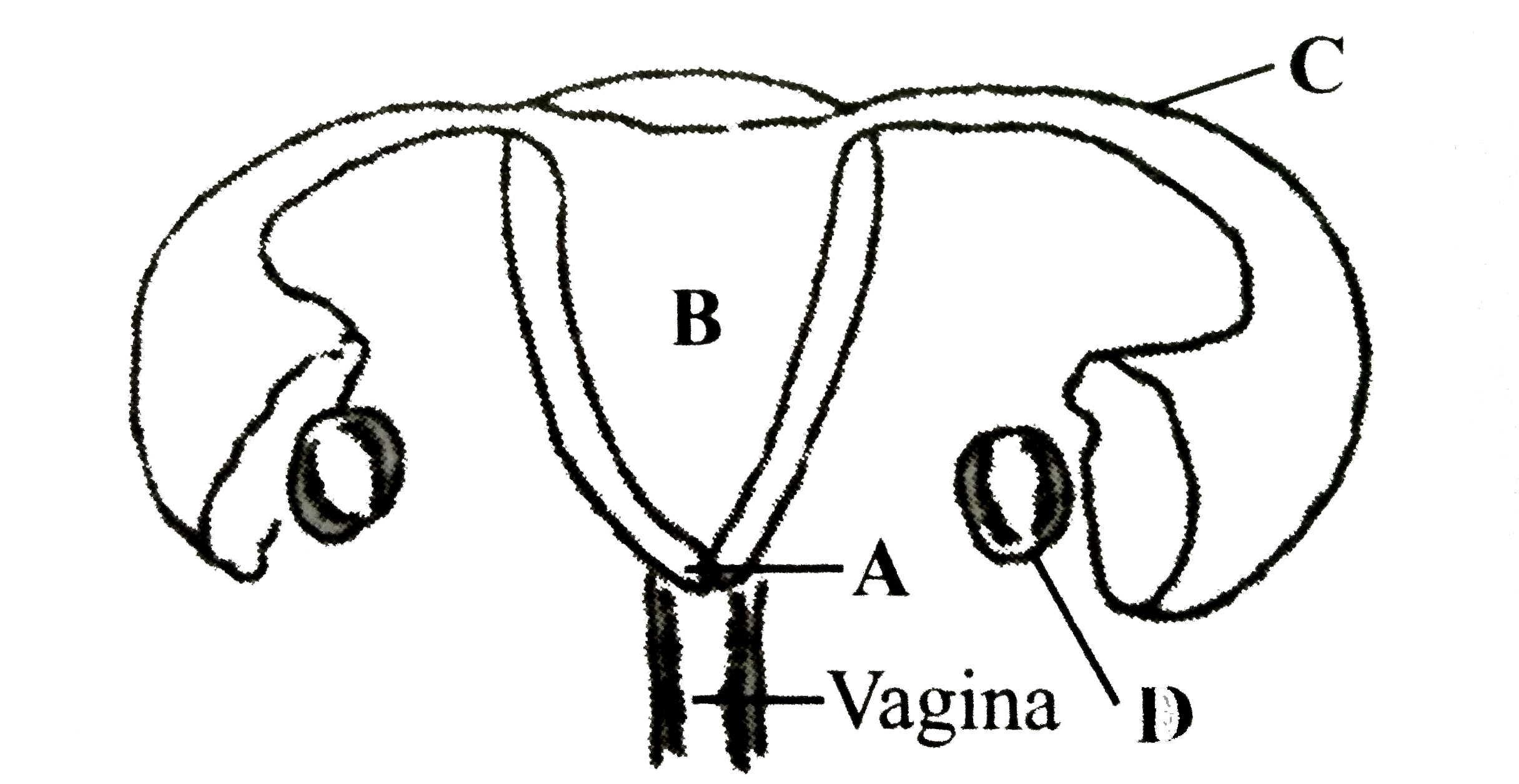 AA-Oviduct, B-Uterus, C-Ovarian ligament, D-OvaryBA-Cervix, B-Uterus, C-Ovary, D-TumourCA-Uterus, B-Uterine cavity, C-Oviducal funnel, D-OvaryDA-Cervix, B-Uterine cavity, C-Fallopian tube, D-Ovary
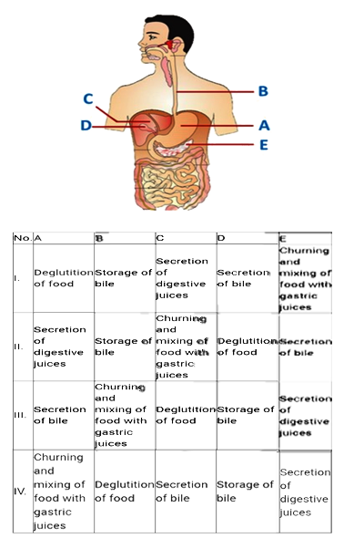
Identify the functions of the parts labelled as A, B,C,D and E in the given figure . Select the correct option after referring to the given table.
 View Solution
(a) Identify the parts A, B, C, D, E and E
(b) What are the major functions of A?
(c) List out the vegetative parts from the given figure.
(d) What is the function of C?View SolutionName the parts 1 to 5 of human female reproductive system.
(b) Name the part in which fertilisation takes place in this system.
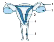 View Solution
The given figure is of human female reproductive system.
Identify the parts labelled as A, B, C and D and select the correct option.
 View Solution
Study the given figure of respiratory passage carefully and identify the parts labelled as A, B, C, D and E.
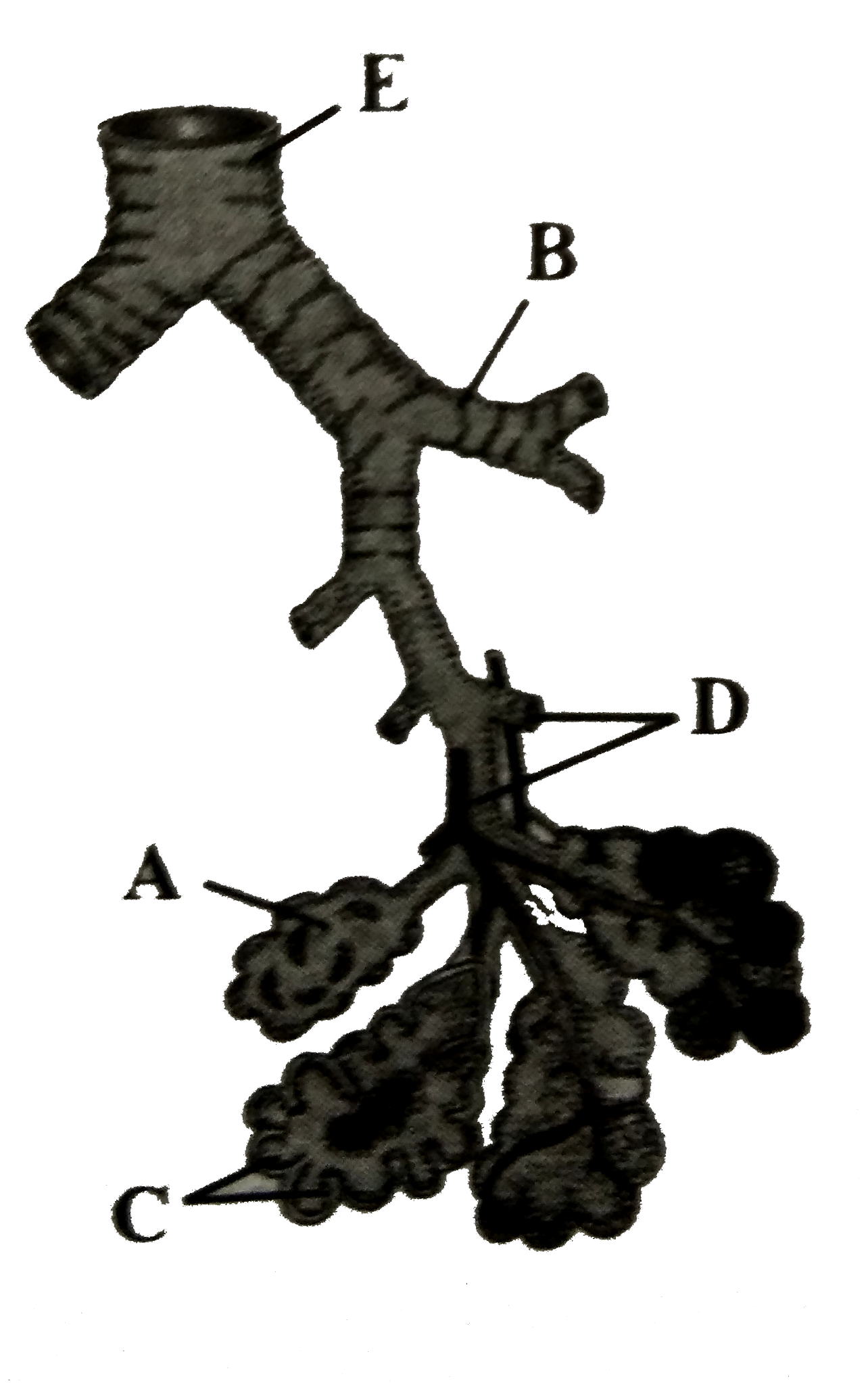 View Solution


