What are foetal membranes. Give their names. Mention the function of each membrane. Solution in Punjabi
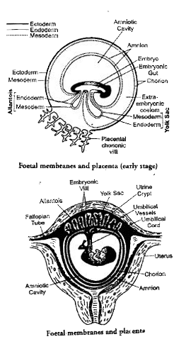
Embryonic or Foetal Membranes. Need for Foetal Membranes. An aquatic embryo is surrounded by water, which protects the Extra embryo, keeps it moist, removes wastes, and permits gas exchange. In land vertebrates (reptiles, birds and mammals), these functions are taken over by the extraembryonic membranes, some of which take Placental part in the formation of placenta in mammals. The placenta develops before the embryo because the embryo depends heavily on the mother.s body for its vital requirements. Four foetal membranes are formed : amnion, chorion allantois and yolk sac.

(i) Amnion. It is formed above the embryo. It Allantois consists of trophoblast inside and mesoderm outside. It encloses a fluid-filled space, the amniotic cavity. The amnion and the amniotic cavity enlarge and nearly surround the embryo. The embryo is suspended in the amniotic cavity by an umbilical cord. The latter is formed of the stalks of the yolk sac and allantois. The main blood vessels from the placenta reach the foetus through the umbilical cord. The amniotic fluid is derived from the foetal kidneys and bladder, i.e., it is foetal urine. It is completely replaced every 24 hours. It serves many functions. (i) It cushions the embryo, protecting it against bumps and against pressure of maternal organs, (ii) permits foetal movement, (iii) prevents bacterial infections, (iv) maintains a constant temperature and pressure, (v) protects the embryo from injury if the mother falls, (vi) allows development to proceed unhampered by the force of gravity, and (vii) the amniotic fluid absorbs shocks and prevents desiccation of the embryo.
Amniocentesis. The amniotic fluid contains foetal cells. These cells are the basis of prenatal test, called amniocentesis, for the sex of the foetus and for checking chromosomal defects in it. For this purpose, it is drawn out with a hypodermic needle and examined.
(ii) Chorion. It completely surrounds the embryo. It is composed of trophoblast outside and mesoderm inside. It protects the embryo and forms placenta for metabolic exchange between the foetus and the mother. In the later stages of pregnancy, the amnion expands to come in contact and partially fuse with the chorion.
(iii) Yolk Sac. It is formed below the embryo. It contains fluid, not yolk. The yolk sac is a vestigial organ inherited from the oviparous reptilian ancestors. Its wall first consists of trophoblast and endoderm. At this stage, it is called primary yolk sac (Fig. ???). Later, the wall of yolk sac comes to consist of endoderm and mesoderm. It is now called secondary yolk sac (Fig. ???). The yolk sac functions as the site of blood cell formation until about the 6th week, when the liver takes over this role. Thereafter, the yolk sac starts to shrink.
(iv) Allantois. It is a small sac that pushes out from the gut of the embryo near the yolk sac by the third week. It remains small and does not reach the chorion. However, it forms umbilical arteries and vein which grow up to the chorion to vascularise it. The allantois also forms blood cells. It consists of endoderm inside and mesoderm outside.
Topper's Solved these Questions
- BOOK - BETTER CHOICE PUBLICATIONCHAPTER - HUMAN REPRODUCTIONEXERCISE - SHORT ANSWER TYPE QUESTIONS (MOAT EXPECTED QUESTIONS)3videos
HUMAN REPRODUCTION
- BOOK - BETTER CHOICE PUBLICATIONCHAPTER - HUMAN HEALTH AND DISEASEEXERCISE - LONG ANSWER TYPE QUESTIONS (MOST EXPECTED QUESTIONS)12videos
HUMAN HEALTH AND DISEASE
- BOOK - BETTER CHOICE PUBLICATIONCHAPTER - MICROBES IN HUMAN WELFAREEXERCISE - LONG ANSWER TYPE QUESTIONS (MOST EXPECTED QUESTIONS)2videos
MICROBES IN HUMAN WELFARE
Similar Questions
What is role played by luteinizing hormone in males and females?
04:10View SolutionWrite the causative organism, mode of infection, symptoms and preventive measures of the disease pertussis.
01:22View SolutionExplain the role of microbes in the treatment of sewage.
02:17View SolutionFoetal membranes produced by trophoblast are
04:19View SolutionNumber of foetal membranes in humans is
05:02View SolutionWhich of the foetal membrane is directly connected with blood ?
01:41View SolutionName the two main foetal membrane?
03:47View SolutionWrite a short note about foetal membrane.
01:34View SolutionFoetal membrane which keeps the embryo shock proof is
02:32View SolutionThe foetal membrane that forms the basis of the umbilical cord is
01:38View Solution(a) Name the foetal membrane that provides a fluid medium to the developing embryo. (b) Mention its two functions.
03:26View SolutionThe inner most foetal membrane around the embryo immediately
02:09View SolutionName the foetal membrane that provides a fluid medium to the developing embryo. Mention its two functions.
02:50View SolutionName the extraembryonic membranes found in mammals. Give function of each.
02:16View SolutionMention the functions of the following: Cell membrane
03:39View SolutionDraw the diagram to show germ layers and foetal membranes.
02:56View Solution

