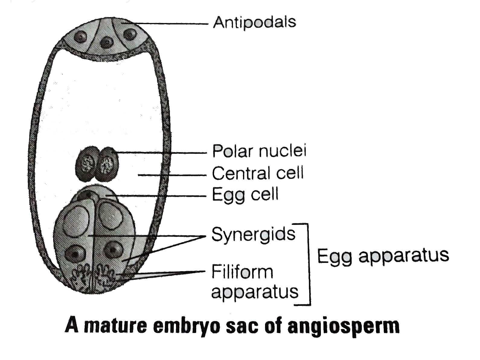
With a neat, labelled diagram, describe the parts of a mature angiosperm embryo sac. Mention the role of synergids.
Video Solution
Text Solution
After the 8-nucleate stage, cell walls are laid down leading to the organisation of the typical female gemetophyte or embryo sac.

Six of the eight nuclei are surrounded by cell walls and organised into cells. Three cells present towards the micropylar end grouped together, constitute the egg apparatus . The egg apperatus , in tum consists of two synergide and one egg cell.
Three cells of the chalazal end are called the antipodals . The large central cell is formed by the fusion of 2-polar nuclal .Thus , a typical angiospermic embryo sac, at maturity consisis of eight nuclei and seven cells . This embryo sac is formed from , the single megaspore, so it is called monosporic embryo sac.
Role of synergids
The synergids have special cellular thickenings at the micropylar tip called filiform apparatus, which play an important role in guiding the pollen tubes into the synergid.
|
Updated on:21/07/2023
Topper's Solved these Questions
Similar Questions
Knowledge Check
- Question 1 - Select One
What is not true for an angiospermic embryo sac?
AOne male gamete is discharged into it during ferilisationBIt is present within an ovuleCIt represents female gametophyteDIts formation is preceded by meiosis - Question 2 - Select One
The largest cell of the mature embryo sac is
Aantipodal cellsBsynergidsCcentral cellDegg cell
With a neat labelled diagram describe Wilfley table method.
View SolutionDraw a neat labelled diagram of nucleus.
View SolutionWith the help of a neat and labelled diagram, describe reflex arc.
View SolutionWith the help of a neat and labelled diagram, describe reflex arc.
View SolutionWith the help of a neat labelled diagram, describe the Geiger-Marsden experiment.
View SolutionDraw a labeled diagram of male reproductive system.
View Solution

