Write short notes on the following :
(a) Neural coordination
(b) Forebrain
(c) Midbrain
(d) Hindbrain
(e) Retina
(f) Ear ossicles
(g) Cochlea
(h) Organ or Corti
(i) Synapse
(a) Neural Coordination
The neural system provies rapid coordination among the orgnas of the body. This coordination is in the form of electric impulses and is quick and short lived. For example, during exercise, our body requires more oxygen and food. Hence, the breathing rate increases automatically and the beats faster. This leads to a faster supply of oxygenated blood to the muscles. Moreover, the cellular functions require regulation continuously. This funcions are carried out by the hormones. Hence, the neural system along with endocrine system control and coordinate the physiological processes.
(b) Forebrain
It is the main thinking party of the brain. It consists of cerebrum, thalamus, and hypothalamus.
Cerebrum : Cerebrum is the largest part of the brain and constitutes about four-fifth of its weight. Cerebrum is dividend into two cerebral hemispheres by a deep longitudinal cerebral fissure. These hemisphere are joined by a tract of nerve fibres known as corpus callosum. The cerebral hemispheres are coverec by a layer of cells known as cerebral cortex or grey matter. The innermost part of cerebrum gives an opaque white appearance to the layer and is known as the white matter.
Thalamus: Thalamus is the main centre of coordination for sensory and motor signalling. It is wrapped by cerebrum.
Hypothalamus: It lies at the base of thalamus and contains a umber of ceteres that regulate body temperature and the urge for eating and drinking along with hypothamamus, region of cerebrum are involved in the regulation of sexual behaviour and expression of emotional reactions such as excitement, pleasure, fear, etx.
(c) Midbrain
It is locate betweeen the thalamus region of the forebrain and pons region of hindbrain. The dorsal suface of midbrain consists of superior and inferior corpora bigemina and 4 round lobes called corpora quadrigemina. A canal known as cerebral aqueduct passes through the midpoint. Midbrain is concerned with the sense of sight and hearing. ltbgt (d) Hinbrain.
It consists of three regions - pons, cere bellum, and medulla oblongata.
Pons is a band of nerve fibres that lies between medulla oblongata and midbrain. It connects the lateral parts of cerebellar hemisphere together.
Cerebellum is a large and well developed part of hindbrain. It is located below the posterior sides of cerebral hemispheres and above the medula oblongata. It is located below the posterior sides of cerebral hemispheres and above the medulla oblongata. It is responsible for maintaining posture and equilibrium of the body.
Medulla oblongata is the posterior and simplest part of the brain. It is located beneath the cerebellum. Its lower end extends in the form of spinal cord and leaves the skull through foramen magnum.
(e) Retina
Retina is the innermost layer. It contains 3 layers of cells - inner ganglion cells, middle bipolar cells, and outermost photoreceptor cells. The receptor cells present in the retina are of two types - rod cells and cone cells.
Rod cells - The rods contain rhodopsin pigment (visul purple ), which is highly sensitive to dim light. It is responsible for twilight vision.
Cone cells - The cones contain iodopsin pigment (visual violet ) and are highly sensitive to high intensity light. They are responsible for daylight and colour visions.
The innermost ganglionic cells give rise to optic nerve fibre that forms optic nerve in each eye and is connected with the brain. In this region, the photoreceptor cells are absent. Hence, it is known as the blind spot. At the posterior part, lateral to blind spot, there is a pigmented spot called macula lutea. This spot has a shallow depression at its middle known as fovea. They are devoid of rod cells. Hence, it is the place of most distinct vision.
(f) Ear Ossicles
The middle ear contains a flexible chain of 3 middle bones called ear ossicles. The three ear ossicles are as follows.
Malleus
Incus
Stapes
The malleus is attached to tympanic membrane on side and to incus on the other side. The incus is connected with stapes. Stapes, in turn, are attached with an oval membrane, fenestra ovalis, of internal ear. The ear ossicles act as a lever that transmits sound waves from extermal ear to internal ear.
(g) Cochlea
Cochlea is a long, coiled outgrowth of sacculus. It is the main hearing organ. The cochlea forms three chambers.
Upper - scala vestibule
Middle - scala media
Lower - scala tympani
The floor of the scala media is basilar membrane while its roof is Reissner's membrane.
Reissner's membrane gives out a projectin called tectorial membrane. The organ of corti, a hearing organ, is located on the basilar membrane. Organ of corti contains receptor hair cells.
(h) Organ of Corti
Organ of corti is the hearing organ. It is located on the basilar membrance that contains hair cells. Hair cells act as auditory receptors. They are present on the internal side of organ of corti.
(i) Synapse
Synapse is jucntion between the axon terminal fo one neuron and the dendrite of next neuron. It is separeted by small gap know synaptic cleft. There are two types of synapses.
Electrical synapse.
chemical synapse.
In electrical synpase, the pre ad post synaptic neurons lie in close proximity to each other. So , the impulse can move directly from one neuron to another across the synapse. It represent a faster method of impulse transmission.
In chemical synapse, the pre and post synaptic neuron are not in close proximity. They are separated by a synaptic cleft. The transmission of nerve impulses is carried out by chemical such as nerurotransmitters.
Topper's Solved these Questions
Similar Questions
Knowledge Check
- Question 1 - Select One
See the following diagram and identify A,B,C,D,E,F and G
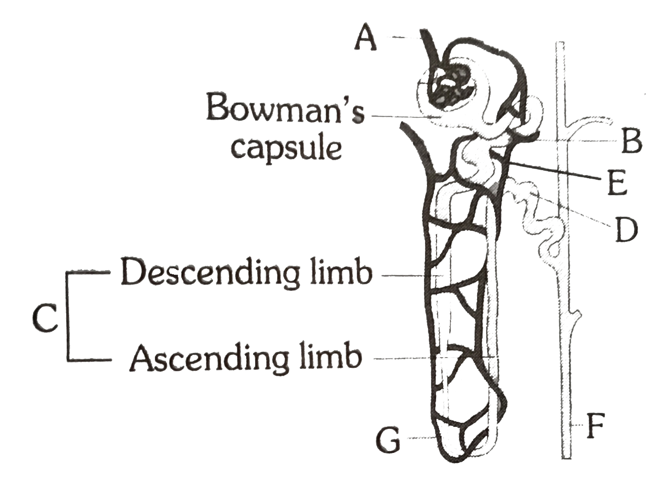 AA-Afferent arteriole, B-Henle' s loop, C-Collecting duct, D-PCT, E-DCT, F-Pertibular capillaries, G- vasa rectaBA-afferent arteriole, B-Peritubular capillaries, C-Helne's loop, D-DCT, E-PCT, F-Collecting duct, G-vasa rectaCA-Afferent arteriole, B- PCT, C-Henle's loop, D-DCT, E-Peritublar capillaries, F-Collecting duct, G-vasa rectaDA-Afferent arteriole, B-Proximal convoluted tubule, C-Henle's loop, D-Distal convoluted tubule, E-Peritbular capillaries, F-Collecting duct, G-vasa recta
Write the symbols for the following elements.
(a) Boron (b) Sulphur
(c ) Argon (d) Calcium
View SolutionWrite the symbols for the following elements.
(a) Boron (b) Sulphur
(c ) Argon (d) Calcium
(e) Selenium (f) Copper
(g) Lead (h) BariumView SolutionList the hormones secreted by the following :
(a) Hypothalamus , (b) Pituitary , (c ) Throid, (d) Parathyroid
(e ) Adrenal, (f) Pancreas, (g) Testis , (h) Ovary
(i) Thymus, (j) Actrium , (k) Kidney, (I) G-I Tract
(M) Transmission of a nerve impulse across a chemical synapseView SolutionComplete the following reactions:
a.
b.
c.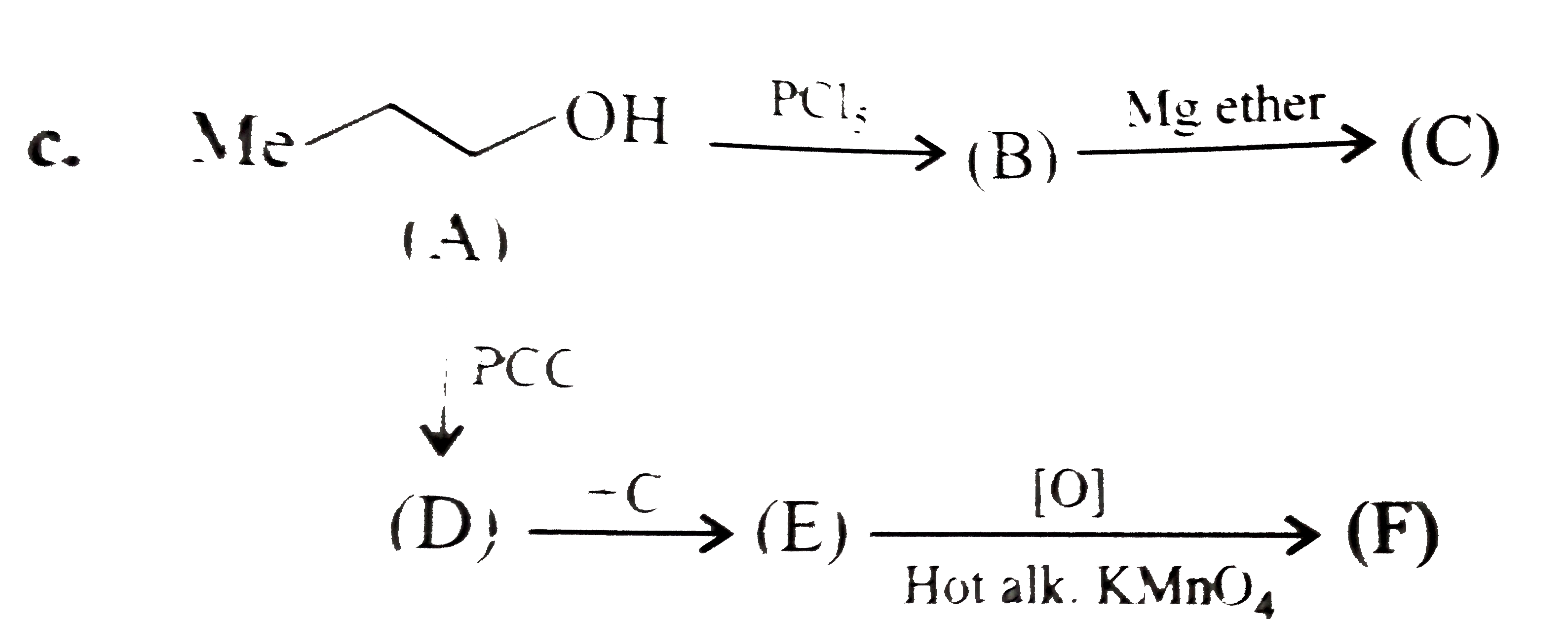
d.
e.
f.
g. View Solution
Complete the following reactions:
a.
b.
c.
d.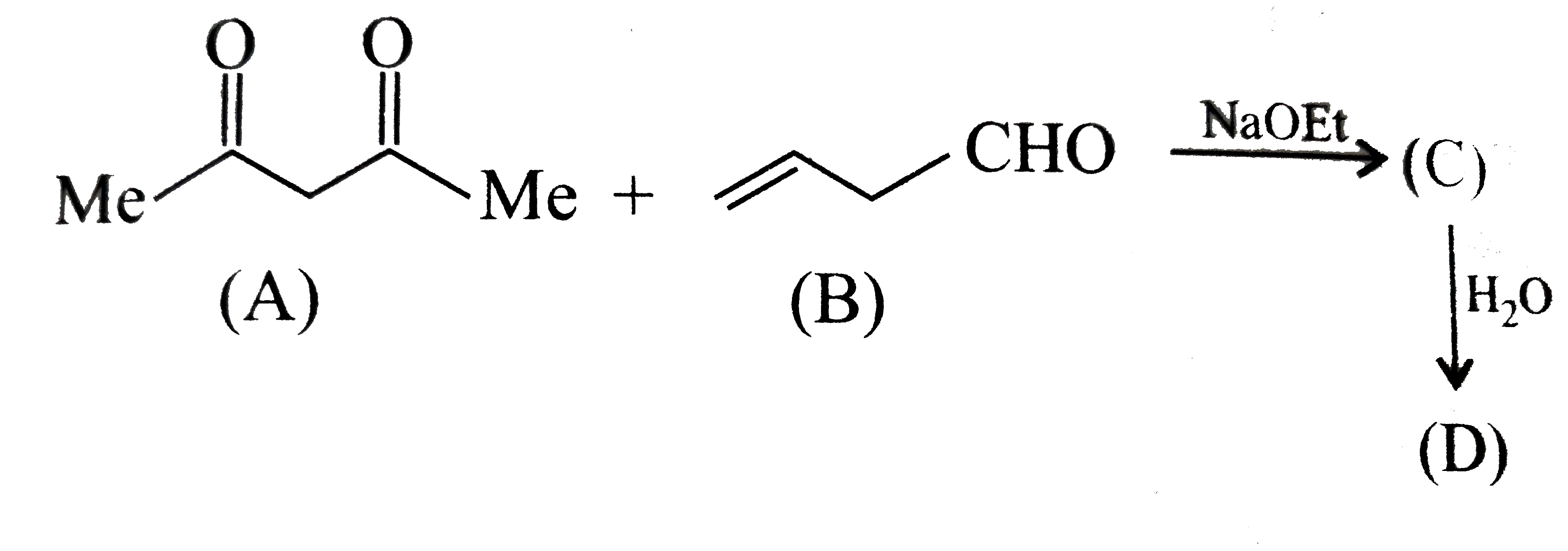
e.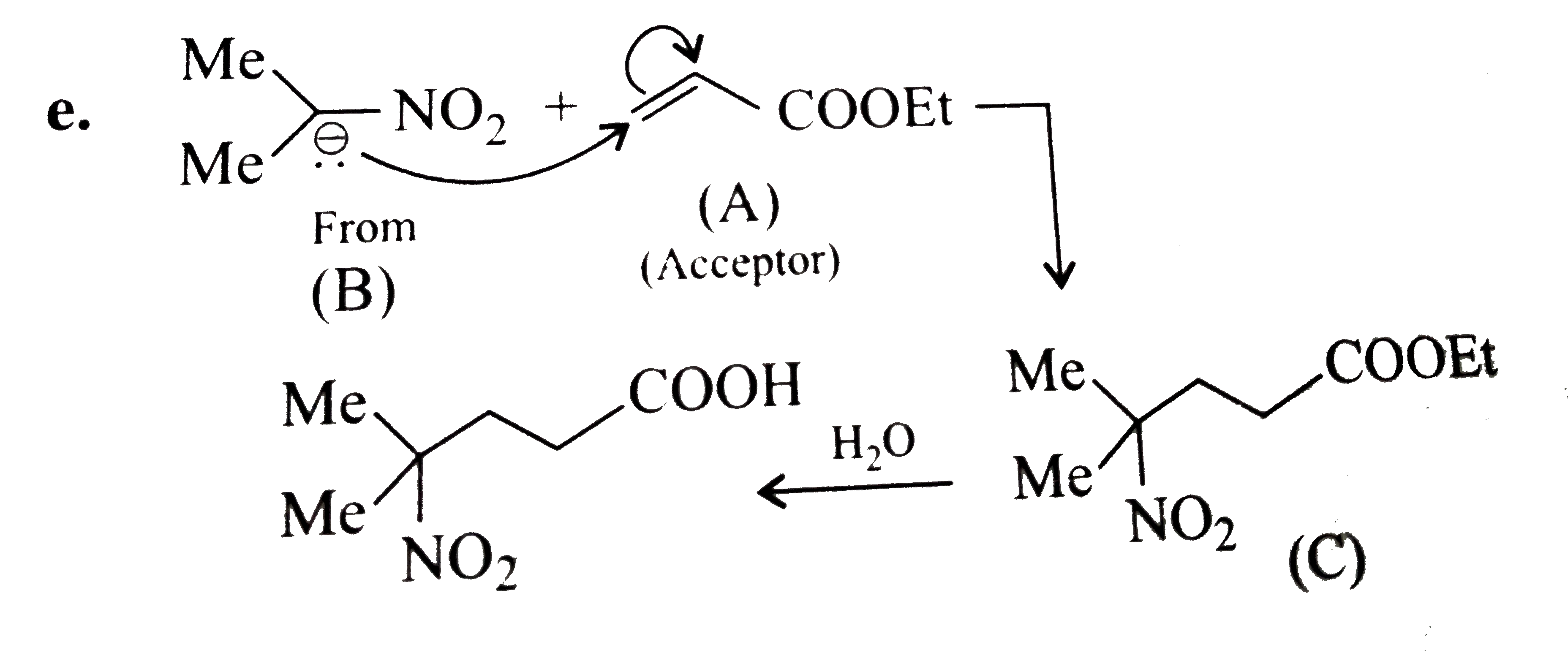
f.
g.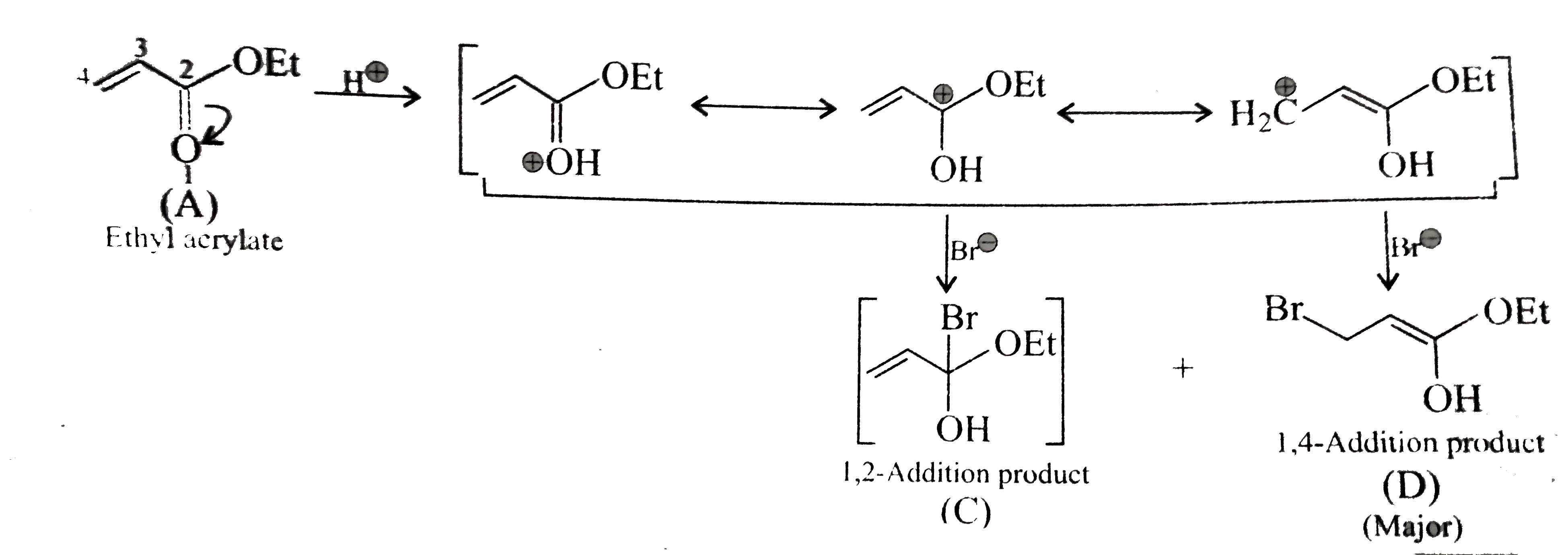 View Solution
Write all the factors of the following numbers : (a) 24 (b) 15 (c) 21(d) 27 (e) 12 (f) 20(g) 18 (h) 23 (i) 36
View SolutionWrite the correct order of acidic strength of following compounds:
(a)H−F (b)H−Cl
(c)H−Br (d)H−IView Solution

