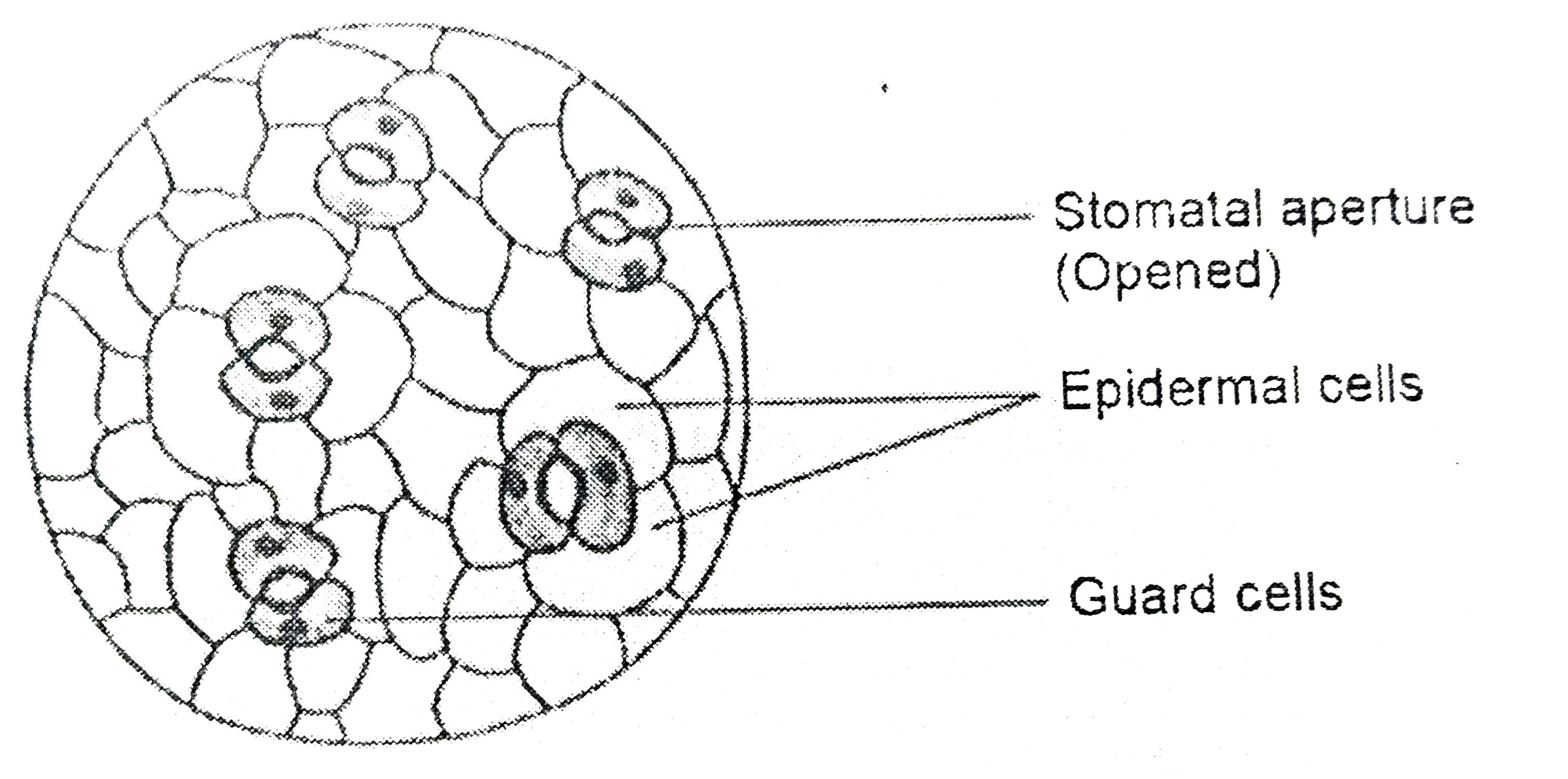
A student is observing the temporary mount of a leaf peel under a microscope. Draw
labelled diageram of the structure (of stomata) as seen under the microscope.
Video Solution
Text Solution
|
Topper's Solved these Questions
- X BOARDSBOOK - X BOARDSCHAPTER - X BOARDSEXERCISE - SET-II SECTION-B
2 Videos
- X BOARDSBOOK - X BOARDSCHAPTER - X BOARDSEXERCISE - SET-II SECTION-C
1 Videos
- X BOARDSBOOK - X BOARDSCHAPTER - X BOARDSEXERCISE - SET-I SECTION-D
3 Videos
- QUESTION PAPER 2023BOOK - X BOARDSCHAPTER - QUESTION PAPER 2023EXERCISE - Question
24 Videos
Similar Questions
Knowledge Check
Study of cell structure under microscope is
ACytologyBCell BiologyCCytochemistryDMicroanatomyMinimum cell size seen under light microscope is
A1μmB0⋅1μmC0⋅25μmD0⋅5μm
Draw a diagram to show the structure and position of stomata on a leaf.
View SolutionHow would you take the sample to prepare temporary stained mount of human cheek cells ? Draw diagram of the structure you would see under the microscope.
View SolutionWhy was the temporary mount of the leaf epidermal peel pinkish-red under the microscope?
View SolutionWhy was the temporary mount of the leaf epidermal peel pinkish-red under the microscope?
View SolutionList the steps of preparation of temporary mount of a leaf peel to observe stomata.
View SolutionThe structure of a cell as seen under an electron microscope is called ultrastructure.
View Solution

