BIOMOLECULES for Class 11 BIOLOGY
INTRODUCTION
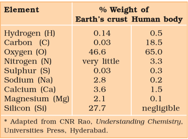
- There is a wide diversity in living organisms in our biosphere. Now a question that arises in our minds is: Are all living organisms made of the same chemicals, i.e., elements and compounds? You have learnt in chemistry how elemental analysis is performed. If we perform such an analysis on a plant tissue, animal tissue or a microbial paste, we obtain a list of elements like carbon, hydrogen, oxygen and several others and their respective content per unit mass of a living tissue. If the same analysis is performed on a piece of earth’s crust as an example of non-living matter, we obtain a similar list. What are the differences between the two lists? In absolute terms, no such differences could be made out. All the elements present in a sample of earth’s crust are also present in a sample of living tissue. However, a closer examination reveals that the relative abundance of carbon and hydrogen with respect to other elements is higher in any living organism than in earth’s crust. A Comparison of Elements Present in Non-living and Living Matter is given below in the tabular form :

INTRODUCTION
- Biochemistry deals in the study of chemicals and their reactions occurring in the living organisms. The term was introduced by German chemist Carl Neuberg (1903).
- The substances of life are both inorganic and organic.
- Inorganic substances include water and salts.
- Organic substances are: carbohydrates, proteins, lipids, vitamins, nucleic acids, etc. both as micromolecule and macromolecules.
- The collection of these molecules in a cell is termed as cellular pool.
-
Relative percentage of these materials in plant and animal protoplasm/cell are as noted below:
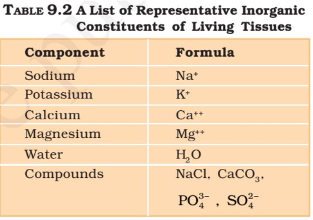
About 93% of the cellular material is made of C, H and O; 2% N, P, S & Cl I, F, Se and Bo are in trace amount. - The metal elements like Ca, K, Na and Mg constitute >5 % while Cu, Co, Zn, Mn, Mb and Cr are in traces.
- Some organic molecules remain in colloidal form in the aqueous intracellular or intercellular fluids. The difference in the constitution of both these fluids is maintained due to selective permeability of the membrane.
- The small molecules (micromolecules) are of low molecular weight and with simple molecular conformation and higher solubility. It includes water, minerals, sugar, amino acids, fatty acids and nucleotides.
- The large molecules (macromolecules) are generally formed by the polymerisation of micromolecules hence are complex in conformations, e.g. Protein, Some lipids, Polysaccharides and Nucleic acids.
Chapter Weightage
- Beta chapter important hai board ke point of view se bhi and competitive exams k point of view se bhi. Plus yeah chapter as a base serve karega class 12 me genetics and molecular biology padhne me bhi. Short answer questions aate plus ek long answer question generally from enzymes
Chapter Weightage
- Beta chapter important hai board ke point of view se bhi and competitive exams k point of view se bhi. Plus yeah chapter as a base serve karega class 12 me genetics and molecular biology padhne me bhi. Iss chapter se NEET mei toh questions aate hi h .Hence it is very important.
Chapter Weightage
- Beta chapter important hai board ke point of view se bhi and competitive exams k point of view se bhi. Plus yeah chapter as a base serve karega class 12 me genetics and molecular biology padhne me bhi. Iss chapter se NEET mei toh questions aate hi h kuch 2-3 , boards k liye bhi kaafi important hai .
How to Analyse Chemical Composition?
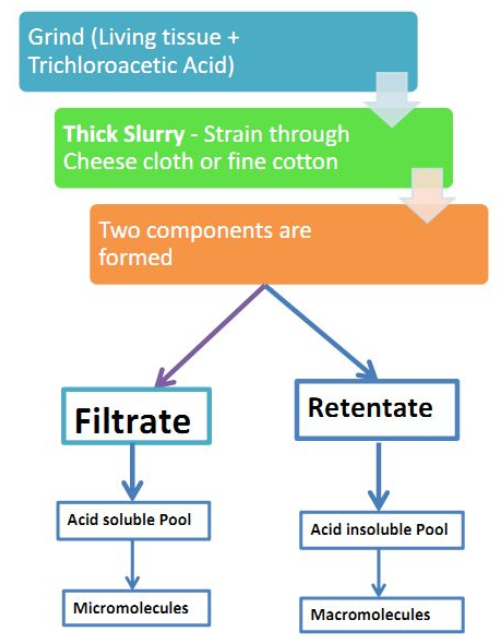
- To analyze biomolecules living tissue (a vegetable or a piece of liver, etc.) is grounded in Trichloroacetic acid (Cl3CCOOH) using a mortar and a pestle in ice. A slurry is made and centrifuged then two fractions are obtained. One is called the filtrate or more technically, the acid-soluble pool, and the second, the retentate or the acid-insoluble fraction.
- Acid soluble fraction has molecules having a molecular weight up to 800 Daltons or inorganic ions and organic molecules which are mainly monomers while, Acid insoluble fraction have macromolecules such as Proteins, Carbohydrates and Nucleic Acids however lipids are present in Acid insoluble fraction though their molecular weight favors Acid soluble fraction.

How to Analyse Chemical Composition?
- Beta, living organisms ke ander jitne molecules milte hai unhi ko hum biomolecules kehte hai jinme se kuch marcomolecules hote hai and kuch micromolecules. Main jo biomacromolecules iss chapter me padhenge wo hai carbohydrates, proteins and fats.
- Koi bhi living tissue lo and usko trichloroacetic acid me ragad do, toh aapke paas kuch slurry/paste jaise aayega and uss tissue me jitni bhi cheezein hongi wo ya toh acid soluble hongi ya fir acid insoluble jinko separate karne ke liye hum uss slurry ko filter kar lete hai jiske baad hume 2 portions milte hai : Acid soluble pool and Acid insoluble pool
- Jaisa ki naam bta rha hai, acid soluble pool me wo sari cheezein aayengi jo trichloroacetic acid me soluble hongi jaise ki simple sugars, amino acids and nucleotides. Inka molecular weight 80-800 Dalton ke beech me hoga.
- Acid insoluble pool yaani ki retentate me hume generally macromolecules jinka molecular weight 1000Da se jyada hota hai, jaise ki polysaccharide and nucleic acids milte hai and sath hi Lipids bhi ( lekin yaad rakhna ki lipids macromolecule nahi hai lekin baki macromolecules ke sath isliye milta hai kyunki yeah acid me insoluble hai) milte hai.
- C, H and O : yeah sabse most common hai and biomolecules me inki presence : O>C>N>H order ko follow karti hai.
- Oily and non polar substances hai jo ki water me insoluble hote hai lekin benzene and chloroform jaise solvents me soluble hai. Simplest example is ghee, oil or wax.
- Lipids ko 3 parts me banta gya hai : Simple lipids, Conjugated lipids and derived lipids.
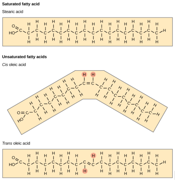
- Simple lipids me aate hai : neutral fats, waxes, cutin and suberin. Simplest fat is a fatty acid. Fatty acid ka matlab hai ek lambi hydrophobic chain ka carboxylic acid hai. Yeah saturated ya unsaturated ho sakte hai. Saturated me koi double bond nahi hota, jaise ki 16 carbon ka palmitic acid ya 18 carbon ka stearic acid. Unsaturated fatty acids me ek ya ek se jyada double bonds hote hai jaise ki 18C ke linoleic acid me 2 double bonds hote hai, vahi 18C ke hi linolenic acid me 3 double hote hai, 20C ke arachidonic acid me 4 double bonds hote hai.
- Fats by definition, fatty acids ke glycerol ke sath esters ko hi hum fats kehte hai. Glycerol me 3 carbon atoms and 3 -OH bonds hote hai. Inhi OH bonds se fatty acids ester form karte hai jinko hum glycerides kehte hai toh glycerides ya toh monoglycerides ho sakte hai ya di ya fir tri jinke structure neeche wale section me given hai.
- Waxes : Fatty acid + monohydric alcohol of high molecular weight waxes bnati hai jaise ki Cuticle is made up of Cutin + wax.
- Conjugate Lipids : jab lipids baki molecules ke sath milkar koi conjugate bnaye usko conjugate lipids kehte hai jaise ki phospholipids me lipids + phosphate groups hote hai. Glycolipids me lipid + glucose/other sugar entity hoti hai. Lipoproteins me lipid+proteins hote hai. Sphingomyelin and lecithin aise hi conjugate lipids hai jinka structure neeche dia hua hai.
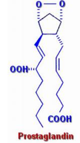
- Derived lipids aise lipids hai jo kisi or molecule se derived hai. Jaise ki prostaglandins arachidonic acid se bante hai.
Next hume ek-ek karke yeah teeno biomacromolecules padhne hai lekin iss section me keval lipids ko detail me discuss karenge.
Lipids
How to Analyse Chemical Composition?
- Take any living tissue (a vegetable or a piece of liver, etc.) and grind it in trichloroacetic acid (Cl3COOH) using a mortar and a pestle. We will obtain a thick slurry. Strain it through a cheesecloth, we would obtain two fractions. One is called the filtrate or more technically, the acid-soluble pool, and the second, the retentate or the acid-insoluble fraction. thousands of organic compounds in the acid-soluble pool. All the carbon compounds that we get from living tissues can be called ‘biomolecules’. However, living organisms have also got inorganic elements and compounds in them.
- From a biological point of view, we can classify biomolecules into amino acids, nucleotide bases, fatty acids etc.
- Amino acids are organic compounds containing an amino group and an acidic group as substituents on the same carbon i.e., the α-carbon. Hence, they are called α-amino acids. They are substituted methanes. There are four substituent groups occupying the four valency positions. These are hydrogen, carboxyl group, amino group and a variable group designated as R group. Based on the nature of R group there are many amino acids. However, those which occur in proteins are only of twenty types. The R group in these proteinaceous amino acids could be a hydrogen (the amino acid is called glycine), a methyl group (alanine), hydroxy methyl (serine), etc. The chemical and physical properties of amino acids are essentially of the amino, carboxyl and the R functional groups. Based on number of amino and carboxyl groups, there are acidic (e.g., glutamic acid), basic (lysine) and neutral (valine) amino acids. Similarly, there are aromatic amino acids (tyrosine, phenylalanine, tryptophan). A particular property of amino acids is the ionizable nature of –NH2 and –COOH groups. Hence in solutions of different pHs, the structure of amino acids changes.
- Lipids are generally water insoluble. They could be simple fatty acids. A fatty acid has a carboxyl group attached to an R group. The R group could be a methyl (–CH3), or ethyl (–C2H5) or higher number of –CH2 groups (1 carbon to 19 carbons). For example, palmitic acid has 16 carbons including carboxyl carbon. Arachidonic acid has 20 carbon atoms including the carboxyl carbon.
 Fatty acids could be saturated (without double bond) or unsaturated (with one or more C=C double bonds).
  Another simple lipid is glycerol which is trihydroxy propane. Many lipids have both glycerol and fatty acids.
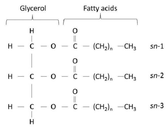
The fatty acids are found esterified with glycerol. They can then be monoglycerides, diglycerides and triglycerides.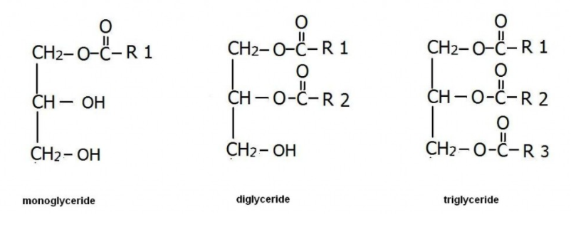
These are also called fats and oils based on melting point. Oils have lower melting point (e.g., gingelly oil) and hence remain as oil in winters.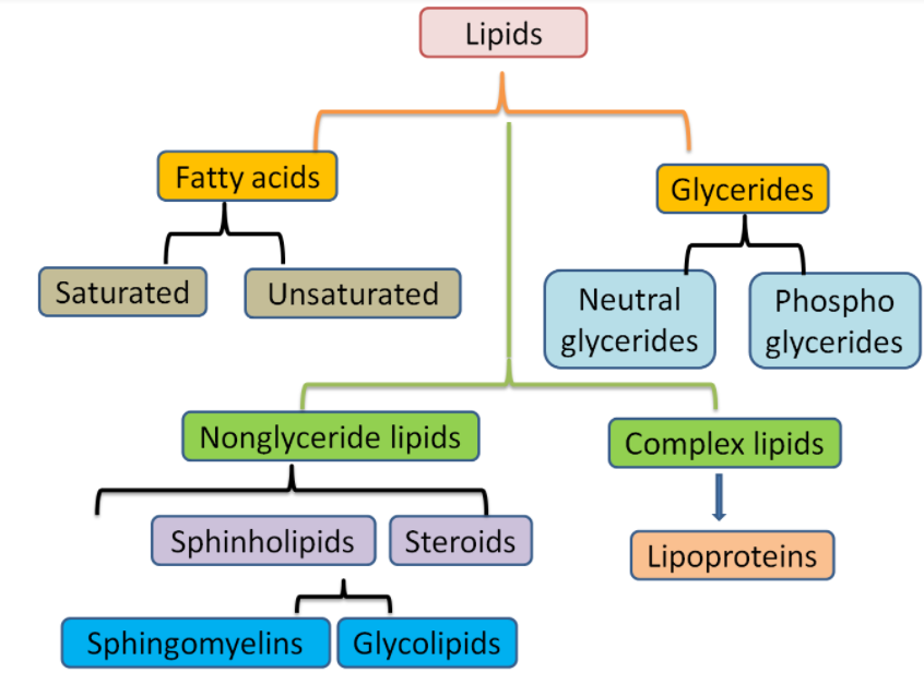
Some lipids have phosphorous and a phosphorylated organic compound in them. These are phospholipids. They are found in cell membrane. Lecithin is one example.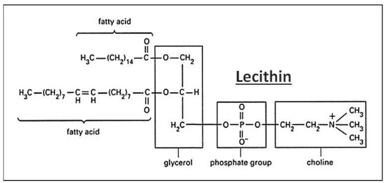
Some tissues especially the neural tissues have lipids with more complex structures. Ex : Sphingomyelin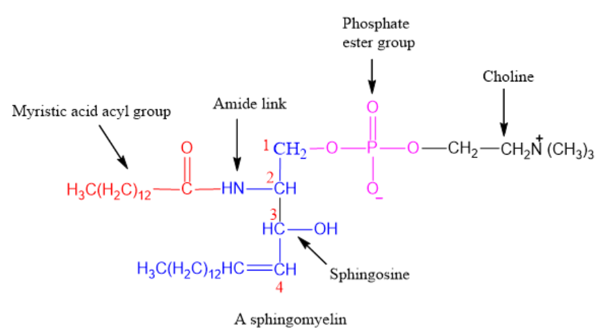
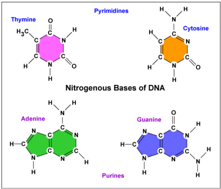
- Nucleic acids : Living organisms have a number of carbon compounds in which heterocyclic rings can be found. Some of these are nitrogen bases – adenine, guanine, cytosine, uracil, and thymine. When found attached to a sugar, they are called nucleosides. If a phosphate group is also found esterified to the sugar they are called nucleotides. Adenosine, guanosine, thymidine, uridine and cytidine are nucleosides. Adenylic acid, thymidylic acid, guanylic acid, uridylic acid and cytidylic acid are nucleotides. Nucleic acids like DNA and RNA consist of nucleotides only. DNA and RNA function as genetic material.

How to Analyse Chemical Composition?
- Take any living tissue (a vegetable or a piece of liver, etc.) and grind it in trichloroacetic acid (Cl3COOH) using a mortar and a pestle. We will obtain a thick slurry. Strain it through a cheesecloth, we would obtain two fractions. One is called the filtrate or more technically, the acid-soluble pool, and the second, the retentate or the acid-insoluble fraction. thousands of organic compounds in the acid-soluble pool. All the carbon compounds that we get from living tissues can be called ‘biomolecules’. However, living organisms have also got inorganic elements and compounds in them.
- From a biological point of view, we can classify biomolecules into amino acids, nucleotide bases, fatty acids etc.
- Amino acids are organic compounds containing an amino group and an acidic group as substituents on the same carbon i.e., the α-carbon. Hence, they are called α-amino acids. They are substituted methanes. There are four substituent groups occupying the four valency positions. These are hydrogen, carboxyl group, amino group and a variable group designated as R group. Based on the nature of R group there are many amino acids. However, those which occur in proteins are only of twenty types. The R group in these proteinaceous amino acids could be a hydrogen (the amino acid is called glycine), a methyl group (alanine), hydroxy methyl (serine), etc. The chemical and physical properties of amino acids are essentially of the amino, carboxyl and the R functional groups. Based on number of amino and carboxyl groups, there are acidic (e.g., glutamic acid), basic (lysine) and neutral (valine) amino acids. Similarly, there are aromatic amino acids (tyrosine, phenylalanine, tryptophan). A particular property of amino acids is the ionizable nature of –NH2 and –COOH groups. Hence in solutions of different pHs, the structure of amino acids changes.
- Lipids are generally water insoluble. They could be simple fatty acids. A fatty acid has a carboxyl group attached to an R group. The R group could be a methyl (–CH3), or ethyl (–C2H5) or higher number of –CH2 groups (1 carbon to 19 carbons). For example, palmitic acid has 16 carbons including carboxyl carbon. Arachidonic acid has 20 carbon atoms including the carboxyl carbon.
 Fatty acids could be saturated (without double bond) or unsaturated (with one or more C=C double bonds).
  Another simple lipid is glycerol which is trihydroxy propane. Many lipids have both glycerol and fatty acids.
The fatty acids are found esterified with glycerol. They can then be monoglycerides, diglycerides and triglycerides.
These are also called fats and oils based on melting point. Oils have lower melting point (e.g., gingelly oil) and hence remain as oil in winters.
Some lipids have phosphorous and a phosphorylated organic compound in them. These are phospholipids. They are found in cell membrane. Lecithin is one example.
Some tissues especially the neural tissues have lipids with more complex structures. Ex : Sphingomyelin
- Nucleic acids : Living organisms have a number of carbon compounds in which heterocyclic rings can be found. Some of these are nitrogen bases – adenine, guanine, cytosine, uracil, and thymine. When found attached to a sugar, they are called nucleosides. If a phosphate group is also found esterified to the sugar they are called nucleotides. Adenosine, guanosine, thymidine, uridine and cytidine are nucleosides. Adenylic acid, thymidylic acid, guanylic acid, uridylic acid and cytidylic acid are nucleotides. Nucleic acids like DNA and RNA consist of nucleotides only. DNA and RNA function as genetic material.


Primary and Secondary Metabolites
- Besides this Biomolecules are also divides into two types depending upon their role in metabolism.
 .
. - A Primary metabolite is a kind of metabolite that is directly involved in normal growth, development, and reproduction. It usually performs a physiological function in the organism (i.e. an intrinsic function). A primary metabolite is typically present in many organisms or cells. Some common examples of primary metabolites include: Lactic acid, and certain amino acids.
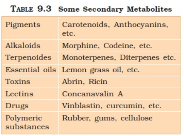
- Secondary metabolites are compounds that are not required for the growth or reproduction of an organism but are produced to confer a selective advantage to the organism. Secondary metabolites commonly mediate antagonistic interactions, such as competition and predation such as Alkaloids, Flavonoids, Rubber, Essential oils, Antibiotics, Gums, Spices etc. .

How to Analyse Chemical Composition?
- Take any living tissue (a vegetable or a piece of liver, etc.) and grind it in trichloroacetic acid (Cl3COOH) using a mortar and a pestle. We will obtain a thick slurry. Strain it through a cheesecloth, we would obtain two fractions. One is called the filtrate or more technically, the acid-soluble pool, and the second, the retentate or the acid-insoluble fraction. thousands of organic compounds in the acid-soluble pool. All the carbon compounds that we get from living tissues can be called ‘biomolecules’. However, living organisms have also got inorganic elements and compounds in them.
- From a biological point of view, we can classify biomolecules into amino acids, nucleotide bases, fatty acids etc.
- Amino acids are organic compounds containing an amino group and an acidic group as substituents on the same carbon i.e., the α-carbon. Hence, they are called α-amino acids. They are substituted methanes. There are four substituent groups occupying the four valency positions. These are hydrogen, carboxyl group, amino group and a variable group designated as R group. Based on the nature of R group there are many amino acids. However, those which occur in proteins are only of twenty types. The R group in these proteinaceous amino acids could be a hydrogen (the amino acid is called glycine), a methyl group (alanine), hydroxy methyl (serine), etc. The chemical and physical properties of amino acids are essentially of the amino, carboxyl and the R functional groups. Based on number of amino and carboxyl groups, there are acidic (e.g., glutamic acid), basic (lysine) and neutral (valine) amino acids. Similarly, there are aromatic amino acids (tyrosine, phenylalanine, tryptophan). A particular property of amino acids is the ionizable nature of –NH2 and –COOH groups. Hence in solutions of different pHs, the structure of amino acids changes.
- Lipids are generally water insoluble. They could be simple fatty acids. A fatty acid has a carboxyl group attached to an R group. The R group could be a methyl (–CH3), or ethyl (–C2H5) or higher number of –CH2 groups (1 carbon to 19 carbons). For example, palmitic acid has 16 carbons including carboxyl carbon. Arachidonic acid has 20 carbon atoms including the carboxyl carbon.
 Fatty acids could be saturated (without double bond) or unsaturated (with one or more C=C double bonds).
  Another simple lipid is glycerol which is trihydroxy propane. Many lipids have both glycerol and fatty acids.
The fatty acids are found esterified with glycerol. They can then be monoglycerides, diglycerides and triglycerides.
These are also called fats and oils based on melting point. Oils have lower melting point (e.g., gingelly oil) and hence remain as oil in winters.
Some lipids have phosphorous and a phosphorylated organic compound in them. These are phospholipids. They are found in cell membrane. Lecithin is one example.
Some tissues especially the neural tissues have lipids with more complex structures. Ex : Sphingomyelin
- Nucleic acids : Living organisms have a number of carbon compounds in which heterocyclic rings can be found. Some of these are nitrogen bases – adenine, guanine, cytosine, uracil, and thymine. When found attached to a sugar, they are called nucleosides. If a phosphate group is also found esterified to the sugar they are called nucleotides. Adenosine, guanosine, thymidine, uridine and cytidine are nucleosides. Adenylic acid, thymidylic acid, guanylic acid, uridylic acid and cytidylic acid are nucleotides. Nucleic acids like DNA and RNA consist of nucleotides only. DNA and RNA function as genetic material.


Primary and Secondary Metabolites
- All metabolites ( intermediate and also end products that are produced during or at the end of cellular metabolism) are biomolecules and are of two types: Primary and Secondary metabolites. Primary metabolites are biomolecules which are required in many cellular functions and metabolic pathways like energy, stimulation, inhibition, protection, etc but compounds other than these primary metabolites e.g. alkaloids, flavonoids, rubber, essential oils, antibiotics, coloured pigments, scents, gums, spices etc are called secondary metabolites.

Carbohydrates
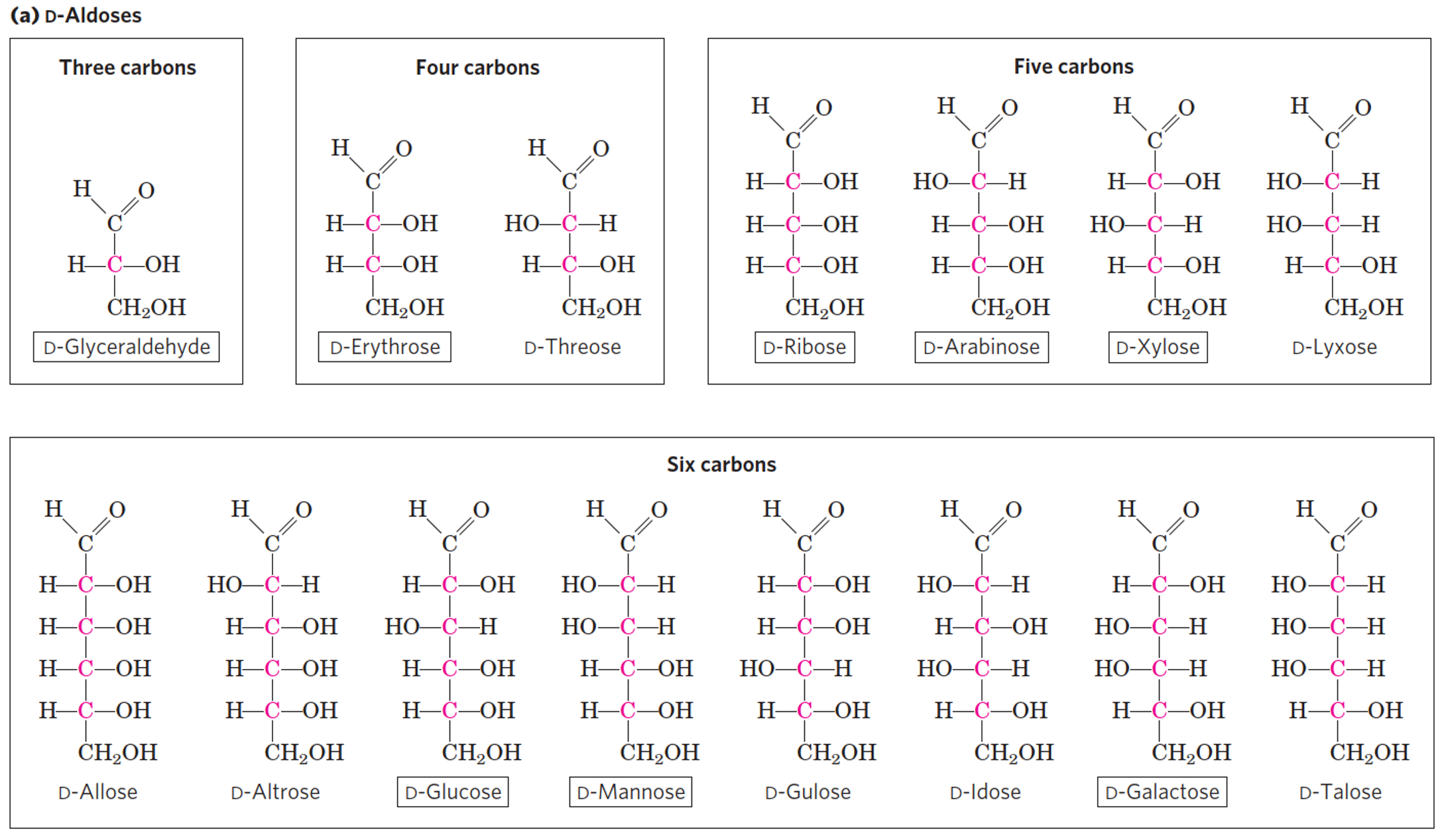
- Carbohydrates represent combinations of carbon, hydrogen and oxygen. Although carbohydrates can be generally represented by the empirical formula (CH2 O)n , some carbohydrates contain additional atoms of sulphur or nitrogen. In these molecules, the carbon forms chains or rings with two or more hydroxyl groups and either an aldehyde or a ketone group, giving them the technical definition of polyhydroxy aldehydes or ketones. These molecules exist in a great variety of configurations. The common term sugar (saccharide) refers to a simple carbohydrate such as a monosaccharide or disaccharide that has a sweet taste. A monosaccharide is a simple polyhydroxy aldehyde or ketone molecule containing 3–7 carbons (trioses, tetroses, pentoses, etc.) A disaccharide is a combination of two monosaccharide molecules, and a polysaccharide is a polymer of many monosaccharides bound in linear or branched chain patterns. Monosaccharides and disaccharides are specified by combining a prefix that describes some characteristics of the sugar with the suffix –ose. For example, hexoses are composed of 6 carbon, and pentoses contain 5 carbon. Glucose is the sugar form of fruits, and xylose a pentose sugar, derives its name from the Greek word (xylon) for wood. .
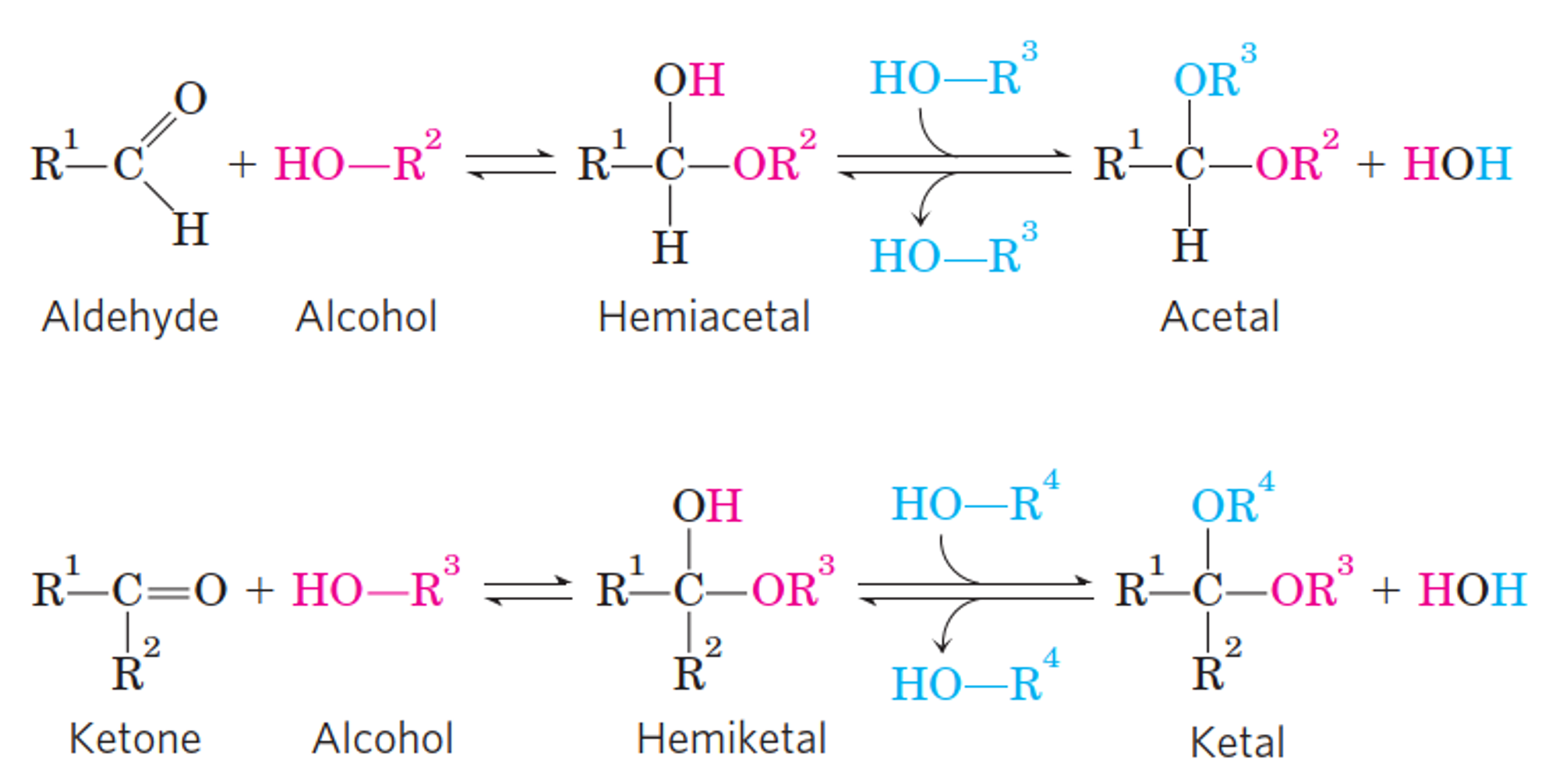
- In aqueous solution, aldotetroses and all monosaccharides with five or more carbon atoms in the backbone occur predominantly as cyclic (ring) structures in which the carbonyl group has formed a covalent bond with the oxygen of a hydroxyl group along the chain. The formation of these ring structures is the result of a general reaction between alcohols and aldehydes or ketones to form derivatives called hemiacetals or hemiketals. .
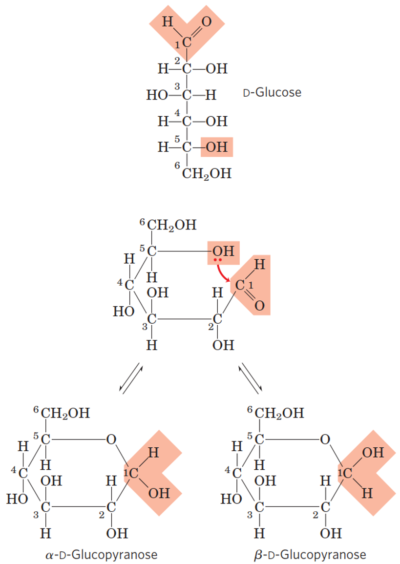
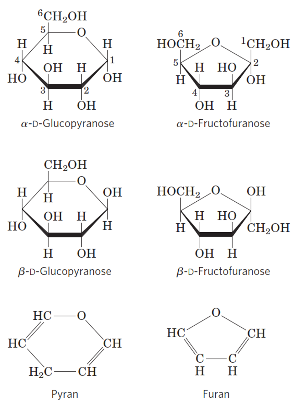
- Two molecules of an alcohol can add to a carbonyl carbon; the product of the first addition is a hemiacetal (for addition to an aldose) or a hemiketal (for addition to a ketose). If the —OH and carbonyl groups are from the same molecule, a five- or six-membered ring results. Addition of the second molecule of alcohol produces the full acetal or ketal, and the bond formed is a glycosidic linkage. When the two molecules that react are both monosaccharides, the acetal or ketal formed is a disaccharide. The reaction with the first molecule of alcohol creates an additional chiral center (the carbonyl carbon). Because the alcohol can add in either of two ways, attacking either the “front” or the “back” of the carbonyl carbon, the reaction can produce either of two stereoisomeric configurations, denoted α and β. For example, D-glucose exists in solution as an intramolecular hemiacetal in which the free hydroxyl group at C-5 has reacted with the aldehydic C-1, rendering the latter carbon asymmetric and producing two possible stereoisomers, designated α and β . Isomeric forms of monosaccharides that differ only in their configuration about the hemiacetal or hemiketal carbon atom are called anomers, and the carbonyl carbon atom is called the anomeric carbon. .
- Six-membered ring compounds are called pyranoses because they resemble the six-membered ring compound pyran. The systematic names for the two ring forms of D-glucose are therefore α-D-glucopyranose and β-D-glucopyranose. Ketohexoses (such as fructose) also occur as cyclic compounds with α and β anomeric forms. In these compounds the hydroxyl group at C-5 (or C-6) reacts with the keto group at C-2, forming a furanose (or pyranose) ring containing a hemiketal linkage. D-Fructose readily forms the furanose ring; the more common anomer of this sugar in combined forms or in derivatives is β -D-fructofuranose.
Oligosaccharides:
- When two monosaccharide join with each other by a chemical bond called Glycosidic bond, by forming glycosidic linkage monomer units can join and form long chains when no of monomer is 2 to 9 it is called Oligosaccharides and if two units join they are called Disaccharides.
Disaccharides
- The simplest oligosaccharides are the disaccharides, which consist of two monosaccharide units linked by a glycosidic bond. As in proteins and nucleic acids, each individual unit in an oligosaccharide is termed a residue. The disaccharides are all commonly found in nature, with sucrose, maltose, and lactose being the most common. Each is a mixed acetal, with one hydroxyl group provided intramolecularly and one hydroxyl from the other monosaccharide. Except for sucrose, each of these structures possesses one free unsubstituted anomeric carbon atom, and thus each of these disaccharides isa reducing sugar. The end of the molecule containing the free anomeric carbon is called the reducing end, and the other end is called the non-reducing end. In the case of sucrose, both of the anomeric carbon atoms are substituted, that is, neither has a free -OH group.
-

Maltose, isomaltose, and cellobiose are all homodisaccharides because they each contain only one kind of monosaccharide, namely, glucose. Maltose is produced from starch (a polymer of α-D-glucose produced by plants) by the action of amylase enzymes and is a component of malt, a substance obtained by allowing grain (particularly barley) to soften in water and germinate. The enzyme diastase, produced during the germination process, catalyzes the hydrolysis of starch to maltose. Maltose is used in beverages (malted milk, for example), and because it is fermented readily by yeast, it is important in the brewing of beer. - In both maltose and cellobiose, the glucose units are 1 4 linked, meaning that the C-1 of one glucose is linked by a glycosidic bond to the C-4 oxygen of the other glucose. The only difference between them is in the configuration at the glycosidic bond. Maltose exists in the α-configuration, whereas cellobiose is in β. Isomaltose is obtained in the hydrolysis of some polysaccharides (such as dextran), and cellobiose is obtained from the acid hydrolysis of cellulose. Isomaltose also consists of two glucose units in a glycosidic bond, but in this case, C-1 of one glucose is linked to C-6 of the other, and the configuration is α.
Trehalose—A Natural Protectant for Bugs.
- Insects use an open circulatory system to circulate hemolymph (insect blood). The “blood sugar” is not glucose but rather Trehalose, an unusual, non-reducing disaccharide.
-
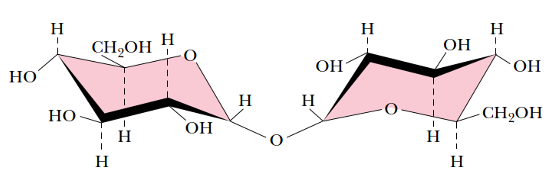
Trehalose is found typically in organisms that are naturally subject to temperature variations and other environmental stresses—bacterial spores, fungi, yeast, and many insects.
β-D-lactose (O-β-D-Galactopyranosyl-(1- 4)-D-glucopyranose)
- It is the principal carbohydrate in milk and is of critical nutritional importance to mammals in the early stages of their lives. It is formed from D-galactose and D-glucose via a β (1 4) link, and because it has a free anomeric carbon, it is capable of mutarotation and is a reducing sugar. It is an interesting quirk of nature that lactose cannot be absorbed directly into the bloodstream. It must first be broken down into galactose and glucose by lactase, an intestinal enzyme that exists in young, nursing mammals but is not produced in significant quantities in the mature mammal. Most humans, with the exception of certain groups in Africa and northern Europe, produce only low levels of lactase. For most individuals, this is not a problem, but some cannot tolerate lactose and experience intestinal pain and diarrhea upon consumption of milk.
- Sucrose, in contrast, is a disaccharide of almost universal appeal and tolerance. Produced by many higher plants and commonly known as table sugar, it is one of the products of photosynthesis and is composed of fructose and glucose. Sucrose has a specific optical rotation, [α]D20, of +66.5°, but an equimolar mixture of its component monosaccharides has a net negative rotation ([α]D20 of glucose is +52.5° and of fructose is -92°).
- For this reason sucrose is also called Invert Sugar. Sucrose is hydrolyzed by the enzyme invertase, so named for the inversion of optical rotation accompanying this reaction. Sucrose is also easily hydrolyzed by dilute acid, apparently because the fructose in sucrose is in the relatively unstable furanose form. Although sucrose and maltose are important to the human diet, they are not taken up directly in the body. In a manner similar to lactose, they are first hydrolyzed by sucrase and maltase, respectively, in the human intestine.
Other Oligosaccharides:
- Raffinose is a trisaccharide composed of galactose, glucose, and fructose. It can be found in beans, cabbage, brussels sprouts, broccoli, asparagus, other vegetables, and whole grains. Humans can not digest it.
- Stachyose is typical of the oligosaccharide components found in substantial quantities in beans, peas, bran, and whole grains. These oligosaccharides are not digested by stomach enzymes, but are metabolized readily by bacteria in the intestines. This is the source of the flatulence that often accompanies the consumption of such foods. Stachyose is a tetrasaccharide consisting of two α-D-galactose units, one α-D-glucose unit, and one β-D-fructose unit.
Derivatives of Monosaccharides
- Sugar Acids
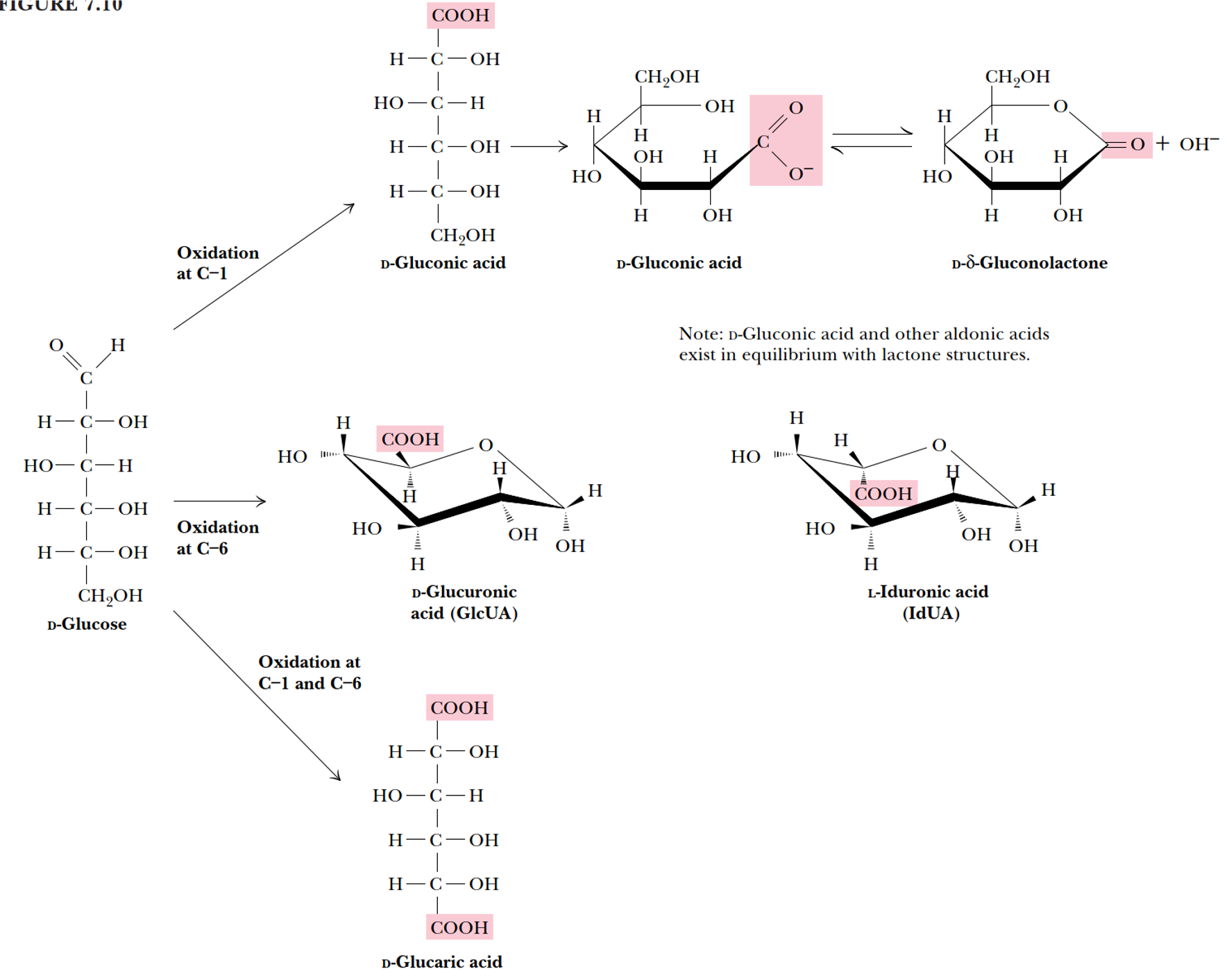
- Monosaccharides can be oxidized enzymatically at C-6, yielding uronic acids, such as D-glucuronic and L-iduronic acids. L --Iduronic acid is similar to D-glucuronic acid, except for having an opposite configuration at C-5. Oxidation at both C-1 and C-6 produces aldaric acids, such as D-glucaric acid.
- Sugar Alcohols
- Sugar alcohols, another class of sugar derivative, can be prepared by the mild reduction (with NaBH4 or similar agents) of the carbonyl groups of aldoses and ketoses. Sugar alcohols, or alditols, are designated by the addition of –itol to the name of the parent sugar. The alditols are linear molecules that cannot cyclize in the manner of aldoses. Nonetheless, alditols are characteristically sweet tasting, and Sorbitol, Mannitol, and Xylitol are widely used to sweeten sugarless gum and mints.
- Deoxy Sugars
- The deoxy sugars are monosaccharides with one or more hydroxyl groups replaced by hydrogens. 2-Deoxy-D-ribose, whose systematic name is 2-deoxy-D-erythropentose, is a constituent of DNA in all living things. Deoxy sugars also occur frequently in glycoproteins and polysaccharides. L-Fucose and L-rhamnose, both 6-deoxy sugars, are components of some cell walls, and rhamnose is a component of
Ouabain, a highly toxic cardiac glycoside found in the bark and root of the ouabaio tree. Ouabain is used by the East African Somalis as an arrow poison. It is also used in very small quantity for cardiac patients to strengthen the contraction of the heart.
- Amino Sugars
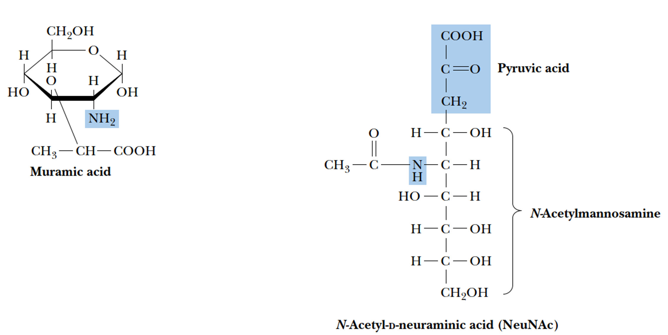
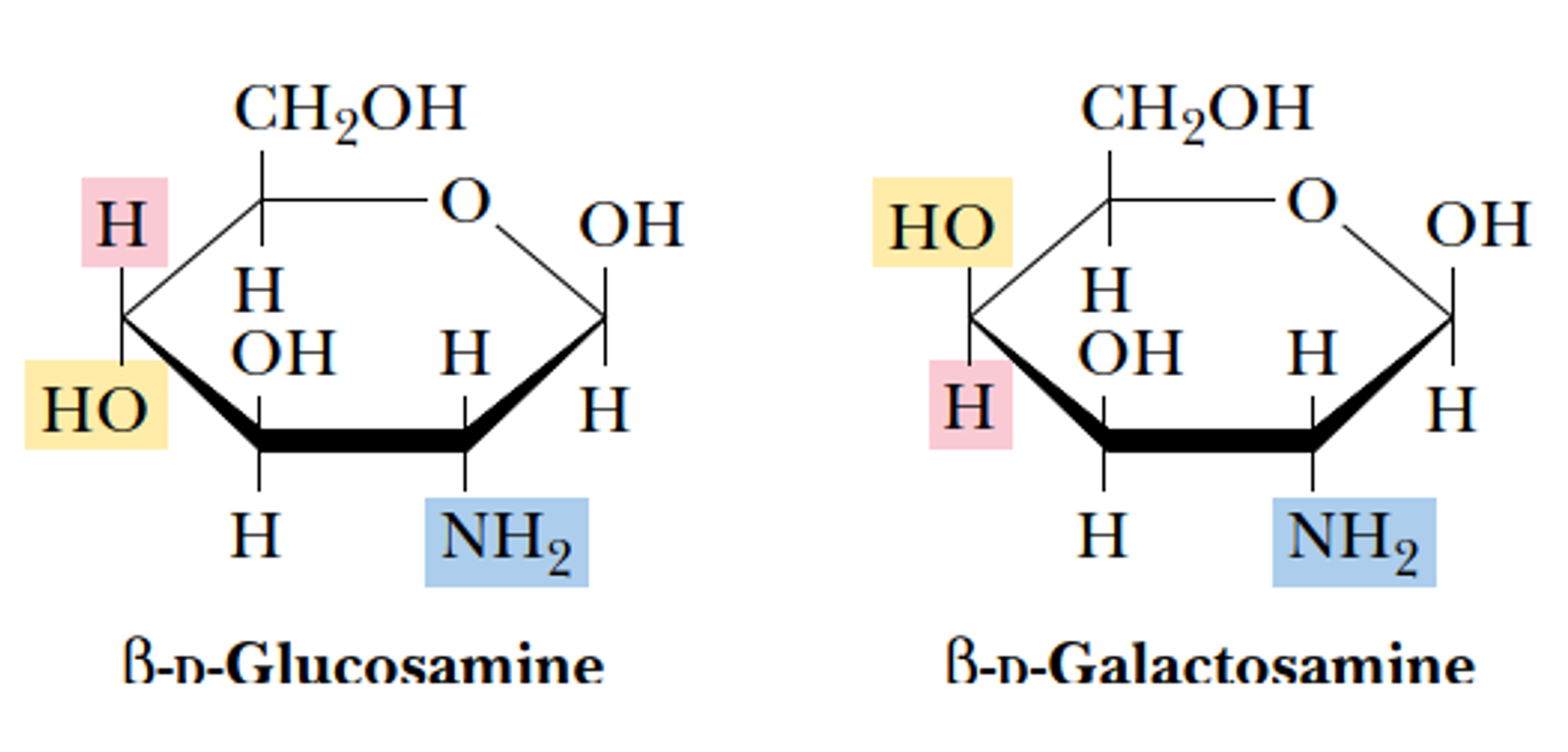
- Amino sugars, including D-glucosamine and D-galactosamine , contain an amino group (instead of a hydroxyl group) at the C-2 position. They are found in many oligo- and polysaccharides, including chitin, a polysaccharide in the exoskeletons of crustaceans and insects. Muramic acid and neuraminic acid, which are components of the polysaccharides of cell membranes of higher organisms and also bacterial cell walls, are.
- Neuraminic acid (an amine isolated from neural tissue) forms a COC bond between the C-1 of N-acetylmannosamine and the C-3 of pyruvic acid. The N-acetyl and N-glycolyl derivatives of neuraminic acid are collectively known as Sialic acids and are distributed widely in bacteria and animal systems.
- Polysaccharides, also called glycans, consist of monosaccharides and their derivatives. If a polysaccharide contains only one kind of monosaccharide molecule, it is a homopolysaccharide, or homoglycan, whereas those containing more than one kind of monosaccharide are heteropolysaccharides. The most common constituent of polysaccharides is D-glucose, but D-fructose, D-galactose, L-galactose, D-mannose, L-arabinose, and D-xylose are also common. Common monosaccharide derivatives in polysaccharides include the amino sugars (D-glucosamine and D-galactosamine), their derivatives (N-acetylneuraminic acid and N-acetylmuramic acid), and simple sugar acids (glucuronic and iduronic acids). Homopolysaccharides are often named for the sugar unit they contain, so that glucose homopolysaccharides are called glucans, while mannose homopolysaccharides are mannans.
- Starch
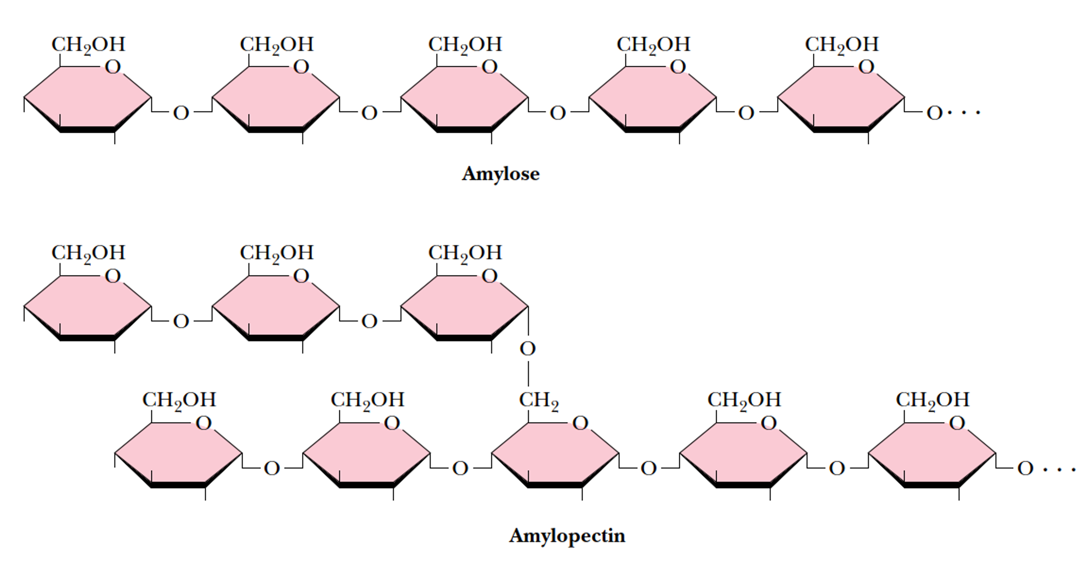
- By far the most common storage polysaccharide in plants is starch, which exists in two forms: α-amylose and amylopectin, the structures of which are shown. Most forms of starch in nature are 10 to 30% α-amylose and 70 to 90% amylopectin. Typical wheat starch produced is about 25% α -amylose and 75% amylopectin. α-Amylose is composed of linear chains of D-glucose in α(1---4) linkages. The chains are of varying length, having molecular weights from several thousand to half a million. The chain has a reducing end and a nonreducing end. Although poorly soluble in water, α-amylose forms micelles in which the polysaccharide chain adopts a helical conformation. Iodine reacts with α -amylose to give a characteristic blue color, which arises from the insertion of iodine into the middle of the hydrophobic amylose helix. In contrast to α-amylose, amylopectin, the other component of typical starches, is a highly branched chain of glucose units. Branches occur in these chains every 12 to 30 residues. The average branch length is between 24 and 30 residues, and molecular weights of amylopectin molecules can range up to 100 million. The linear linkages in amylopectin are α(1---4), whereas the branch linkages are α(1----6). As is the case for α-amylose, amylopectin forms micellar suspensions in water; iodine reacts with such suspensions to produce a red-violet color.
- Glycogen
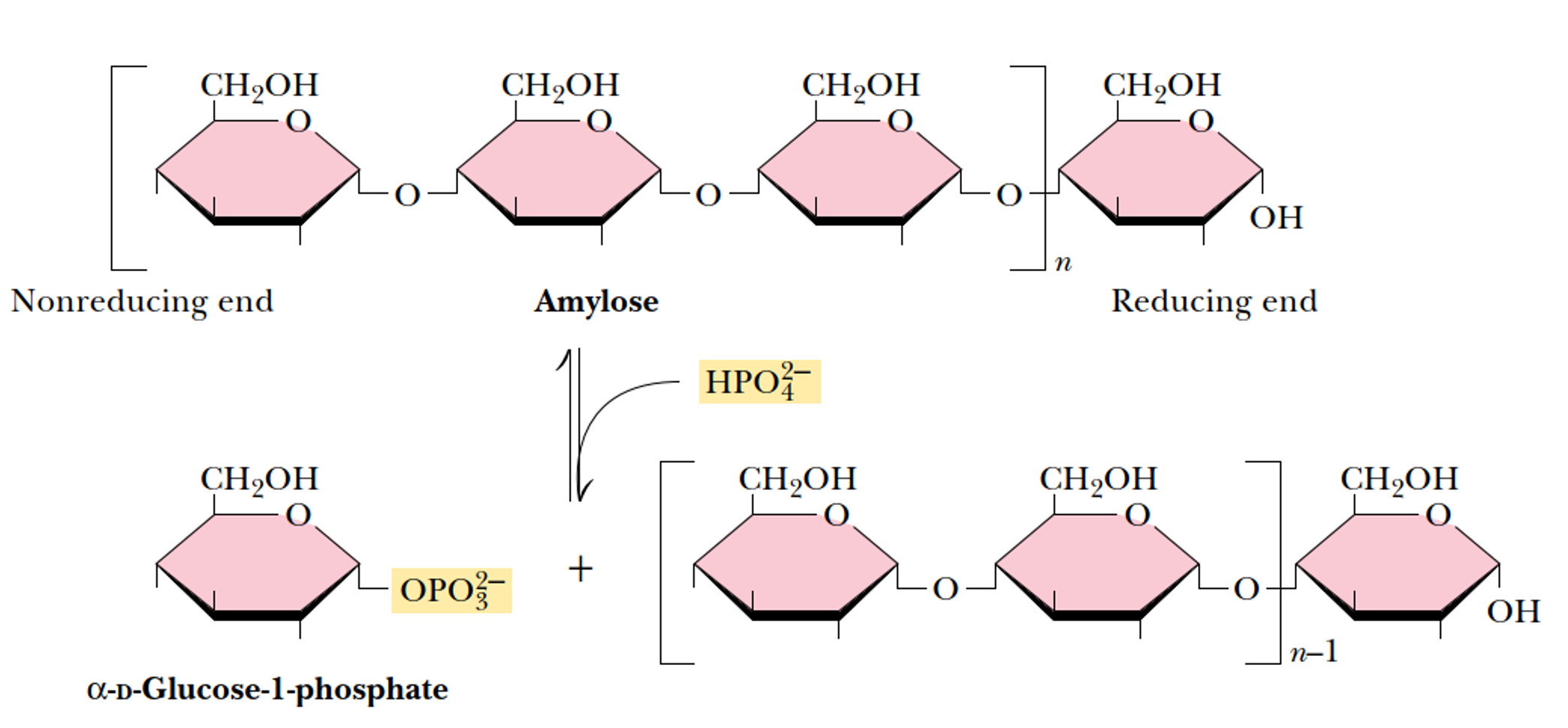
- The major form of storage polysaccharide in animals is glycogen. Glycogen is found mainly in the liver (where it may amount to as much as 10% of liver mass) and skeletal muscle (where it accounts for 1 to 2% of muscle mass). Liver glycogen consists of granules containing highly branched molecules, with α(1---6) branches occurring every 8 to 12 glucose units. Like amylopectin, glycogen yields a red-violet color with iodine. Glycogen can be hydrolyzed by both α- and β-amylases, yielding glucose and maltose, respectively, as products and can also be hydrolyzed by glycogen phosphorylase, an enzyme present in liver and muscle tissue, to release glucose-1-phosphate.
- Cellulose, a fibrous, tough, water-insoluble substance, is found in the cell walls of plants, particularly in stalks, stems, trunks, and all the woody portions of the plant body. Cellulose constitutes much of the mass of wood, and cotton is almost pure cellulose. Like amylose, the cellulose molecule is a linear, unbranched homopolysaccharide, consisting of 10,000 to 15,000 D-glucose units. But there is a very important difference: in cellulose the glucose residues have the β-configuration.
-
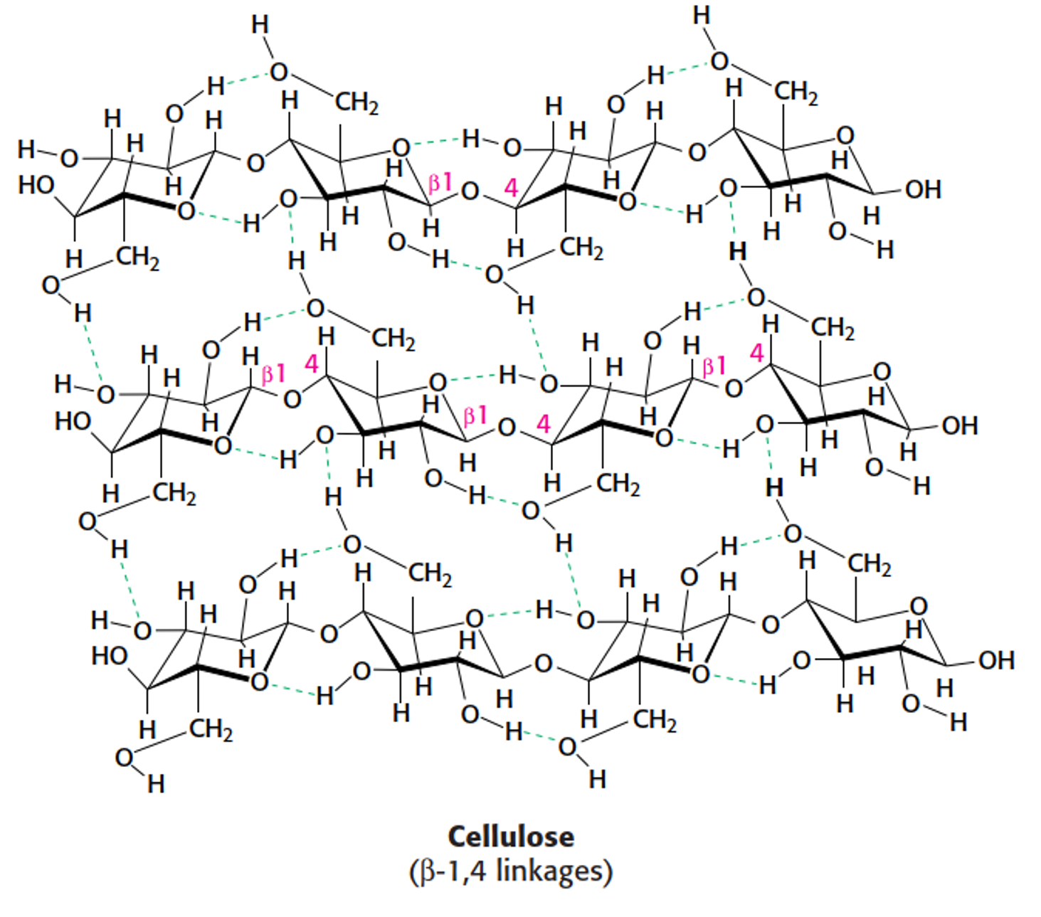
Commercially, cellulose is extremely important. e.g. used to make cotton goods and is a constituent of paper and Cellotape. Rayon and Cellophane are mainly formed of Cellulose xanthate. - Cellulose Acetate fibres yield wrinkle-proof, moth proof Tericot and double knits. Cigarette filters are also formed from these fibres. Other uses of cellulose acetate is in plastics, shatter proof glass and transparent photographic films.
- Cellulose Nitrate (Nitrocellulose) yields propellant explosives. Gun cotton is fully nitrated cellulose. Smokeless ammunition powder is a mixture of higher and lower nitrates of cellulose.
- Carboxymethyl Cellulose : It is semisynthetic gum used in detergents for removing dust (hence extra brightening effect), sizing of paper and textiles, stabilisation of latex paints, smoothening reagent in cosmetics, medicines, ice creams and other foods.
-
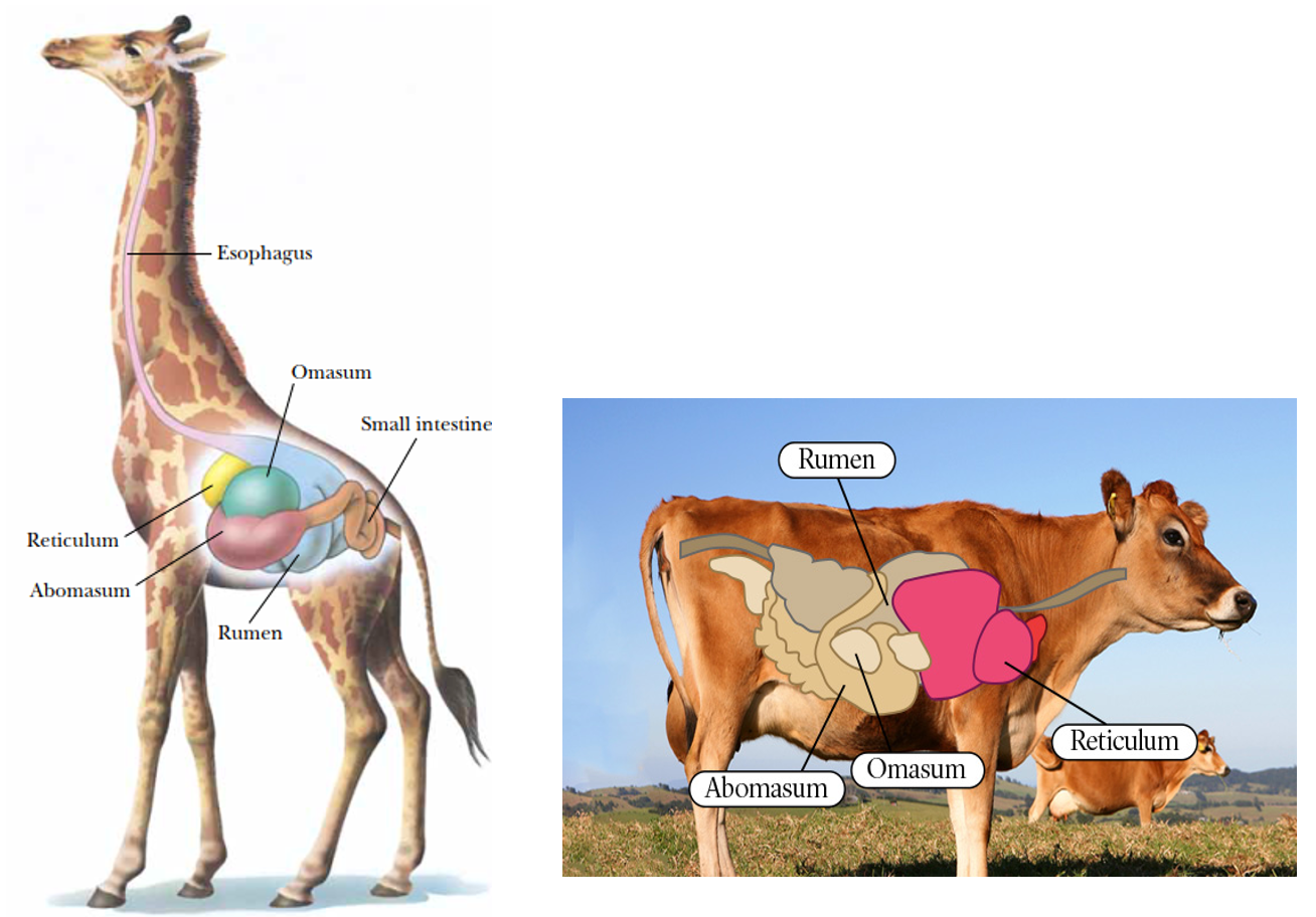
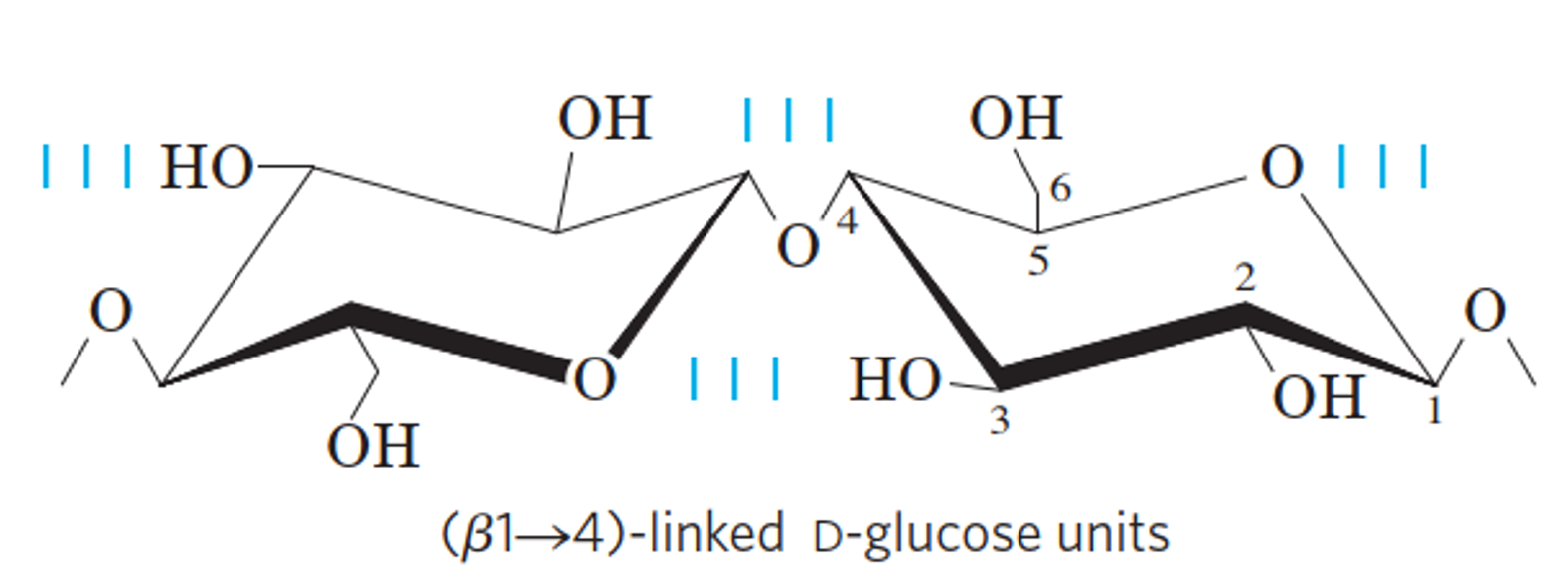
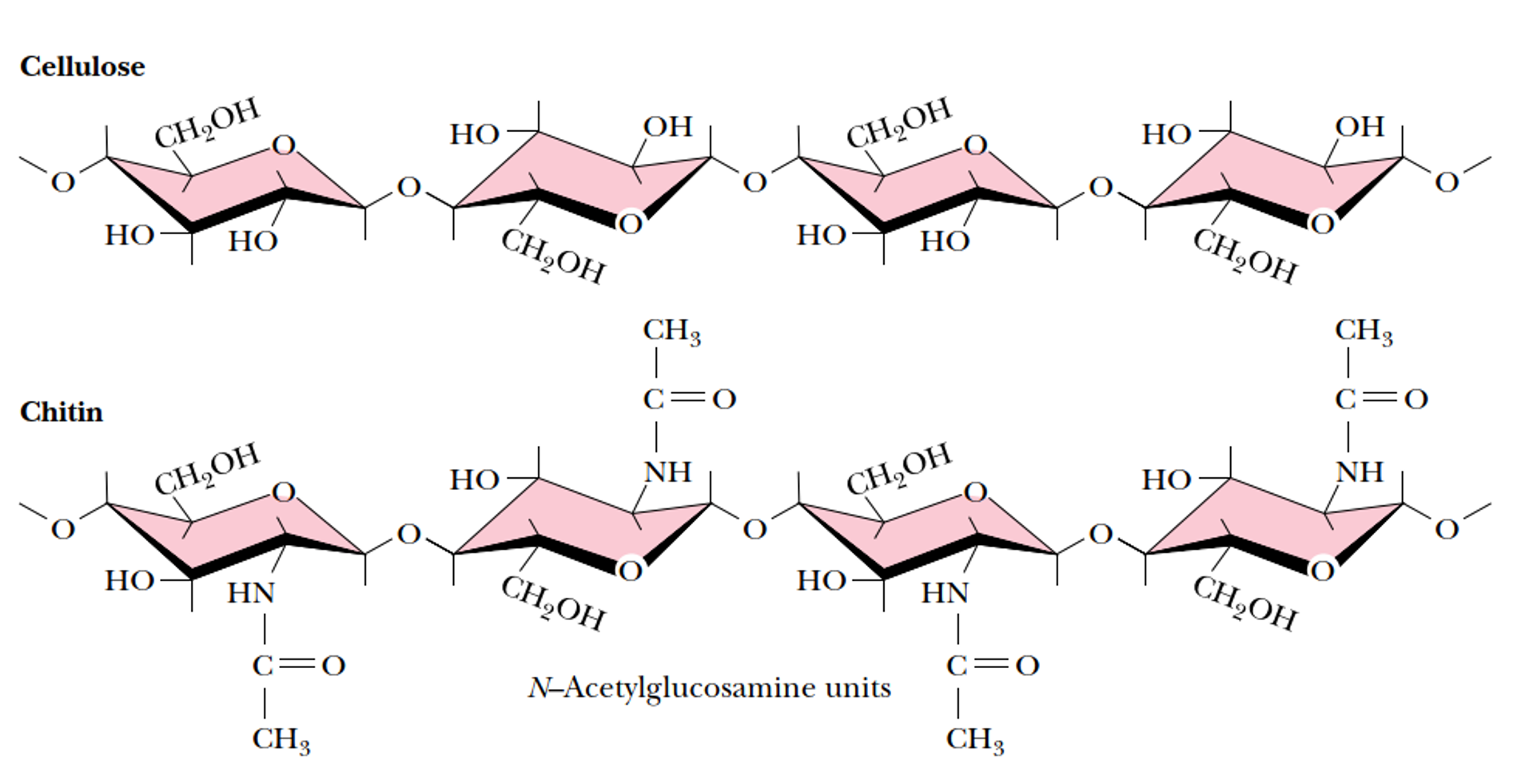
Most vertebrate animals cannot use cellulose as a fuel source, because they lack an enzyme to hydrolyze the (β1--4) linkages. Termites readily digest cellulose (and therefore wood), but only because their intestinal tract harbors a symbiotic microorganisms, such as Entodinium and Trichonympha, that secretes β-glucosidase, which hydrolyzes the (β1--4) linkages. However grazers have cellulose digesting bacteria in the ruminants which digest the cellulose while Abomasum is similar like human stomach and secrete HCl. - Chitin is the earth’s second most abundant carbohydrate polymer (after cellulose). Chitin is present in the cell walls of fungi and is the fundamental material in the exoskeletons of crustaceans, insects, and spiders. The structure of chitin, an extended ribbon, is identical to cellulose, except that the --OH group on each C-2 is replaced by –NHCOCH3, so that the repeating units are N-acetyl-D-glucosamines in β(1---4) linkage. Chitin-based coatings can extend the shelf life of fruits, and a chitin derivative that binds to iron atoms in meat has been found to slow the reactions that cause rancidity and flavor loss.
-
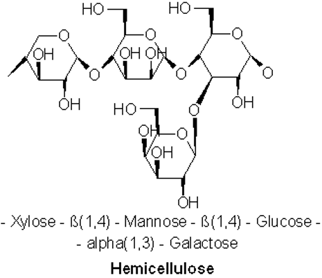
A Hemicellulose (also known as polyose) is one of a number of heteropolymers (matrix polysaccharides), such as arabinoxylans, present along with cellulose in almost all terrestrial plant cell walls. While cellulose is crystalline, strong, and resistant to hydrolysis, hemicelluloses have random, amorphous structure with little strength. They are easily hydrolyzed by dilute acid or base as well as a myriad of hemicellulase enzymes. Hemicelluloses are polysaccharides often associated with cellulose, but with distinct compositions and structures. - Whereas cellulose is derived exclusively from glucose, hemicelluloses are composed of diverse sugars, and can include the five-carbon sugars xylose and arabinose, the six-carbon sugars glucose, mannose and galactose, and the six-carbon deoxy sugar rhamnose. Hemicelluloses contain most of the D-pentose sugars, and occasionally small amounts of L-sugars as well. Xylose is in most cases the sugar monomer present in the largest amount, although in softwoods mannose can be the most abundant sugar.
- Mucopolysaccharides
- These are Heteropolysaccharides of high molecular weight that are gelatinous in consistency, functioning in lubrication and components of sticky substances in organisms. Variety of mucopolysaccharides are present ranging from pectins of cell wall, calcium pectate of middle lamella, gums mucilages, slimes and phycocolloids to hyaluronic acid, chondroitin sulphate, keratin sulphate, heparin, etc. They often contain amino sugars and sugar acids.
- They perform various types of function as follows :
-Mucilage Sheaths occur over aquatic plants, cyanobacteria (blue-green algae) and bacteria and protect the organisms from desiccation, rotting effect of water, epiphytic growth and toxic chemicals. Mucilage also occurs in the unripe fruits of Okra (Lady finger, vern Bhindi) especially around the seeds.
- Pectin occurs in cell wall. Calcium pectate is found in middle lamella. Pectin is made of galactose, galacturonic acid and arabinose. Pectin compounds of middle lamella help in cementing two adjacent cells, pectins of cell wall determine its hydration and orientation of cellulose microfibrils during growth. Pectin compounds are commercial jellying agents.
- Psyllium (Isbgol) powdered colloidal mucilage obtained from the seed husks of Plantago ovata (Plantain, vern, Isbgol), used as a purgative.
- Aloe Gel is gelatinous pulp obtained from fleshy leaves of Aloe barbadensis. Gives relief when applied over inflamed areas and also added in shampoos, conditioners, hand lotions and sun screen creams.
- Agar-Agar is cell wall mucopolysaccharide of some red algae (e.g. Gelidium, Gelidiella, Gracilaria). The phycocolloid is formed of D-galactose 3-6 anhydro L-galactose with sulphate at every tenth position. Agar is used as culture medium, thickener, stabilizer and as laxative. Related compound is Carrageenin (from Chondrus) as emulsifier and clearing agent.
- Glycosaminoglycans
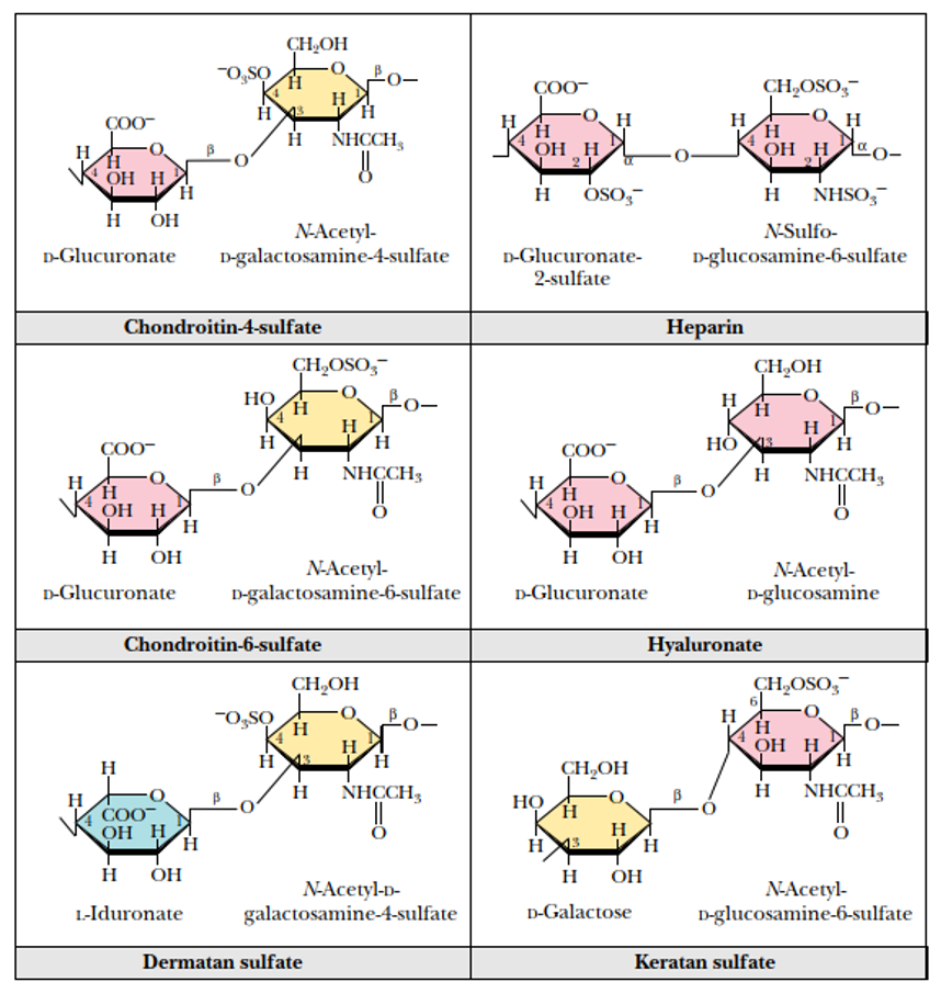
- The extracellular space in the tissues of multicellular animals is filled with a gel-like material, the extracellular matrix (ECM), also called ground substance, which holds the cells together and provides a porous pathway for the diffusion of nutrients and oxygen to individual cells. The ECM that surrounds fibroblasts and other connective tissue cells is composed of an interlocking meshwork of heteropolysaccharides and fibrous proteins such as fibrillar collagens, elastins, and fibronectins. Basement membrane is a specialized ECM that underlies epithelial cells; it consists of specialized collagens, laminins, and heteropolysaccharides. These heteropolysaccharides, the glycosaminoglycans, are a family of linear polymers composed of repeating disaccharide units. Glycosaminoglycans is involved in a variety of extracellular (and sometimes intracellular) functions. Glycosaminoglycans consist of linear chains of repeating disaccharides in which one of the monosaccharide units is an amino sugar and one (or both) of the monosaccharide units contains at least one negatively charged sulfate or carboxylate group. They are of various types and gerneally bind to water.
- Hyaluronic Acid is a thread-like heteropolysaccharide formed by alternating unit of D-glucuronic acid and N-acetylglucosamine. Hyaluronic acid acts as cement between adjacent animal cells. It occurs in the matrix of connective tissue, around ovum and in various lubricating fluids like cerebrospinal fluid, synovial fluid of bone joints and viscous solution in vitreous humour.
- Keratosulphate (Keratin sulphate) is formed from N-acetylglucosamine, galactose and sulphate. It provides both strength and flexibility to skin, connective tissues, cartilage and cornea. It is also complexed with hyaluronic acid in forming proteoglycan.
- Chondroitin Sulphate is a linear polymer of sulphated N-acetylgalactosamine alternating with glucuronic or iduronic acid. It is able to bind water and provide support to connective tissue against compression. The complex also occurs in skin, tendon and cartilage.
- Heparin is a highly sulphated mucopolysaccharide made of alternate units of glucosamine and glucuronic acid. Heparin is mainly produced by liver and mast cells. It is most abundant in blood and connective tissue. Inhibits blood coagulation inside the blood vessels and used as anticoagulant.


Primary and Secondary Metabolites
- Beta, har living cell me kuch reactions chalte hai jo uss cell ko live bnate hai hai. Matlab jeevan ki basic ikaie hi wo chemical reactions hai jinko hum metabolic reactions kehte hai and inhi reaction se ek reactant se koi product bante waqt kafi sare intermediate compounds hote hai jinko hum metabolites kehte hai.
Jo compounds cell directly use kare apne metabolism me jaise ki carbohydrates, lipids ya nucleic acids vgarah primary metabolites kehlate hai. Inke alawa jitne bhi chemical compounds ek cell me bante hai wo secondary metabolites kehlate hai jaise ki pigments hogye, toxins, drugs, essential oils and other polymeric substances like gums etc. yeah secondary metabolites plants ke kisi kaam ke nahi hote lekin hum human ke kaam ke zarur hai.
Biomacromolecules
- one feature common to all those compounds found in the acid soluble pool. They have molecular weights ranging from 18 to around 800 daltons (Da) approximately. The acid insoluble fraction has only four types of organic compounds i.e., proteins, nucleic acids, polysaccharides and lipids. These classes of compounds with the exception of lipids, have molecular weights in the range of ten thousand daltons and above. For this very reason, biomolecules, i.e., chemical compounds found in living organisms are of two types. One, those which have molecular weights less than one thousand dalton and are usually referred to as micromolecules or simply biomolecules while those which are found in the acid insoluble fraction are called macromolecules or biomacromolecules. The molecules in the insoluble fraction with the exception of lipids are polymeric substances. Lipids are indeed small molecular weight arranged into structures like cell membrane and other membranes. When we grind a tissue, we are disrupting the cell structure. Cell membrane and other membranes are broken into pieces, and form vesicles which are not water soluble. Therefore, these membrane fragments in the form of vesicles get separated along with the acid insoluble pool and hence in the macromolecular fraction. Lipids are not strictly Macromolecules. The acid soluble pool represents roughly the cytoplasmic composition. The macromolecules from cytoplasm and organelles become the acid insoluble fraction. Together they represent the entire chemical composition of living tissues or organisms.

Proteins
- Proteins are polymers of amino acids, with each amino acid residue joined to its neighbor by a specific type of covalent bond. (The term “residue” reflects the loss of the elements of water when one amino acid is joined to another.) Proteins can be broken down (hydrolyzed) to their constituent amino acids by a variety of methods, and the earliest studies of proteins naturally focused on the free amino acids derived from them. Around more than 300 amino acids are found, which divide into two types,
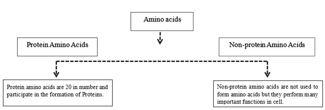
- Amino acids are the organic acids; containing at least one –NH2 (amine) and one –COOH(carboxylic acids) groups as active groups attached to α-carbon (Chiral carbon).
- The other valencies of carbon are satisfied by one -H and one R- group (or alkyl group) as side chain which may be polar or non- polar; aliphatic or aromatic; simple or heterocyclic compound.
- ‘R’ group may vary from a H-atom to any large aromatic compound; hence the type of amino acids depends upon R-group.
- These are ‘building blocks’ of protein.
- These are more than 200 types in the body, but only 20 types are recognised, which take part in the synthesis of protein.
- These are synthesized by plants and microorganism as only they can utilize inorganic nitrogen. Therefore, animals have to depend upon the plants for their supply.
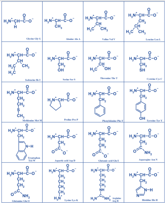
- The 20 types are: Glycine, Alanine, Valine, Leucin, Isoleucine, Serine, Tyrosine, Methionine, Cysteine, Phenylalanine, Tryptophan, Proline, Threonine, Aspartic acid, Glutamic acid, Asparagine, Glutamine, Lysine, Histidine, Arginine.
Amino Acids)
Protein (Amino Acids )
- Twenty different amino acids are commonly found in proteins. The first to be discovered was asparagine, in 1806. The last of the 20 to be found, threonine, was not identified until 1938. All the amino acids have trivial or common names, in some cases derived from the source from which they were first isolated. Asparagine was first found in asparagus, and glutamate in wheat gluten; tyrosine was first isolated from cheese (its name is derived from the Greek tyros, “cheese”); and glycine (Greek glykos, “sweet”) was so named because of its sweet taste. All 20 of the common amino acids are α-amino acids. They have a carboxyl group and an amino group bonded to the same carbon atom (the α carbon). They differ from each other in their side chains, or R groups, which vary in structure, size, and electric charge, and which influence the solubility of the amino acids in water.
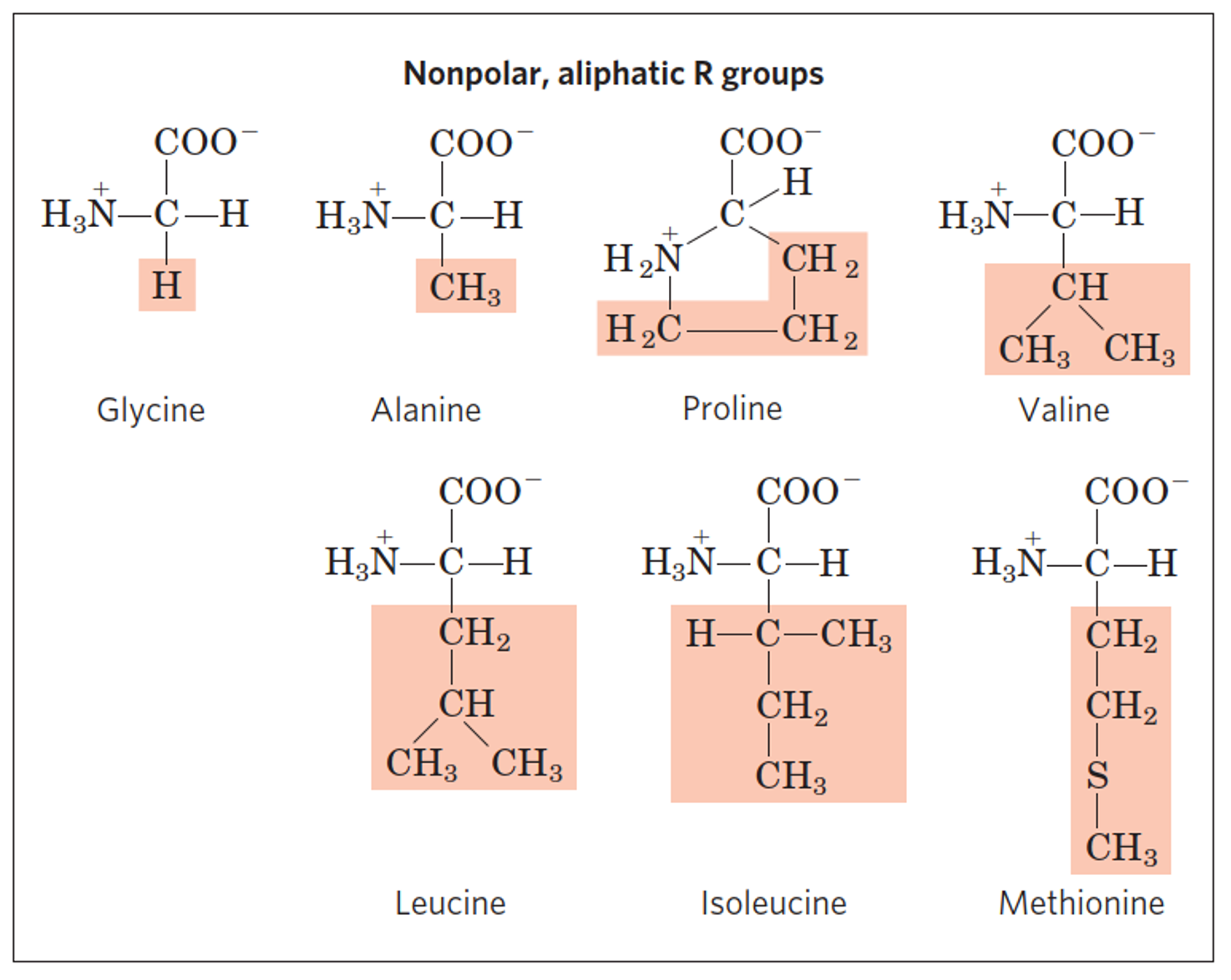
- R group is Non-polar
- R group is aromatic
- R group is polar
- R group is positively charged
- R group is negatively charged.
- R group is non-polar: The R groups in this class of amino acids are nonpolar and hydrophobic. The side chains of alanine, valine, leucine, and isoleucine tend to cluster together within proteins, stabilizing protein structure by means of hydrophobic interactions. Glycine has the simplest structure. Although it is most easily grouped with the nonpolar amino acids, its very small side chain makes no real contribution to hydrophobic interactions.
Methionine, one of the two sulfur containing amino acids, has a slightly nonpolar thioether group in its side chain. Proline has an aliphatic side chain with a distinctive cyclic structure. The secondary amino (imino) group of proline residues is held in a rigid conformation that reduces the structural flexibility of polypeptide regions containing proline. - R group is aromatic: Phenylalanine, tyrosine, and tryptophan, with their aromatic side chains, are relatively nonpolar (hydrophobic). All can participate in hydrophobic interactions. The hydroxyl group of tyrosine can form hydrogen bonds, and it is an important functional group in some enzymes. Tyrosine and tryptophan are significantly more polar than phenylalanine, because of the tyrosine hydroxyl group and the nitrogen of the tryptophan indole ring.
-
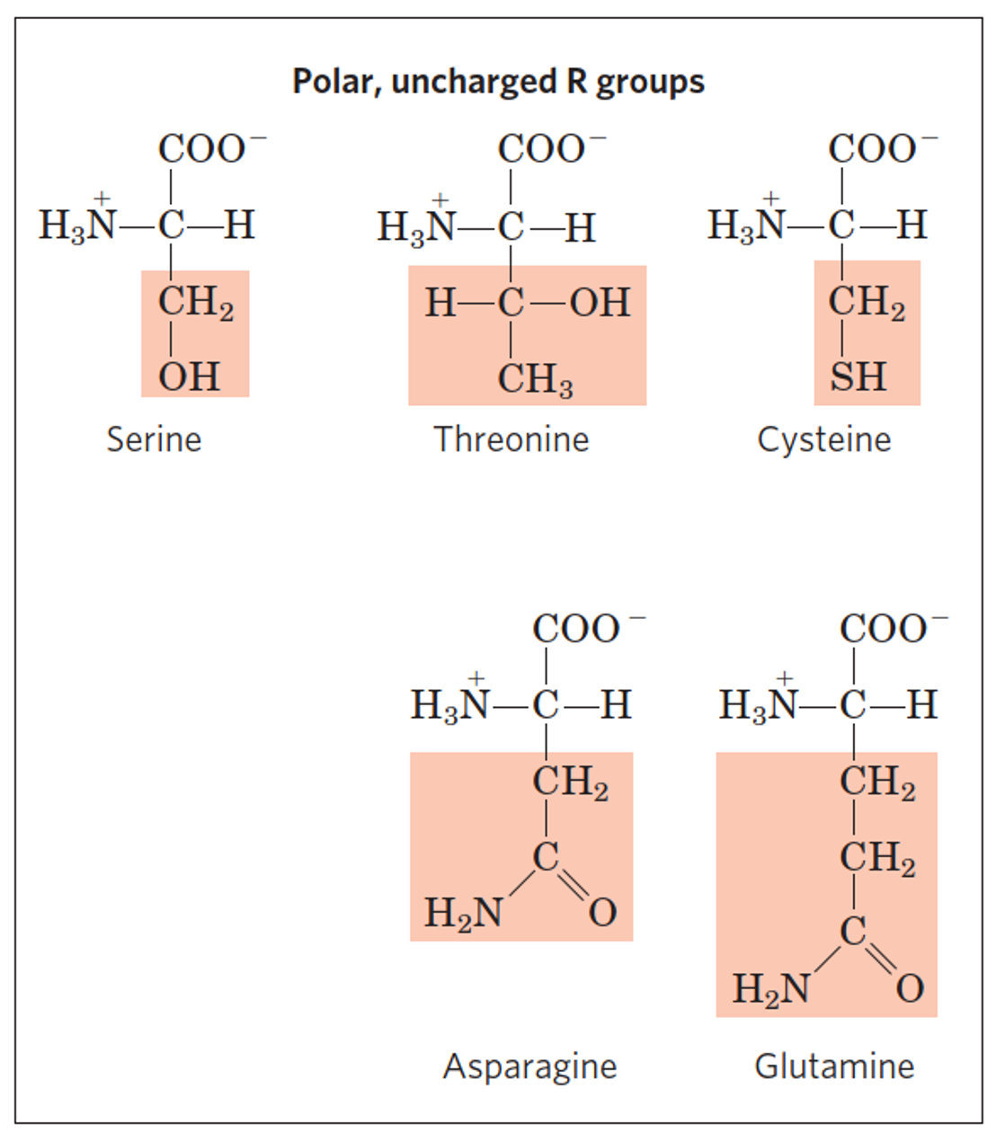
R group is non-polar but hydrophilic The R groups of these amino acids are more soluble in water, or more hydrophilic, than those of the nonpolar amino acids, because they contain functional groups that form hydrogen bonds with water. This class of amino acids includes serine, threonine, cysteine, asparagine, and glutamine. The polarity of serine and threonine is contributed by their hydroxyl groups, and that of asparagine and glutamine by their amide groups. Cysteine is readily oxidized to form a covalently linked dimeric amino acid called cystine, in which two cysteine molecules or residues are joined by a disulfide bond. Asparagine and glutamine are the amides of two other amino acids also found in proteins—aspartate and glutamate, respectively.
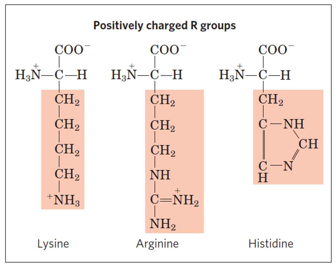
- R group is Positively Charged (Basic): The most hydrophilic R groups are those that are either positively or negatively charged. The amino acids in which the R groups have significant positive charge at pH 7.0 are lysine, which has a second primary amino group at the α position on its aliphatic chain; arginine, which has a positively charged guanidinium group; and histidine, which has an aromatic imidazole group.
- R group is Negatively charged: The two amino acids having R groups with a net negative charge at pH 7.0 are aspartate and glutamate, each of which has a second carboxyl group. Both aspartic acid and Glutamic acid can act as neurotransmitters, and participate in many important physiological functions for example they participate in visual pathways.

Protein amino acids are 20 in number and can be classified on the basis of their R group depending upon the nature of R groups amino acids divide into 5 groups.
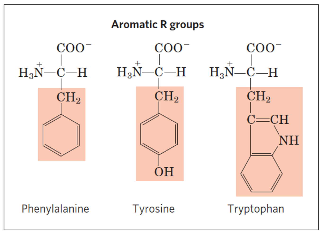
Tryptophan is the most essential amino acid and it is a precursor of many important hormones and neurotransmitters such as Melatonin and serotonin, while both tyrosine and Phenylalanine are used to form Catecholamines and Melanin.
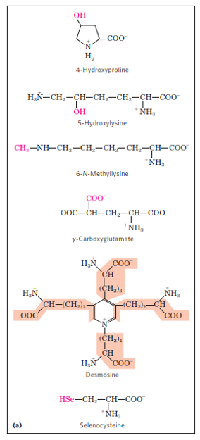
In addition to the 20 common amino acids, proteins may contain residues created by modification of common residues already incorporated into a polypeptide. Among these uncommon amino acids are 4-hydroxyproline, a derivative of proline, and 5-hydroxylysine, derived from lysine. The former is found in plant cell wall proteins, and both are found in collagen, a fibrous protein of connective tissues. 6-N-Methyllysine is a constituent of myosin, a contractile protein of muscle. Another important uncommon amino acid is γ-carboxyglutamate, found in the blood-clotting protein prothrombin and in certain other proteins that bind Ca+2 as part of their biological function. More complex is desmosine, a derivative of four Lys residues, which is found in the fibrous protein elastin. Selenocysteine is a special case. This rare amino acid residue is introduced during protein synthesis rather than created through a post synthetic modification. It contains selenium rather than the sulfur of cysteine. Actually derived from serine, selenocysteine is a constituent of just a few known proteins. Some amino acid residues in a protein may be modified transiently to alter the protein’s function. The addition of phosphoryl, methyl, acetyl, adenylyl, ADP-ribosyl, or other groups to particular amino acid residues can increase or decrease a protein’s activity. Phosphorylation is a particularly common regulatory modification.
Some 300 additional amino acids have been found in cells. They have a variety of functions but are not all constituents of proteins. Ornithine and citrulline deserve special note because they are key intermediates (metabolites) in the biosynthesis of arginine and in the urea cycle. γ-Aminobutyric acid, or GABA, is produced by the decarboxylation of glutamic acid and is a potent neurotransmitter. Histamine, which is synthesized by decarboxylation of histidine, and serotonin, which is derived from tryptophan, similarly function as neurotransmitters and regulators. -Alanine is found in nature in the peptides carnosine and anserine and is a component of pantothenic acid (a vitamin), which is a part of coenzyme A.
Epinephrine (also known as adrenaline), derived from tyrosine, is an important hormone. Penicillamine is a constituent of the penicillin antibiotics.
Amino Acids Can Act as Acids and Bases:
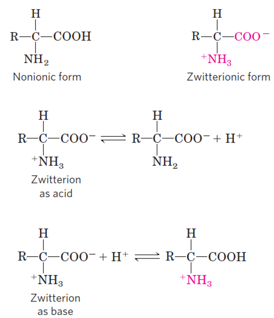
- The amino and carboxyl groups of amino acids, along with the ionizable R groups of some amino acids, function as weak acids and bases. When an amino acid lacking an ionizable R group is dissolved in water at neutral pH, it exists in solution as the dipolar ion, or zwitterion (German for “hybrid ion”), which can act as either an acid or a base. Substances having this dual (acid-base) nature are amphoteric and are often called Ampholytes (from “amphoteric electrolytes”). A simple monoamino monocarboxylic α- amino acid, such as alanine, is a diprotic acid when fully protonated; it has two groups, the —COOH group and the —NH+3 group, that can yield protons:

Peptides Are Chains of Amino Acids:
- Two amino acid molecules can be covalently joined through a substituted amide linkage, termed a peptide bond, to yield a dipeptide. Such a linkage is formed by removal of the elements of water (dehydration) from the α-carboxyl group of one amino acid and the α-amino group of another. Peptide bond formation is an example of a condensation reaction. Three amino acids can be joined by two peptide bonds to form a tripeptide; similarly, four amino acids can be linked to form a tetrapeptide, five to form a pentapeptide, and so forth. When a few amino acids are joined in this fashion, the structure is called an oligopeptide.
When many amino acids are joined, the product is called a polypeptide. Proteins may have thousands of amino acid residues. Although the terms “protein” and “polypeptide” are sometimes used interchangeably, molecules referred to as polypeptides generally have molecular weights below 10,000, and those called proteins have higher molecular weights. In a peptide, the amino acid residue at the end with a free α-amino group is the amino-terminal (or N-terminal) residue; the residue at the other end, which has a free carboxyl group, is the carboxyl-terminal (C-terminal) residue.
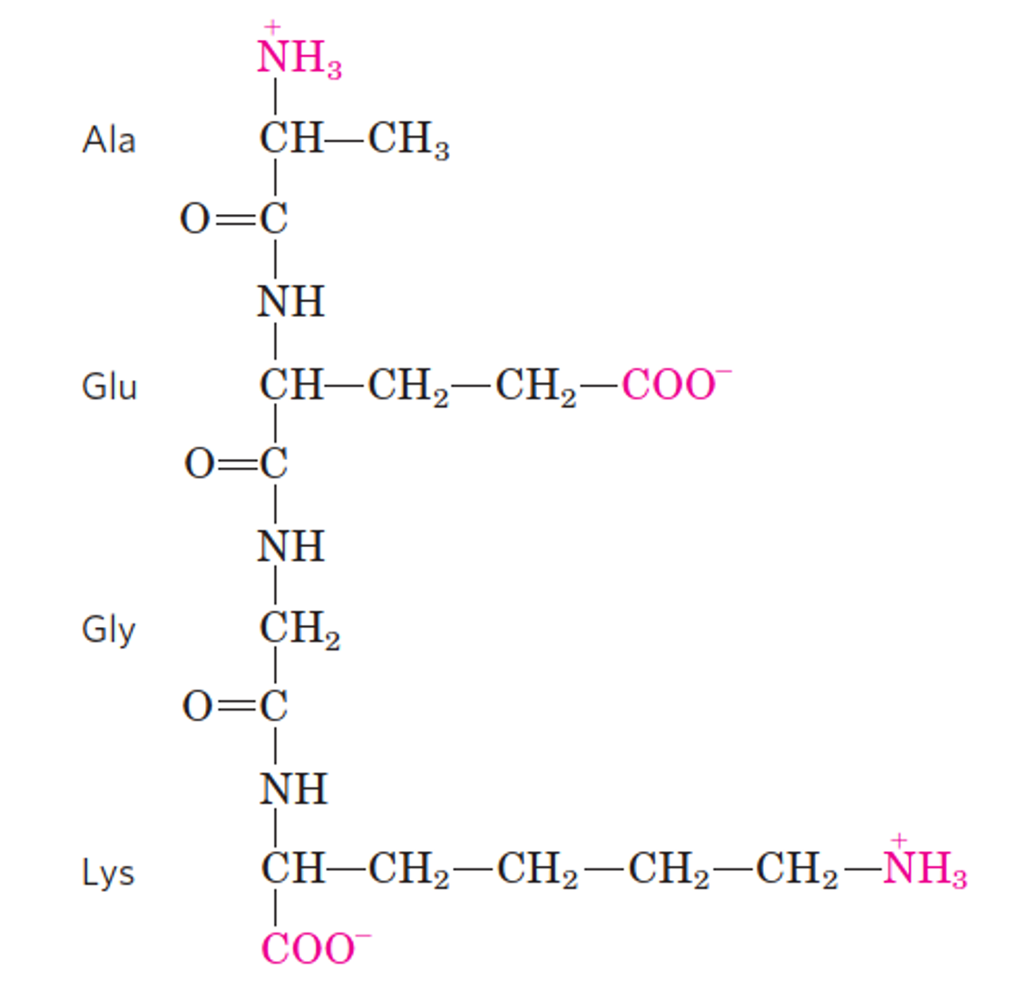

Naturally occurring peptides :
- Range in length from two to many thousands of amino acid residues. Even the smallest peptides can have biologically important effects. Consider the commercially synthesized dipeptide L-aspartyl-L-phenylalanine methyl ester, the artificial sweetener better known as Aspartame or NutraSweet.
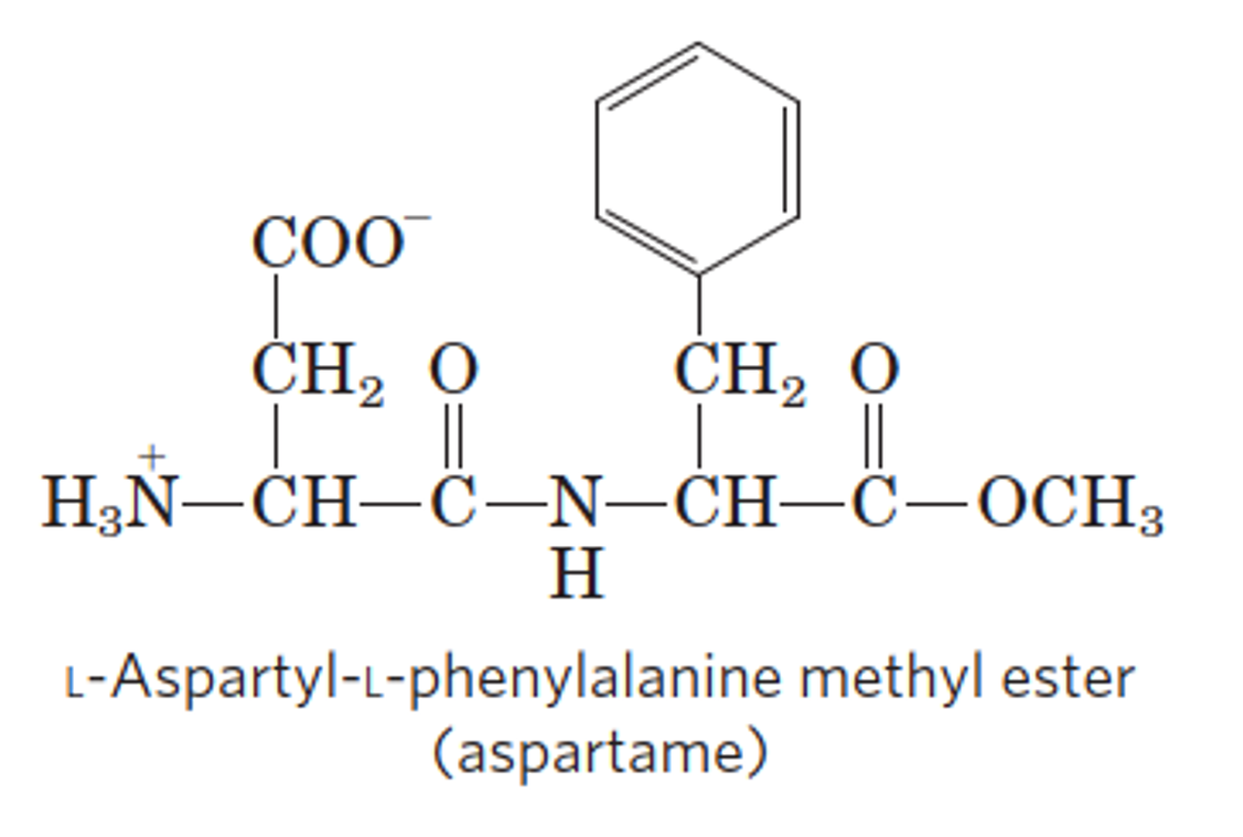
Many kinds of peptides are known. They have been classified or categorized according to their sources and functions. According to the Handbook of Biologically Active Peptides, some groups of peptides include plant peptides, bacterial/antibiotic peptides, fungal peptides, invertebrate peptides, amphibian/skin peptides, venom peptides, cancer/anticancer peptides, vaccine peptides, immune/inflammatory peptides, brain peptides, endocrine peptides, digestive peptides, gastrointestinal peptides, cardiovascular peptides, renal peptides, respiratory peptides, opiate peptides, neurotrophic peptides, and blood–brain peptides. Endorphins consist of a large group of peptides. They are produced by the central nervous system and the pituitary gland. Since endorphins act on the opiate receptors in our brains, they reduce pain and boost pleasure, resulting in a feeling of well-being.
Enkephalin, naturally occurring pentapeptide that has potent painkilling effects and is released by neurons in the central nervous system and by cells in the adrenal medulla.
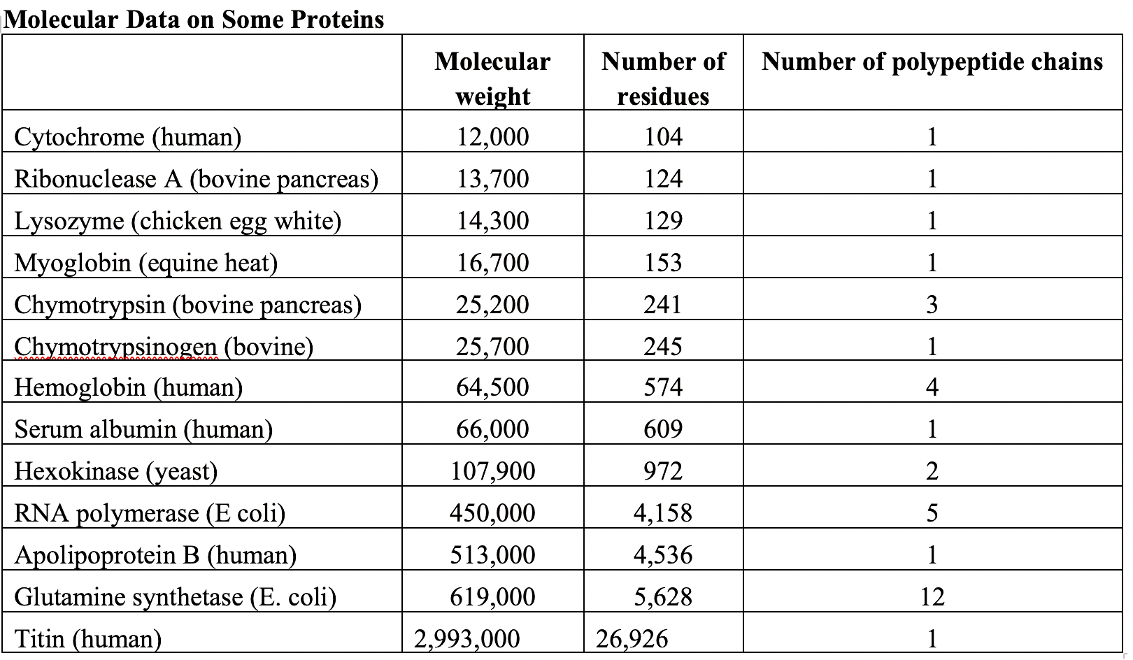
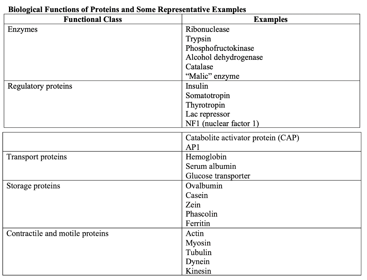
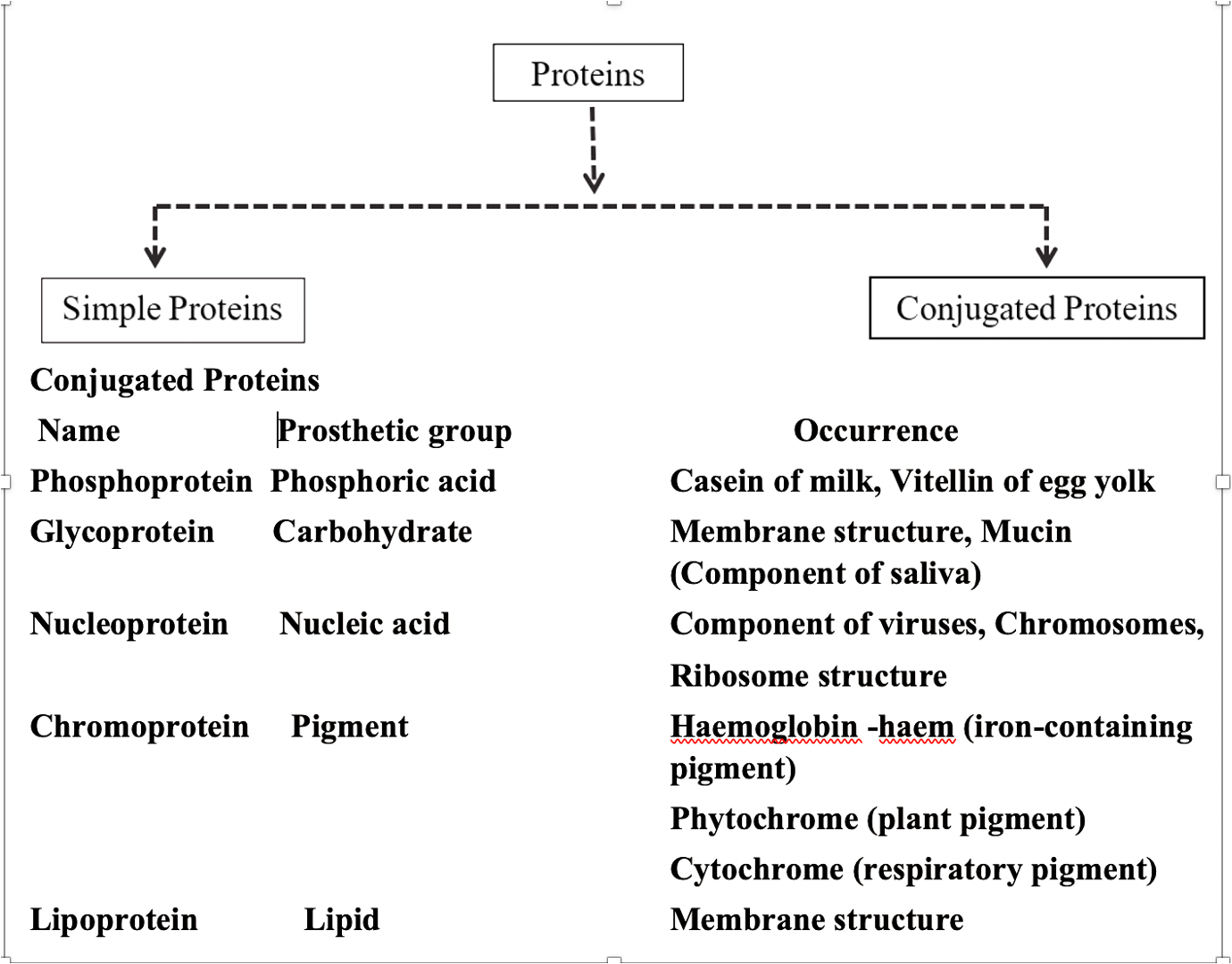
Proteins: Proteins are polymers of amino acids. They are most abundant molecules in the cell. Their molecular weight varies from protein to protein. The smallest is cytochrome c while largest is Titin.Proteins are the agents of biological function. Virtually every cellular activity is dependent on one or more particular proteins. Thus, a convenient way to classify the enormous number of proteins is by the biological roles they fill.
Enzymes:
- By far the largest class of proteins is enzymes. More than 3000 different enzymes are listed in Enzyme Nomenclature, the standard reference volume on enzyme classification. Enzymes are catalysts that accelerate the rates of biological reactions. Each enzyme is very specific in its function and acts only in a particular metabolic reaction.
Regulatory Proteins:
- A number of proteins do not perform any obvious chemical transformation but nevertheless can regulate the ability of other proteins to carry out their physiological functions. Such proteins are referred to as regulatory proteins. A well-known example is insulin, the hormone regulating glucose metabolism in animals.
Transport Proteins:
- A third class of proteins is the transport proteins. These proteins function to transport specific substances from one place to another. One type of transport is exemplified by the transport of oxygen from the lungs to the tissues by hemoglobin.
Storage Proteins:
- Proteins whose biological function is to provide a reservoir of an essential nutrient are called storage proteins eg. Albumin, Oval-albumin, Glidin Glutein etc., some of the storage protein are extremely toxic such as,
- Abrin is an extremely toxic toxalbumin found in the seeds of the rosary pea (or jequirity pea), Abrus precatorius.
- Ricin is a lectin (a carbohydrate-binding protein) and a highly potent toxin produced in the seeds of the castor oil plant, Ricinus communis.
Contractile and Motile Proteins:
- Certain proteins endow cells with unique capabilities for movement. Cell division, muscle contraction, and cell motility represent some of the ways in which cells execute motion. The contractile and motile proteins underlying these motions share a common property: they are filamentous or polymerize to form filaments. Examples include actin and myosin, the filamentous proteins forming the contractile systems of cells, and tubulin, the major component of microtubules (the filaments involved in the mitotic spindle of cell division as well asin flagella and cilia).

Structural Proteins:
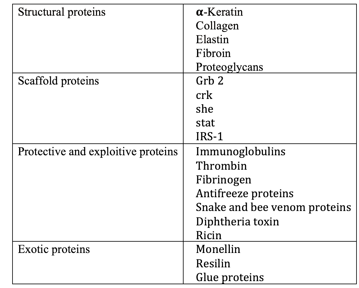
- Structural proteins provide strength and protection to cells and tissues. Monomeric units of structural proteins typically polymerize to generate long fibers (as in hair) or protective sheets of fibrous arrays, as in cowhide (leather). α-Keratins are insoluble fibrous proteins making up hair, horns, and fingernails. Collagen, another insoluble fibrous protein, is found in bone, connective tissue, tendons, cartilage, and hide, where it forms inelastic fibrils of great strength. One-third of the total protein in a vertebrate animal is collagen. A structural protein having elastic properties is, appropriately, elastin, an important component of ligaments. Because of the way elastin monomers are cross-linked in forming polymers, elastin can stretch in two dimensions. Certain insects make a structurally useful protein, Fibroin (a β-keratin), the major constituent of cocoons (silk) and spider webs. An important protective barrier for animal cells is the extracellular matrix containing collagen and proteoglycans, covalent protein–polysaccharide complexes that cushion and lubricate.
Protective and Exploitive Proteins:
- In contrast to the passive protective nature of some structural proteins, another group can be more aptly classified as protective or exploitive proteins because of their biologically active role in cell defense, protection, or exploitation. Prominent among the protective proteins are the immunoglobulins or antibodies produced by the lymphocytes of vertebrates. Antibodies have the remarkable ability to “ignore” molecules that are an intrinsic part of the host organism, yet they can specifically recognize and neutralize “foreign” molecules resulting from the invasion of the organism by bacteria, viruses, or other infectious agents. Another group of protective proteins is the blood-clotting proteins, thrombin and fibrinogen, which prevent the loss of blood when the circulatory system is damaged. Arctic and Antarctic fishes have antifreeze proteins to protect their blood against freezing in the below-zero temperatures of high latitude seas. In addition, various proteins serve defensive or exploitive roles for organisms, including the lytic and neurotoxic proteins of snake and bee venoms and toxic plant proteins, such as ricin, whose apparent purpose is to thwart predation by herbivores. Another class of exploitive proteins includes the toxins produced by bacteria, such as diphtheria toxin and cholera toxin.
According to Structure:
- Fibrous : Fibrous proteins have secondary structure. They are insoluble in water and physically tough. They consist of long parallel polypeptide chains cross-linked at intervals forming long fibres or sheets and perform structural functional in cells and organisms e.g. collagen (tendons, bone, connective tissue), myosin (in muscle), fibroin silk (spiders’ webs), keratin (hair, horn, nails, feathers).
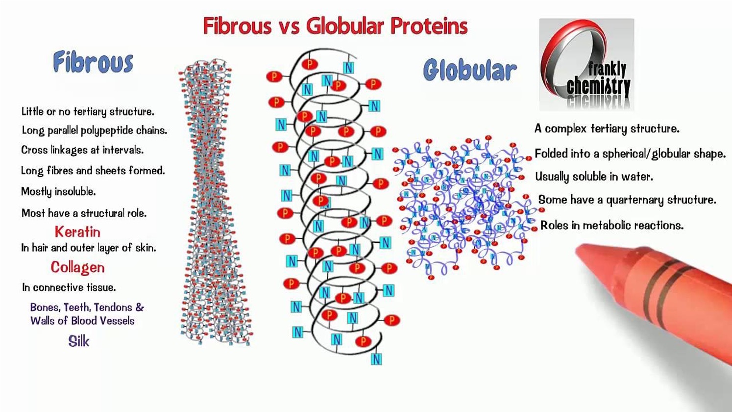
Globular: These proteins have tertiary structure. Polypeptide chains are tightly folded to form spherical shape. They are easily soluble and form enzymes, antibodies and some hormones, e.g. Insulin. Other important roles.
According to Constitution:
Primary Structure (1° structure) :
The primary structure is the sequence of amino acids in a polypeptide chain. Fred Sanger was first person to work out the complete amino acid sequence of hormone insulin and was awarded the Nobel prize for his work in 1958. Insulin is a protein of 51 amino acids. It is made of two polypeptide chains held together by disulphide bridges.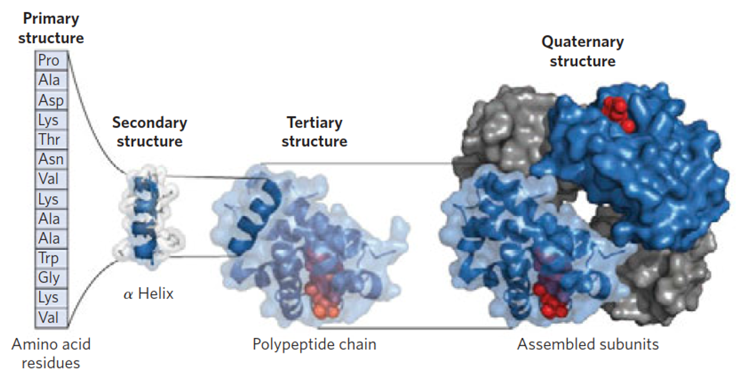
Secondary Structure (2° structure) :
Secondary structure is the local spatial arrangement of the main-chain atoms in a selected segment of a polypeptide chain. The most common regular secondary structures are the α-helix, the β conformation, and β-turns, The most common regular secondary structures are the α-helix, the β conformation, and β-turns, all most all fibrous proteins have secondary structures such as Keratin, Fibroin collagen etc,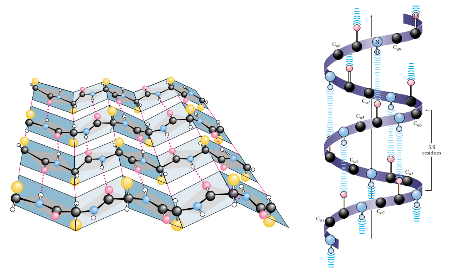
Tertiary Structure (3° structure) :
The overall three-dimensional arrangement of all atoms in a protein is referred to as the protein’s tertiary structure. Whereas the term “secondary structure” refers to the spatial arrangement of amino acid residues that are adjacent in a segment of a polypeptide, tertiary structure includes longer-range aspects of amino acid sequence. Amino acids that are far apart in the polypeptide sequence and are in different types of secondary structure may interact within the completely folded structure of a protein. Some proteins contain two or more separate polypeptide chains, or subunits, which may be identical or different. The arrangement of these protein subunits in three-dimensional complexes constitutes Quaternary structure.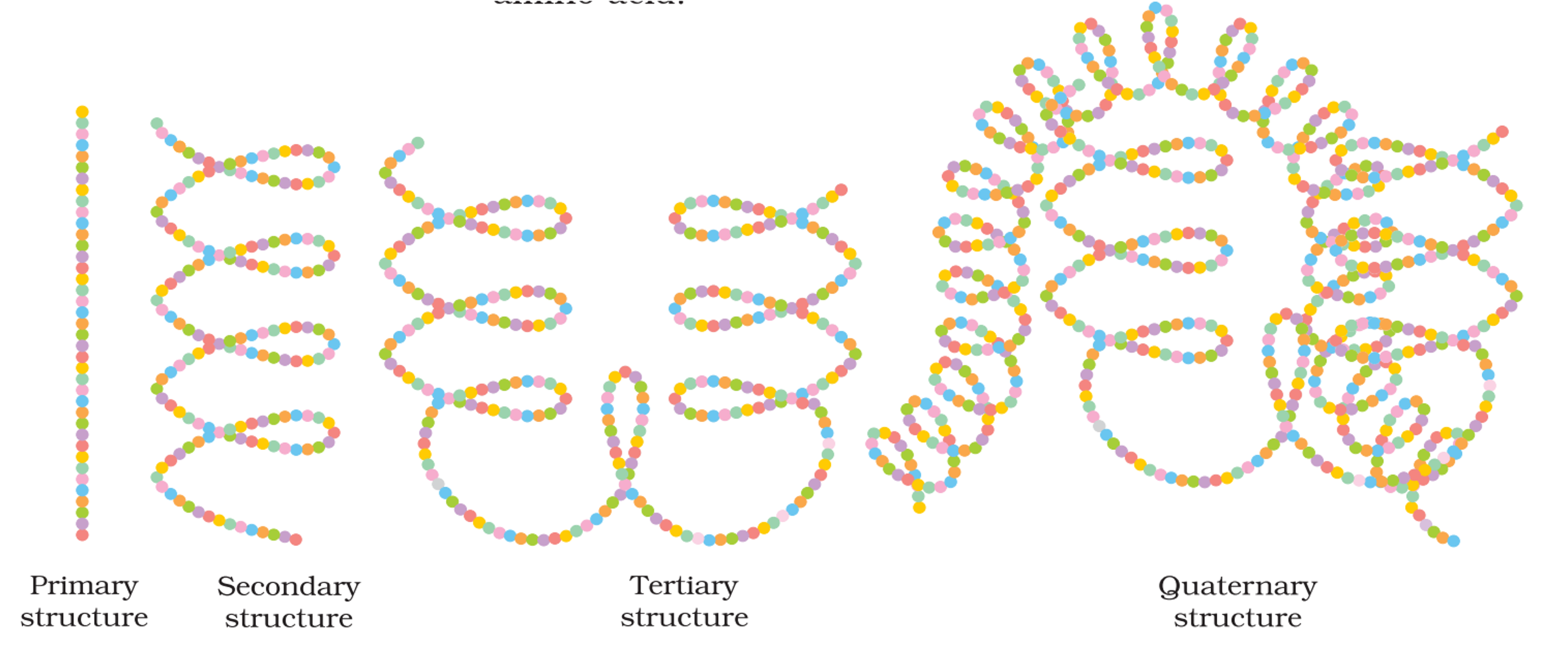
The structure of large molecules such as proteins can be described at several levels of complexity, arranged in a kind of conceptual hierarchy. Four levels of protein structure are commonly defined,
Primary and Secondary Metabolites
- All metabolites ( intermediate and also end products that are produced during or at the end of cellular metabolism) are biomolecules and are of two types: Primary and Secondary metabolites. Primary metabolites are biomolecules which are required in many cellular functions and metabolic pathways like energy, stimulation, inhibition, protection, etc but compounds other than these primary metabolites e.g. alkaloids, flavonoids, rubber, essential oils, antibiotics, coloured pigments, scents, gums, spices etc are called secondary metabolites.

Proteins
- Proteins are polypeptides. They are linear chains of amino acids linked by peptide bonds.
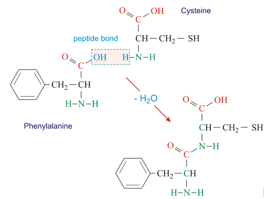
Each protein is a polymer of amino acids. As there are 20 types of amino acids (e.g., alanine, cysteine, proline, tryptophan, lysine, etc.), a protein is a heteropolymer and not a homopolymer. A homopolymer has only one type of monomer repeating ‘n’ number of times.
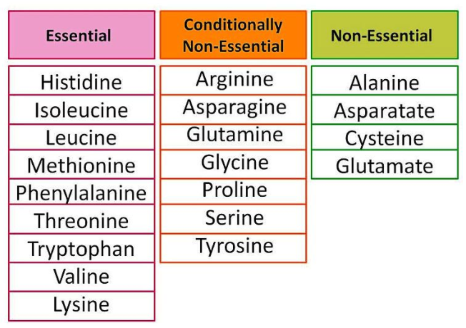
Amino acids can be essential or non-essential. The latter are those which our body can make, while we get essential amino acids through our diet/food.
Proteins carry out many functions in living organisms, some transport nutrients across cell membrane, some fight infectious organisms, some are hormones, some are enzymes.
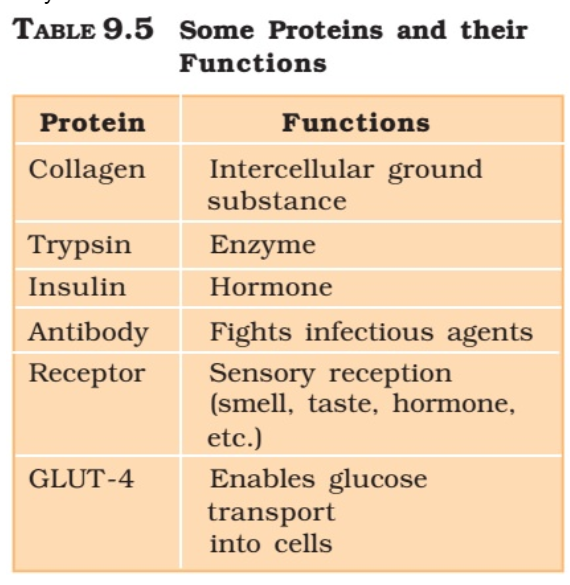
Structure of Proteins
Primary Structure : Simple linear chain of amino acids joined together by peptide bonds.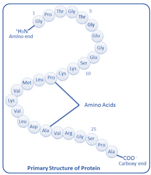
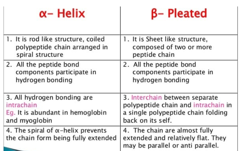
Secondary Structure : New interactions like hydrogen bonds between different groups arise in the primary structure giving rise to a folded structure which is a bit more complex than the previous linear structure known as secondary structure. Types of secondary structures are ‘alpha’-helix, β-sheet and collagen helix.
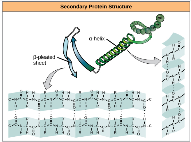
Common features of α-helical structure :
Tertiary Structure : this is the 3D structure of a protein which forms due to interactions (like hydrophobic or van der waals force and/or electrostatic interactions) between α-helix and β-pleated sheets. It is of 2 types : globular and fibrous.
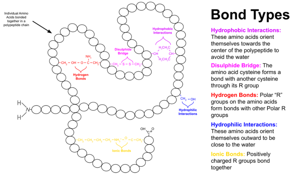
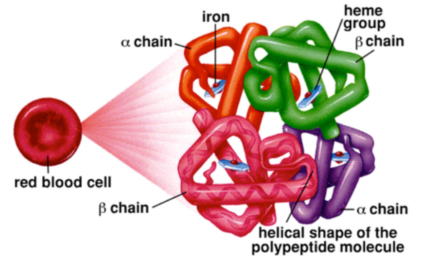
Quaternary Structure : some proteins are multimeric so the interactions between different polypeptide chains of a protein give rise to a quaternary structure. Ex : hemoglobin molecule.
Proteins
- Proteins are polypeptides. They are linear chains of amino acids linked by peptide bonds.

Each protein is a polymer of amino acids. As there are 20 types of amino acids (e.g., alanine, cysteine, proline, tryptophan, lysine, etc.), a protein is a heteropolymer and not a homopolymer. A homopolymer has only one type of monomer repeating ‘n’ number of times.

Amino acids can be essential or non-essential. The latter are those which our body can make, while we get essential amino acids through our diet/food.

Proteins carry out many functions in living organisms, some transport nutrients across cell membrane, some fight infectious organisms, some are hormones, some are enzymes.

Structure of Proteins
Primary Structure : Simple linear chain of amino acids joined together by peptide bonds.
Secondary Structure : New interactions like hydrogen bonds between different groups arise in the primary structure giving rise to a folded structure which is a bit more complex than the previous linear structure known as secondary structure. Types of secondary structures are ‘alpha’-helix, β-sheet and collagen helix.

Common features of α-helical structure :

Tertiary Structure : this is the 3D structure of a protein which forms due to interactions (like hydrophobic or van der waals force and/or electrostatic interactions) between α-helix and β-pleated sheets. It is of 2 types : globular and fibrous.

Quaternary Structure : some proteins are multimeric so the interactions between different polypeptide chains of a protein give rise to a quaternary structure. Ex : hemoglobin molecule.

Enzymes
- Much of the history of biochemistry is the history of enzyme research. Biological catalysis was first recognized and described in the late 1700s, in studies on the digestion of meat by secretions of the stomach. In 1752 Remur proposed that digestive reactions are impossible at room temperature. In 1809 Kirchoff found that wheat extract can digest starch.
French chemist Anselme Payen was the first to discover an enzyme, diastase, in 1833. A few decades later, when studying the fermentation of sugar to alcohol by yeast, Louis Pasteur concluded that this fermentation was caused by a vital force contained within the yeast cells called "ferments", which were thought to function only within living organisms. He wrote that "alcoholic fermentation is an act correlated with the life and organization of the yeast cells, not with the death or putrefaction of the cells. In 1877, German physiologist Wilhelm Kühne (1837–1900) first used the term enzyme, which comes from "in yeast", to describe this process. Eduard Buchner submitted his first paper on the study of yeast extracts in 1897. In a series of experiments at the University of Berlin, he found that sugar was fermented by yeast extracts even when there were no living yeast cells in the mixture. He named the enzyme that brought about the fermentation of sucrose “zymas”.
In 1912 Kinsbury proposed that enzymes are small carbohydrates like molecules but in 1928 Summner isolated Urease from Jack bean or Canavalia ensiformis and crystalized it to find that Urease or Summner enzyme is a protein. In 1930 John Northop crystallized Pepsin and Trypin and found out that they are also proteins.
Properties of Enzymes:
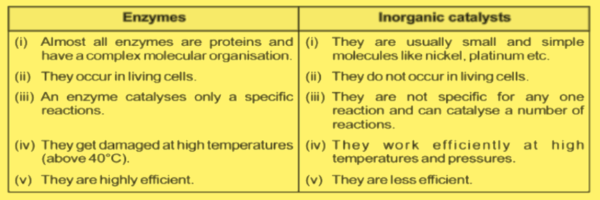
All enzymes are large specialized proteins having a molecular weight ranging from 12000 daltons to many millions. However in 1967, Carl Woese, Francis Crick, and Leslie Orgel were the first to suggest that RNA could act as a catalyst. This idea was based upon the discovery that RNA can form complex secondary structures. These ribozymes were found in the intron of an RNA transcript, which removed itself from the transcript, as well as in the RNA component of the RNase P complex, which is involved in the maturation of pre-tRNAs. In 1989, Thomas R. Cech and Sidney Altman shared the Nobel Prize in chemistry for their "discovery of catalytic properties of RNA.
Specificity of Enzymatic reaction:
A given enzyme is very selective, both in the substances with which it interacts and in the reaction that it catalyzes. The substances upon which an enzyme acts are traditionally called Substrates. In an enzyme-catalyzed reaction, none of the substrate is diverted into nonproductive side reactions, so no wasteful by-products are produced. It follows then that the products formed by a given enzyme are also very specific. The selective qualities of an enzyme are collectively recognized as its specificity.
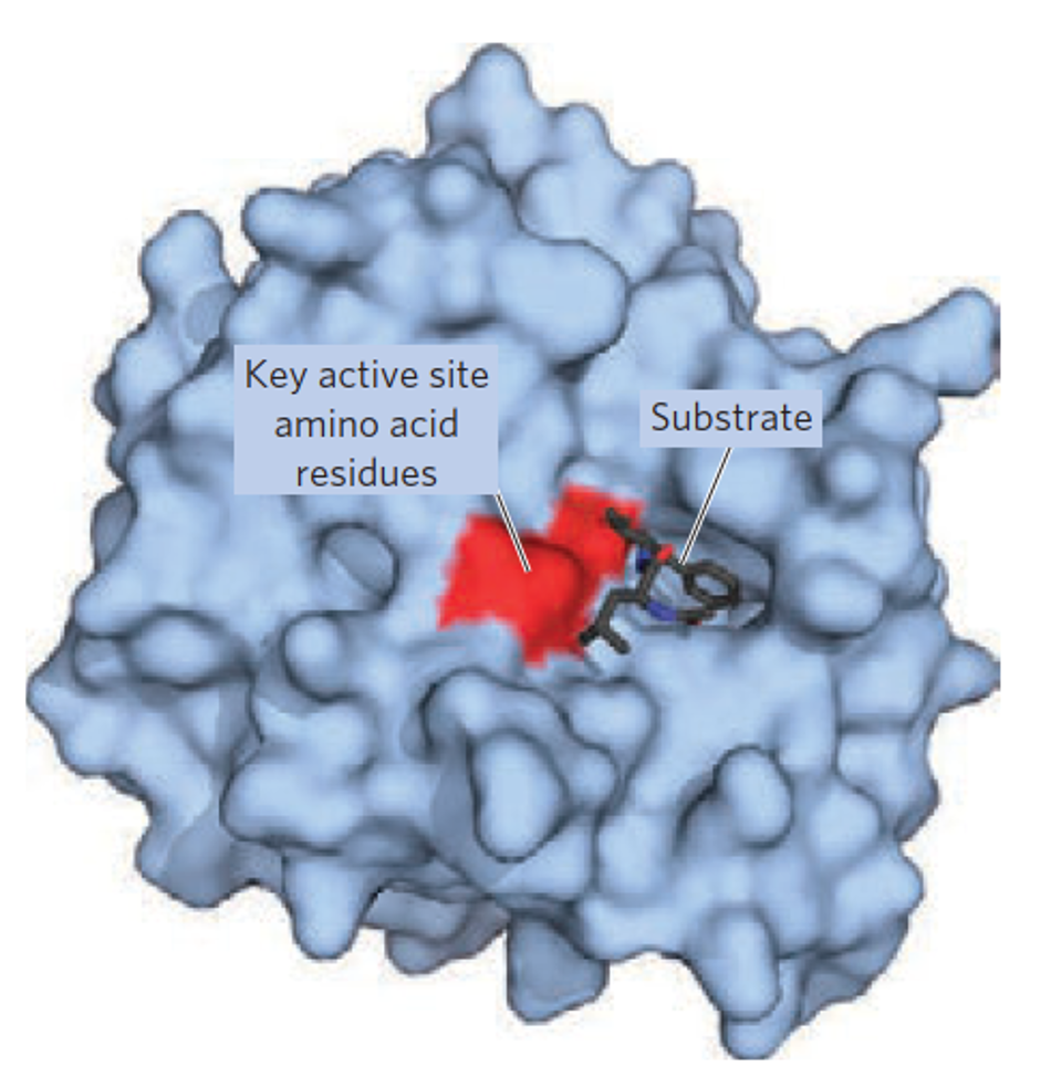
Intimate interaction between an enzyme and its substrates occurs through molecular recognition based on structural complementarity; such mutual recognition is the basis of specificity. The specific site on the enzyme where substrate binds and catalysis occurs is called the Active site. Enzymes Affect Reaction Rates, Not Equilibria
A simple enzymatic reaction might be written as,

where E, S, and P represent the enzyme, substrate, and product; ES and EP are transient complexes of the enzyme with the substrate and with the product.
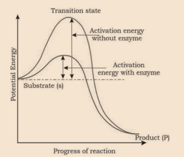
In the coordinate diagram, the free energy of the system is plotted against the progress of the reaction (the reaction coordinate). The starting point for either the forward or the reverse reaction is called the ground state, the contribution to the free energy of the system by an average molecule (S or P) under a given set of conditions.
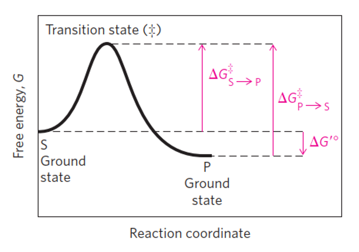
The difference between the energy levels of the ground state and the transition state is the activation energy, G‡. The rate of a reaction reflects this activation energy: a higher activation energy corresponds to a slower reaction. Reaction rates can be increased by raising the temperature and/or pressure, thereby increasing the number of molecules with sufficient energy to overcome the energy barrier. Alternatively, the activation energy can be lowered by adding a catalyst . Catalysts enhance reaction rates by lowering activation energies.
Coenzymes:
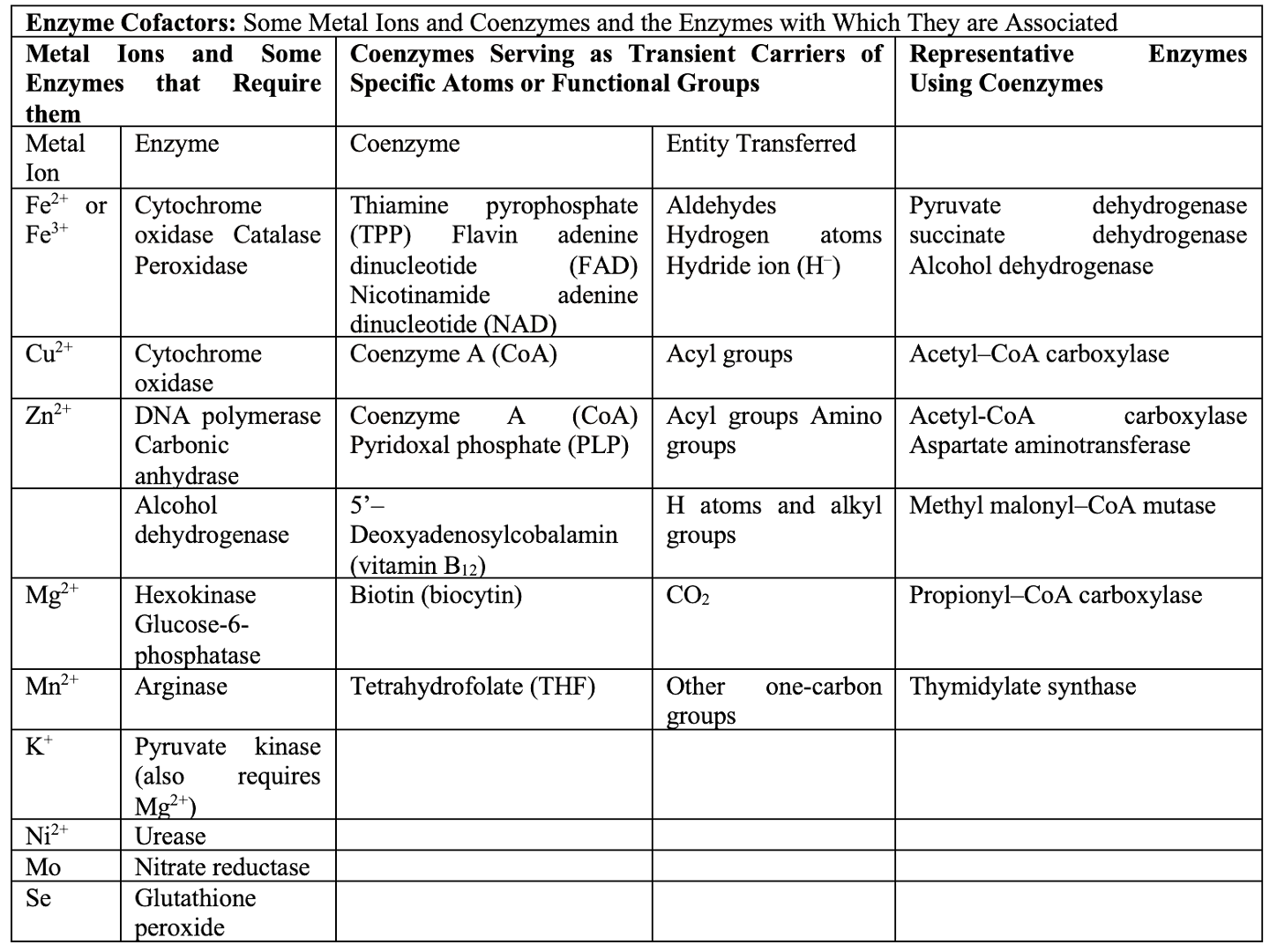
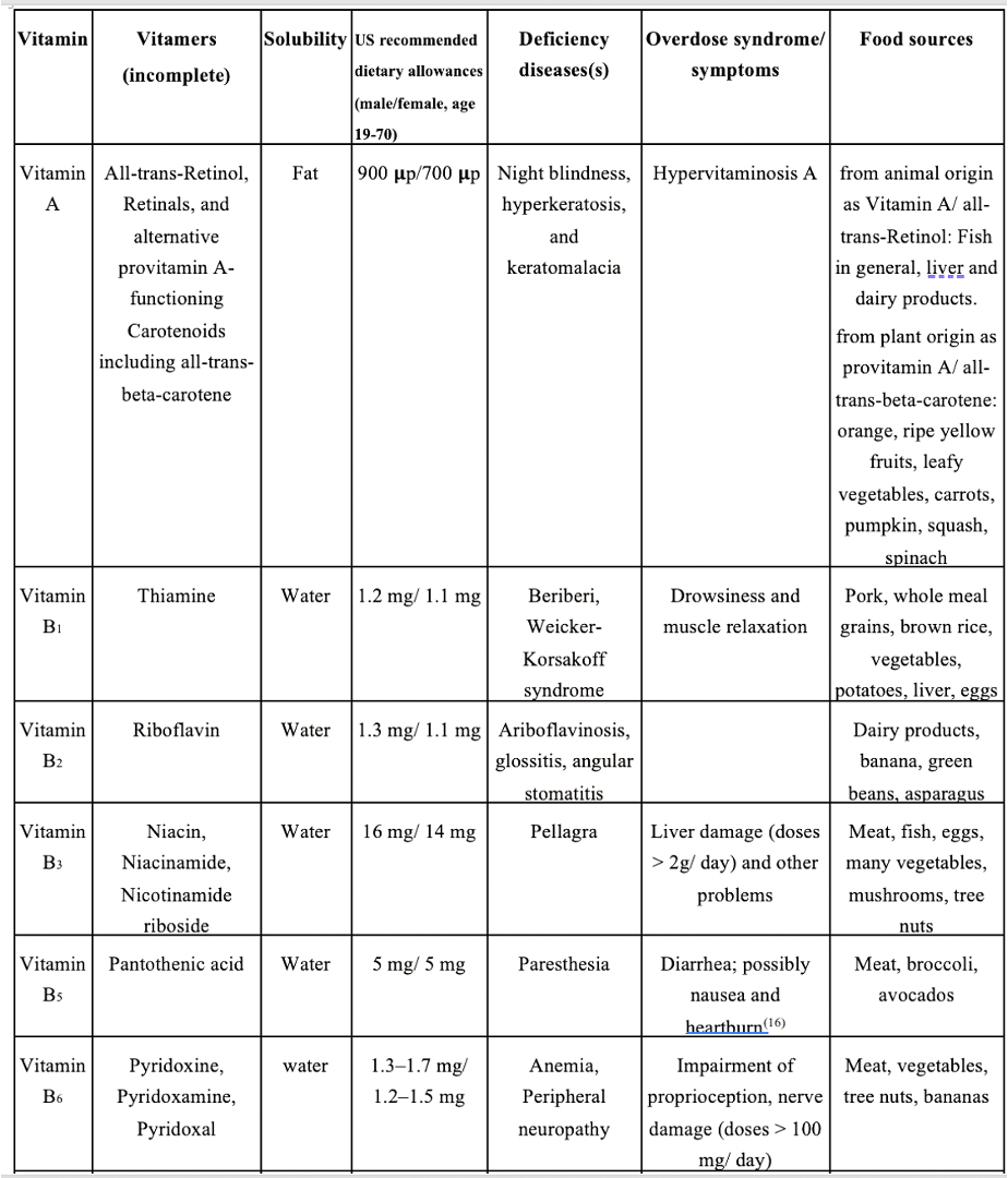
- Many enzymes carry out their catalytic function relying solely on their protein structure. Many others require non protein components, called cofactors. Cofactors may be metal ions or organic molecules referred to as coenzymes. Cofactors, because they are structurally less complex than proteins, tend to be stable to heat (incubation in a boiling water bath). Typically, proteins are denatured under such conditions. Many coenzymes are vitamins or contain vitamins as part of their structure. Usually coenzymes are actively involved in the catalytic reaction of the enzyme, often serving as intermediate carriers of functional groups in the conversion of substrates to products. In most cases, a coenzyme is firmly associated with its enzyme, perhaps even by covalent bonds, and it is difficult to separate the two. Such tightly bound coenzymes are referred to as prosthetic groups of the enzyme.
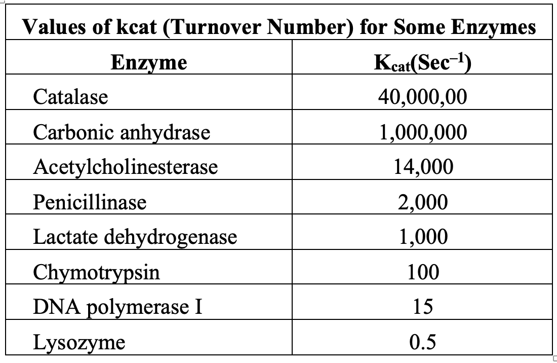
- Lock and Key Hypothesis: When the enzyme binds to substrate, it forms an intermediate complex before the final product is formed. The reaction takes place in two ways as given below,
-Step1: Combining of enzyme and the reactant/substrate.
E + S → [ES]
-Step 2: Disintegration of the complex molecule to give the product.
[ES]→ E + P
Thus, the whole catalyst action of enzymes is summarized as:
E + S → [ES] → [EP] → E + P
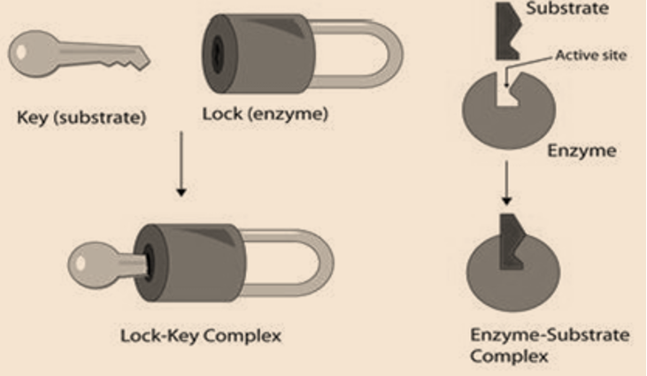
Fischer has developed a Lock and Key theory to describe the mode of action of the enzyme. According to this principle, if the right key fits inside the right lock, the lock will be opened otherwise it will not. Likewise, if the right enzyme fits into the right substrate, the drug will form, otherwise, it won’t. Enzyme shape provides surface configurations that can fit with the other molecules. The molecules on which the enzymes act are known as substrates of enzymes. Substrates that have the correct geometric shape can fit within the active site of the enzyme.
The active site of the enzyme is highly specific about the surface of its substrates. The enzyme and substrate fit like a lock and key, making it a lock and key enzyme action model. Sometimes, however, certain molecules close to the substrate may also interact with an enzyme’s active site. The molecule completes with the substrate and can either slow down or stop the reaction. Such substance is called the competitive inhibitor because it acts to prevent a product from being created.
-
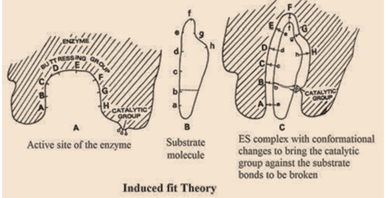
Induced fit Hypothesis : The induced fit model is a model for enzyme-substrate interaction. It describes that only the proper substrate is capable of inducing the proper alignment of the active site that will enable the enzyme to perform its catalytic function. It also suggests that the active site continues to change until the substrate is completely bound to it, at which point the final shape and charge is determined. The induced fit model suggested by Daniel Koshland in 1958.
It is the more accepted model for enzyme-substrate complex than the lock-and-key model. In the lock-and-key model, the interaction of the substrate and the enzyme is likened to a key (the substrate) that is highly specific to the lock (the active site of the enzyme).
Koshland (1959) believes that each active site of an enzyme contains two regions, buttressing and catalytic. The buttressing region provides attachment site to the substrate molecules. For this it has various types of weak bonds or linkages. The catalytic group which lies at a distance from buttressing group is specialised to weaken bonds of reactant or substrate molecules through electrophilic and nucleophilic forces. When the substrate molecules come in contact with buttressing group, the active site undergoes conformational change which brings the catalytic group opposite those bonds of the substrate which are to be weakened.
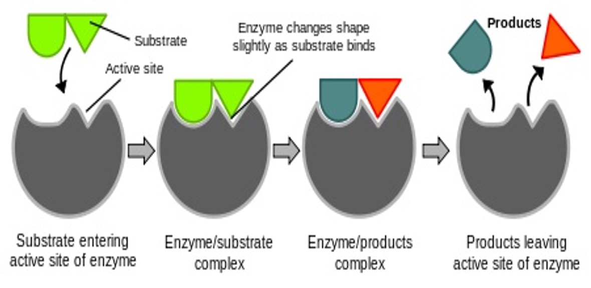
The catalytically active complex of protein and prosthetic group is called the Holoenzyme. The protein without the prosthetic group is called the Apoenzyme; it is catalytically inactive.
Effect of pH on Enzymatic Activity:
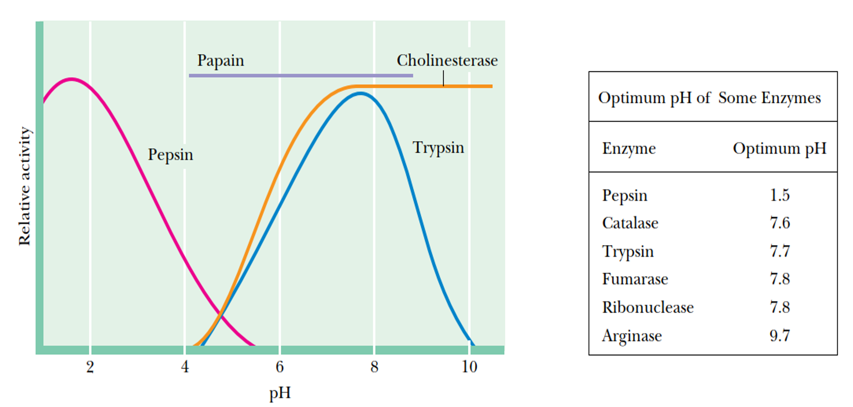
- Enzyme-substrate recognition and the catalytic events that ensue are greatly dependent on pH. An enzyme possesses an array of ionizable side chains and prosthetic groups that not only determine its secondary and tertiary structure but may also be intimately involved in its active site. Further, the substrate itself often has ionizing groups, and one or another of the ionic forms may preferentially interact with the enzyme. Enzymes in general are active only over a limited pH range and most have a particular pH at which their catalytic activity is optimal. These effects of pH may be due to effects on Km or Vmax or both.
Effect of Temperature on Enzymatic Activity:
- Like most chemical reactions, the rates of enzyme-catalyzed reactions generally increase with increasing temperature. However, at temperatures above 50°to 60°C, enzymes typically show a decline in activity. Two effects are operating here:
-
(a) The characteristic increase in reaction rate with temperature, and
(b) Thermal denaturation of protein structure at higher temperature. Most enzymatic reactions double in rate for every 10°C rise in temperature (that is, Q10=2, where Q10 is defined as the ratio of activities at two temperatures 10° apart) as long as the enzyme is stable and fully active. Some enzymes, those catalyzing reactions having very high activation energies, show proportionally greater Q10 values. The increasing rate with increasing temperature is ultimately offset by the instability of higher orders of protein structure at elevated temperatures, where the enzyme is inactivated. Not all enzymes are quite so thermally labile. For example, the enzymes of thermophilic bacteria (thermophilic = “heat-loving”) found in geothermal springs retain full activity at temperatures in excess of 85°C.
Enzyme Nomenclature:
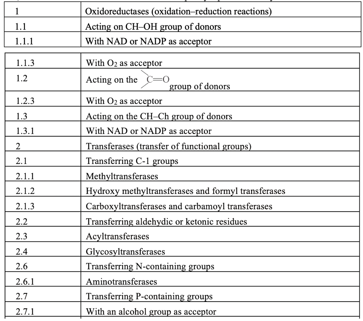
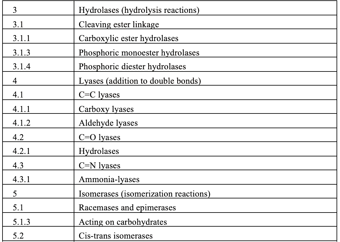
- Traditionally, enzymes often were named by adding the suffix -ase to the name of the substrate upon which they acted, as in urease for the urea-hydrolyzing enzyme or phosphatase for enzymes hydrolyzing phosphoryl groups from organic phosphate compounds. Other enzymes acquired names bearing little resemblance to their activity, such as the peroxide-decomposing enzyme catalase or the proteolytic enzymes (proteases) of the digestive tract, trypsin and pepsin. Because of the confusion that arose from these trivial designations, an International Commission on Enzymes was established in 1956 to create a systematic basis for enzyme nomenclature. Although common names for many enzymes remain in use, all enzymes now are classified and formally named according to the reaction they catalyze. Six classes of reactions are recognized. Within each class are subclasses, and under each subclass are sub-subclasses within which individual enzymes are listed. Classes, subclasses, sub-subclasses, and individual entries are each numbered, so that a series of four numbers serves to specify a particular enzyme.

Chemical kinetics is the study of the rates of chemical reactions. Consider a reaction of overall stoichiometry.
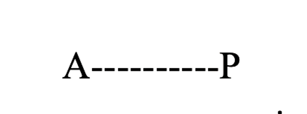
Although we treat this reaction as a simple, one-step conversion of A to P, it more likely occurs through a sequence of elementary reactions, each of which is a simple molecular process, as in

where I and J represent intermediates in the reaction. Precise description of all of the elementary reactions in a process is necessary to define the over all reaction mechanism for A------P.
Let us assume that A -----P is an elementary reaction and that it is spontaneous and essentially irreversible. Irreversibility is easily assumed if the rate of P conversion to A is very slow or the concentration of P (expressed as [P]) is negligible under the conditions chosen. The velocity, v, or rate, of the reaction A----P is the amount of P formed or the amount of A consumed per unit time, t. That is,
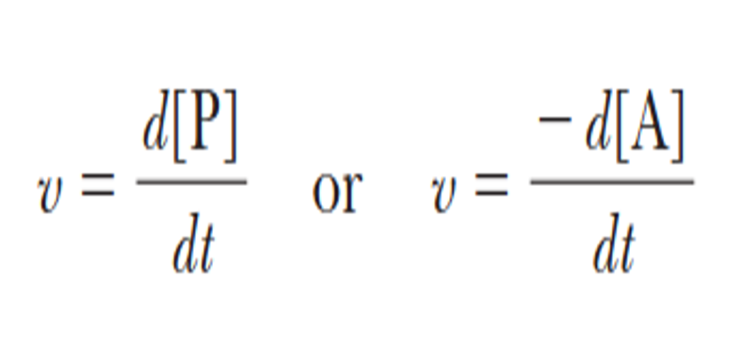
The mathematical relationship between reaction rate and concentration of reactant(s) is the rate law. For this simple case, the rate law is
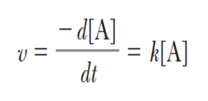
From this expression, it is obvious that the rate is proportional to the concentration of A, and k is the proportionality constant, or rate constant. k has the units of (time)-1, usually sec-1. v is a function of [A] to the first power, or, in the terminology of kinetics, v is first-order with respect to A. For an elementary reaction, the order for any reactant is given by its exponent in the rate equation. The number of molecules that must simultaneously interact is defined as the molecularity of the reaction. Thus, the simple elementary reaction of A----P is a first-order reaction. If we draw a plot of the reaction A----P, a plot of the reaction rate as a function of the concentration of A yields a straight line whose slope is k . The more A that is available, the greater the rate of the reaction, v. Similar analyses of enzyme-catalyzed reactions involving only a single substrate yield remarkably different results. At low concentrations of the substrate S, v is proportional to [S], as expected for a first-order reaction.
However, v does not increase proportionally as [S] increases, but instead begins to level off. At high [S], v becomes virtually independent of [S] and approaches a maximal limit. The value of v at this limit is written Vmax. Because rate is no longer dependent on [S] at these high concentrations, the enzyme-catalyzed reaction is now obeying zero-order kinetics; that is, the rate is independent of the reactant (substrate) concentration. This behavior is a saturation effect: when v shows no increase even though [S] is increased, the system is saturated with substrate. Such plots are called substrate saturation curves. The physical interpretation is that every enzyme molecule in the reaction mixture has its substrate-binding site occupied by S. Indeed, such curves were the initial clue that an enzyme interacts directly with its substrate by binding it.
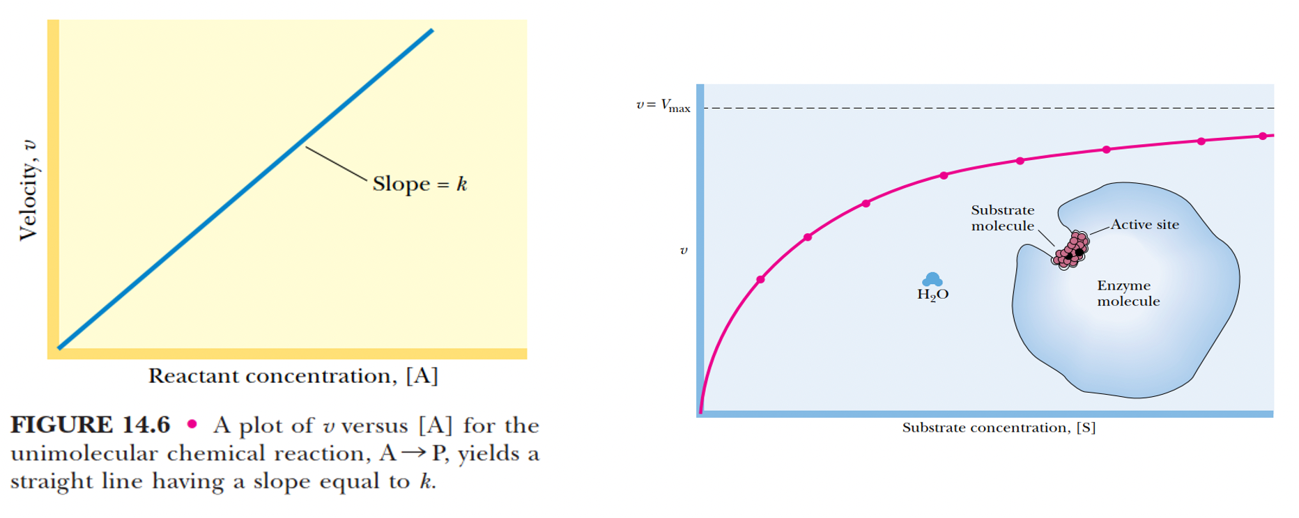
The Michaelis–Menten Equation :
- Lenore Michaelis and Maud L. Menten proposed a general theory of enzyme action in 1913 consistent with observed enzyme kinetics. Their theory was based on the assumption that the enzyme, E, and its substrate, S, associate reversibly to form an enzyme-substrate complex, ES:
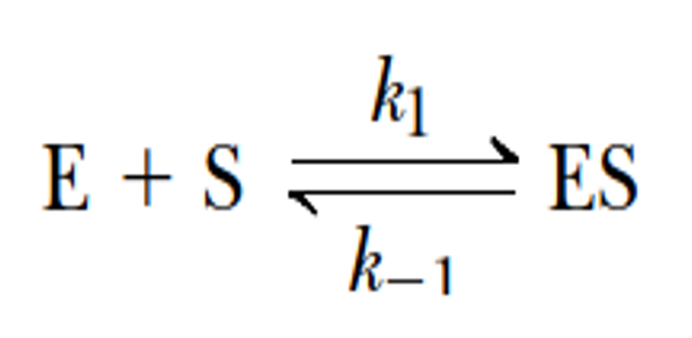
This equation says that the rate of an enzyme-catalyzed reaction, v, at any moment is determined by two constants, Km and Vmax, and the concentration of substrate at that moment.
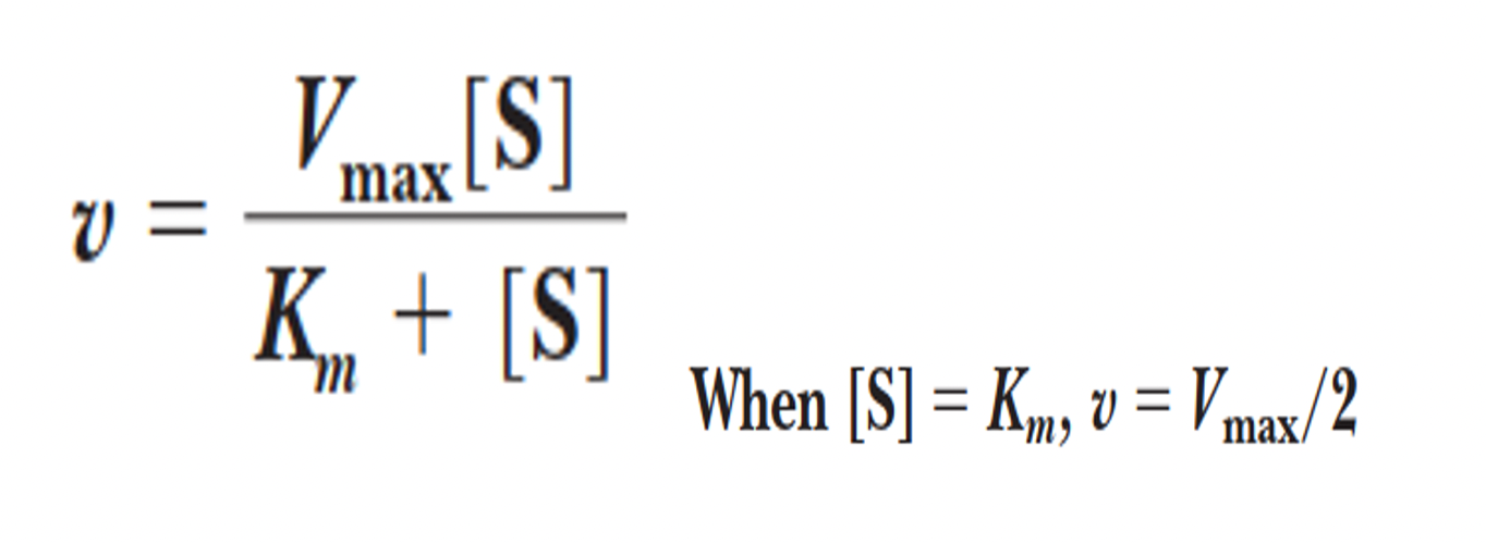
Turnover Number :
- The turnover number of an enzyme, kcat, is a measure of its maximal catalytic activity. kcat is defined as the number of substrate molecules converted into product per enzyme molecule per unit time when the enzyme is saturated with substrate. The turnover number is also referred to as the molecular activity of the enzyme.
 Mechanism of enzymatic action: Many theories have been given to explain the enzymatic action but two theories are most accepted,
-
1. Lock and Key Hypothesis
2. Induced fit Hypothesis
Enzyme Inhibition:
- If the function of the enzyme is inhibited by the use of chemicals, then phenomenon is called Enzymatic inhibition and the chemical is called Chemical Inhibitor.
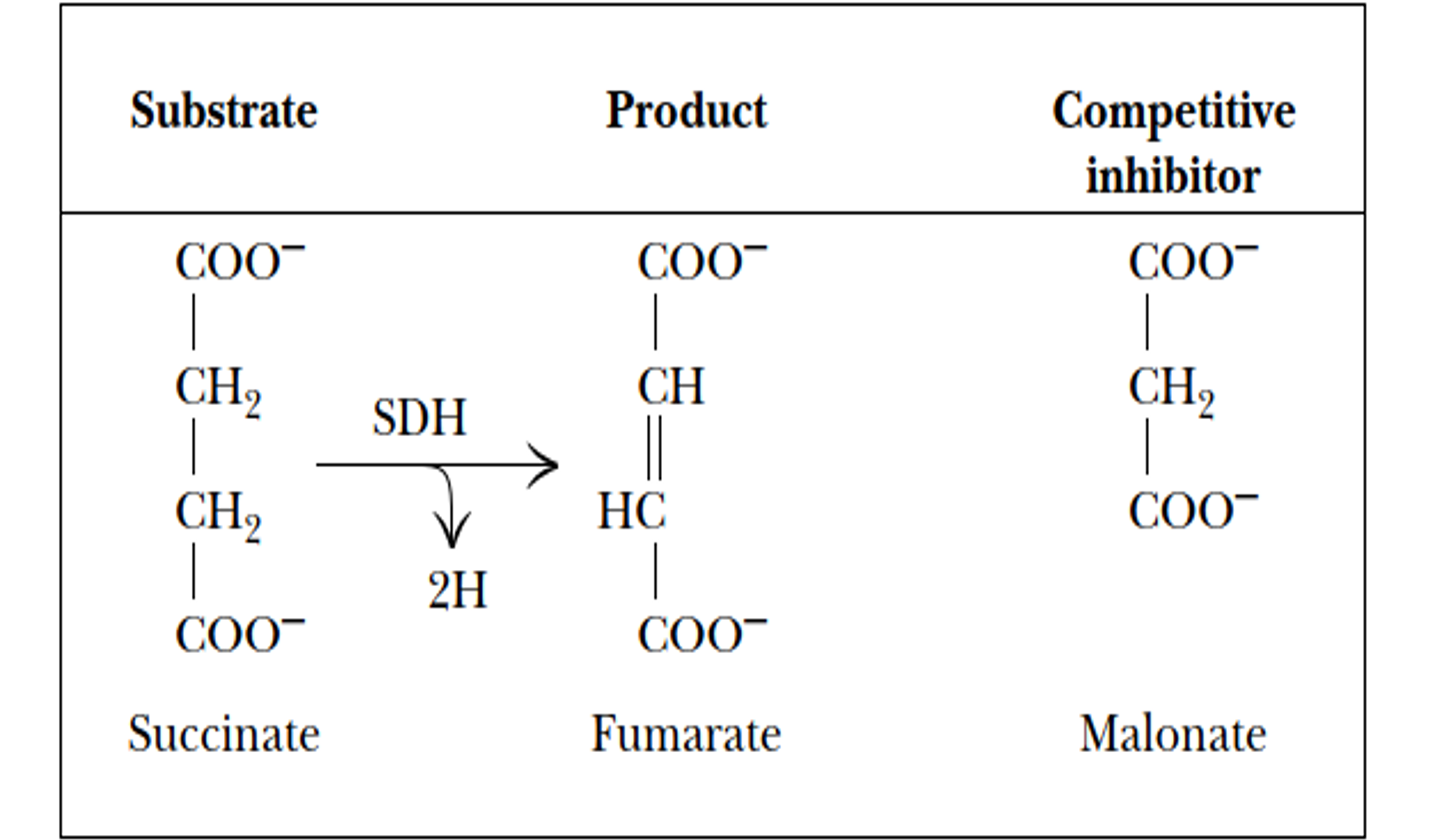
A medical therapy based on competition at the active site is used to treat patients who have ingested methanol, a solvent found in gas-line anti-freeze. The liver enzyme alcohol dehydrogenase converts methanol to formaldehyde, which is damaging to many tissues. Blindness is a common result of methanol ingestion, because the eyes are particularly sensitive to formaldehyde. Ethanol competes effectively with methanol as an alternative substrate for alcohol dehydrogenase. The effect of ethanol is much like that of a competitive inhibitor, with the distinction that ethanol is also a substrate for alcohol dehydrogenase and its concentration will decrease over time as the enzyme converts it to acetaldehyde. The therapy for methanol poisoning is slow intravenous infusion of ethanol, at a rate that maintains a controlled concentration in the bloodstream for several hours. This slows the formation of formaldehyde, lessening the danger while the kidneys filter out the methanol to be excreted harmlessly in the urine.
Isoenzymes (or isozymes) are a group of enzymes that catalyze the same reaction but have different enzyme forms and catalytic efficiencies.
Isozymes are usually distinguished by their electrophoretic mobilities.
The enzyme Lactate dehydrogenase is a tetramer made of two different sub-units, the H-form and the M-form. These combine in different combinations depending on the tissue.
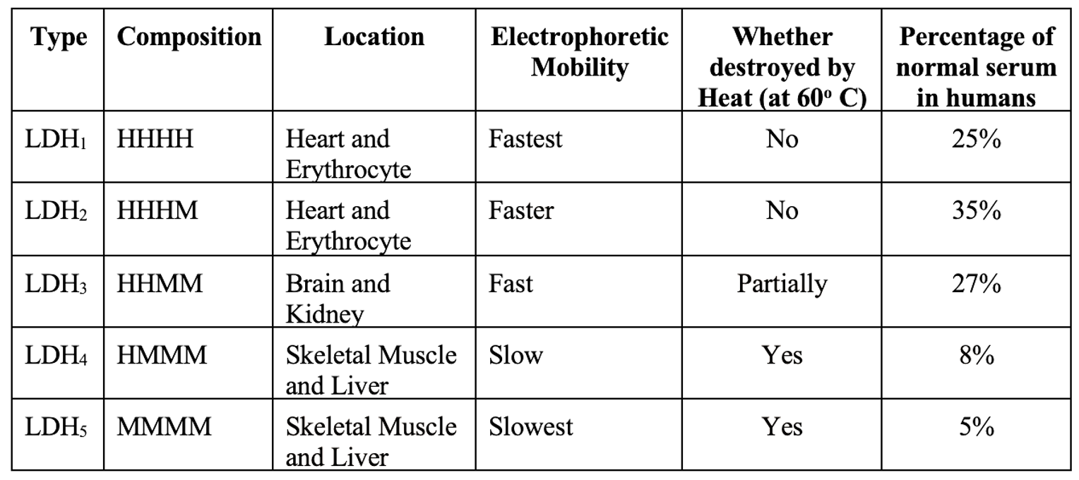 Similarly there are isoenzymes of Creatine phosphokinase and there are six Isoenzymes of alkaline phosphatase and finally α-amylase of wheat have 12 isoenzymatic forms.
Biomacromolecules
- one feature common to all those compounds found in the acid soluble pool. They have molecular weights ranging from 18 to around 800 daltons (Da) approximately. The acid insoluble fraction has only four types of organic compounds i.e., proteins, nucleic acids, polysaccharides and lipids. These classes of compounds with the exception of lipids, have molecular weights in the range of ten thousand daltons and above. For this very reason, biomolecules, i.e., chemical compounds found in living organisms are of two types. One, those which have molecular weights less than one thousand dalton and are usually referred to as micromolecules or simply biomolecules while those which are found in the acid insoluble fraction are called macromolecules or biomacromolecules. The molecules in the insoluble fraction with the exception of lipids are polymeric substances. Lipids are indeed small molecular weight arranged into structures like cell membrane and other membranes. When we grind a tissue, we are disrupting the cell structure. Cell membrane and other membranes are broken into pieces, and form vesicles which are not water soluble. Therefore, these membrane fragments in the form of vesicles get separated along with the acid insoluble pool and hence in the macromolecular fraction. Lipids are not strictly Macromolecules. The acid soluble pool represents roughly the cytoplasmic composition. The macromolecules from cytoplasm and organelles become the acid insoluble fraction. Together they represent the entire chemical composition of living tissues or organisms.

Polysaccharides
- Polysaccharides are long chains of sugars. They are threads (literally a cotton thread) containing different monosaccharides (mono=single, saccharide=sugar unit) as building blocks.
- For example, cellulose is a polymeric polysaccharide consisting of only one type of monosaccharide i.e., glucose. Cellulose is a homopolymer.
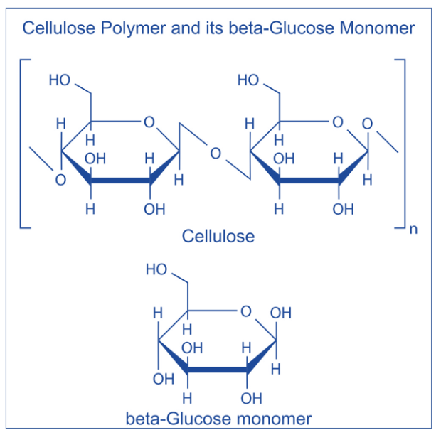
- Starch is a variant of this but present as a store house of energy in plant tissues.
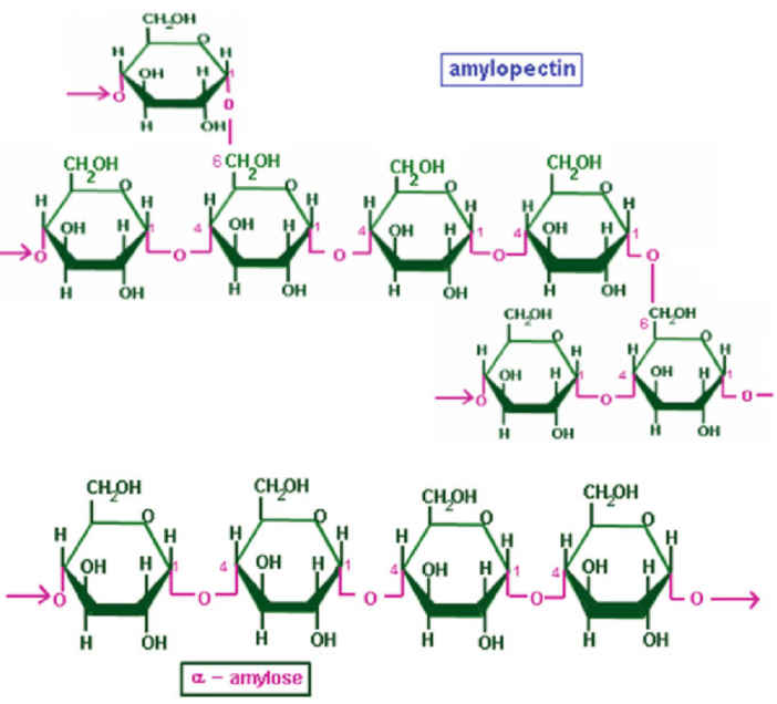
- Animals have another variant called glycogen.
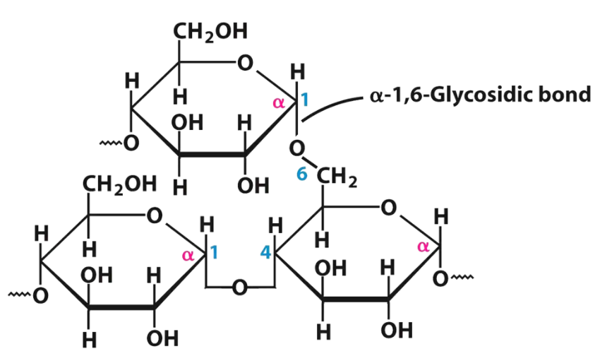
- Inulin is a polymer of fructose.
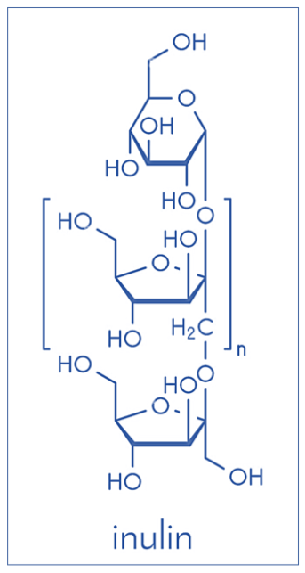
- In a polysaccharide chain (say glycogen), the right end is called the reducing end and the left end is called the non-reducing end.
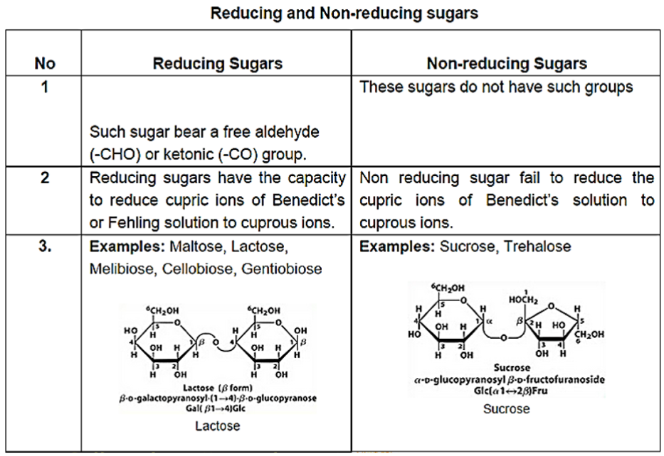
- Starch forms helical secondary structures. In fact, starch can hold I2 molecules in the helical portion. The starch-I2 is blue in colour. Cellulose does not contain complex helices and hence cannot hold I2.
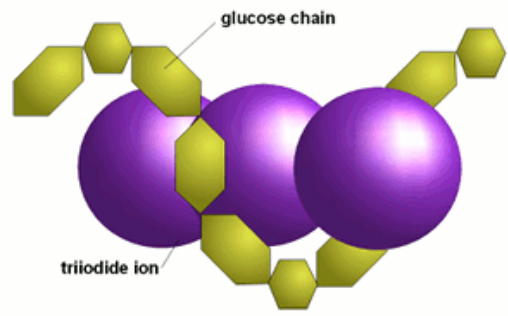
- There are more complex polysaccharides in nature. They have as building blocks, amino-sugars and chemically modified sugars (e.g., glucosamine, N-acetyl galactosamine, etc.).
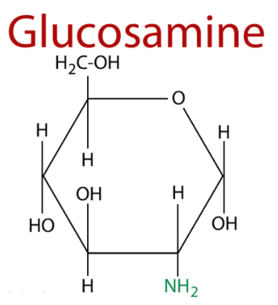
- Exoskeletons of arthropods, for example, have a complex polysaccharide called chitin.
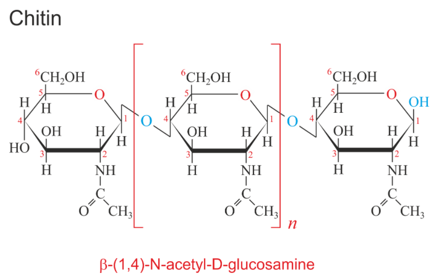
Polysaccharides
- Polysaccharides are long chains of sugars. They are threads (literally a cotton thread) containing different monosaccharides (mono=single, saccharide=sugar unit) as building blocks.
- For example, cellulose is a polymeric polysaccharide consisting of only one type of monosaccharide i.e., glucose. Cellulose is a homopolymer.

- Starch is a variant of this but present as a store house of energy in plant tissues.

- Animals have another variant called glycogen.

- Inulin is a polymer of fructose.

- In a polysaccharide chain (say glycogen), the right end is called the reducing end and the left end is called the non-reducing end.

- Starch forms helical secondary structures. In fact, starch can hold I2 molecules in the helical portion. The starch-I2 is blue in colour. Cellulose does not contain complex helices and hence cannot hold I2.

- There are more complex polysaccharides in nature. They have as building blocks, amino-sugars and chemically modified sugars (e.g., glucosamine, N-acetyl galactosamine, etc.).

- Exoskeletons of arthropods, for example, have a complex polysaccharide called chitin.

Lipids
- Biological lipids are a chemically diverse group of compounds, the common and defining feature of which is their insolubility in water. The biological functions of the lipids are as diverse as their chemistry. Fats and oils are the principal stored forms of energy in many organisms. Phospholipids and sterols are major structural elements of biological membranes. Other lipids, although present in relatively small quantities, play crucial roles as enzyme cofactors, electron carriers, light-absorbing pigments, hydrophobic anchors for proteins, “chaperones” to help membrane proteins fold, emulsifying agents in the digestive tract, hormones, and intracellular messengers.
- The simplest lipids constructed from fatty acids are the triacylglycerols, also referred to as triglycerides, fats, or neutral fats. Triacylglycerols are composed of three fatty acids each in ester linkage with a single glycerol . Those containing the same kind of fatty acid in all three positions are called simple triacylglycerols and are named after the fatty acid they contain. Simple triacylglycerols of 16:0, 18:0, and 18:1, for example, are tripalmitin, tristearin, and triolein, respectively. Most naturally occurring triacylglycerols are mixed; they contain two or three different fatty acids.
- In most eukaryotic cells, triacylglycerols form a separate phase of microscopic, oily droplets in the aqueous cytosol, serving as depots of metabolic fuel. In vertebrates, specialized cells called adipocytes, or fat cells, store large amounts of triacylglycerols as fat droplets that nearly fill the cell. Triacylglycerols are also stored as oils in the seeds of many types of plants, providing energy and biosynthetic precursors during seed germination. Adipocytes and germinating seeds contain lipases, enzymes that catalyze the hydrolysis of stored triacylglycerols, releasing fatty acids for export to sites where they are required as fuel.
- Biological waxes are esters of long-chain (C14 to C36) saturated and unsaturated fatty acids with long-chain (C16 to C30) alcohols. Their melting points (60 to 100o C) are generally higher than those of triacylglycerols. In plankton, the free-floating microorganisms at the bottom of the food chain for marine animals, waxes are the chief storage form of metabolic fuel. Waxes also serve a diversity of other functions related to their water-repellent properties and their firm consistency. Certain skin glands of vertebrates secrete waxes to protect hair and skin and keep it pliable, lubricated, and waterproof. Birds, particularly waterfowl, secrete waxes from their preen glands to keep their feathers water-repellent. The shiny leaves of holly, rhododendrons, poison ivy, and many tropical plants are coated with a thick layer of waxes, which prevents excessive evaporation of water and protects against parasites. Biological waxes find a variety of applications in the pharmaceutical, cosmetic, and other industries. Lanolin (from lamb’s wool), Beeswax, carnauba wax (from a Brazilian palm tree), and wax extracted from Spermaceti oil (from whales) are widely used in the manufacture of lotions, ointments, and polishes.
-
There are various types of waxes present in the biological world,
- Lanolein: It is also called Wool Fat and formed the esterification of cholesterol and Palmitic, Stearic or Oleic acid.
- Spermaceti: Spermaceti (from Greek sperma meaning "seed", and ceti, the genitive form of "whale”) is a waxy substance found in the head cavities of the sperm whale (and, in smaller quantities, in the oils of other whales). Spermaceti is created in the spermaceti organ inside the whale's head. This organ may contain as much as 1,900 litres (500 US gal) of spermaceti. Spermaceti consists principally of cetyl palmitate (the ester of cetyl alcohol and palmitic acid), C15H31COO-C16H33. Simple triglycerides are seen as well. A botanical alternative to spermaceti is a derivative of jojoba oil, jojoba esters, C19H41COO-C20H41, a solid wax which is chemically and physically very similar to spermaceti and may be used in many of the same applications. It is obtained from Gout nut or Simmondsia chinensis.
- Bee wax : Beeswax (cera alba) is a natural wax produced by honey bees of the genus Apis. The wax is formed into scales by eight wax-producing glands in the abdominal segments of worker bees, which discard it in or at the hive. It is formed by the esterification. .
- Sterols are structural lipids present in the membranes of most eukaryotic cells. The characteristic structure of this fifth group of membrane lipids is the steroid nucleus, consisting of four fused rings, three with six carbons and one with five. The steroid nucleus is almost planar and is relatively rigid; the fused rings do not allow rotation about COC bonds. Cholesterol, the major sterol in animal tissues, is amphipathic, with a polar head group (the hydroxyl group at C-3) and a non-polar hydrocarbon body (the steroid nucleus and the hydrocarbon side chain at C-17), about as long as a 16-carbon fatty acid in its extended form. Similar sterols are found in other eukaryotes: Stigmasterol in plants and Ergosterol in fungi, for example. Bacteria cannot synthesize sterols; a few bacterial species, however, can incorporate exogenous sterols into their membranes. The sterols of all eukaryotes are synthesized from simple five-carbon isoprene subunits, as are the fat-soluble vitamins, quinones, and dolichols.
- In addition to their roles as membrane constituents, the sterols serve as precursors for a variety of products with specific biological activities. Steroid hormones, for example, are potent biological signals that regulate gene expression. Bile acids are polar derivatives of cholesterol that act as detergents in the intestine, emulsifying dietary fats to make them more readily accessible to digestive lipases.
- The terpenes are a class of lipids formed from combinations of two or moremolecules of 2-methyl-1,3-butadiene, better known as isoprene (a five-carbon unit that is abbreviated (C5). A monoterpene (C10) consists of two isoprene units, a sesquiterpene (C15) consists of three isoprene units, a diterpene (C20) has four isoprene units, and so on. Isoprene units can be linked in terpenes to form straight chain or cyclic molecules, and the usual method of linking isoprene units is head to tail. Monoterpenes occur in all higher plants, while sesquiterpenes and diterpenes are less widely known. The triterpenes are C30 terpenes and include squalene and lanosterol, two of the precursors of cholesterol and other steroids. Tetraterpenes (C40) are less common but include the carotenoids, a class of colorful photosynthetic pigments. -Carotene is the precursor of vitamin A, while lycopene, similar to Carotene but lacking the cyclopentene rings, is a pigment found in tomatoes.
- It provides energy to the cells.
- ⁕ It is an essential part of cell membranes.
- ⁕ It helps in determining the blood group of an individual.
- ⁕ It acts as receptors at the surface of the red blood cells.
- ⁕ It also functions by assisting the immune system.
Depending upon Saponification of formation of soaps Lipids are of two types,
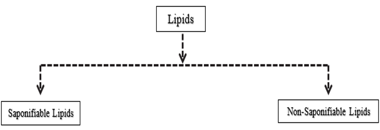
Saponifiable Lipids
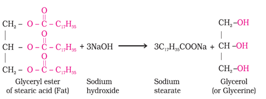
Fatty Acids:
- A fatty acid is composed of a long hydrocarbon chain (“tail”) and a terminal carboxyl group (or “head”). The carboxyl group is normally ionized under physiological conditions. Fatty acids occur in large amounts in biological systems, but rarely in the free, uncomplexed state. They typically are esterified to glycerol or other backbone structures. Most of the fatty acids found in nature have an even number of carbon atoms (usually 14 to 24). Certain marine organisms, however, contain substantial amounts of fatty acids with odd numbers of carbon atoms. Fatty acids are either saturated (all carbon–carbon bonds are single bonds) or unsaturated (with one or more double bonds in the hydrocarbon chain). If a fatty acid has a single double bond, it is said to be monounsaturated, and if it has more than one, polyunsaturated. Fatty acids can be named or described in at least three ways. For example, a fatty acid composed of an 18-carbon chain with no double bonds can be called by its systematic name (octadecanoic acid), its common name (stearic acid), or its shorthand notation, in which the number of carbons is followed by a colon and the number of double bonds in the molecule (18:0 for stearic acid). Stearic acid(18:0) and palmitic acid (16:0) are the most common saturated fatty acids in nature.
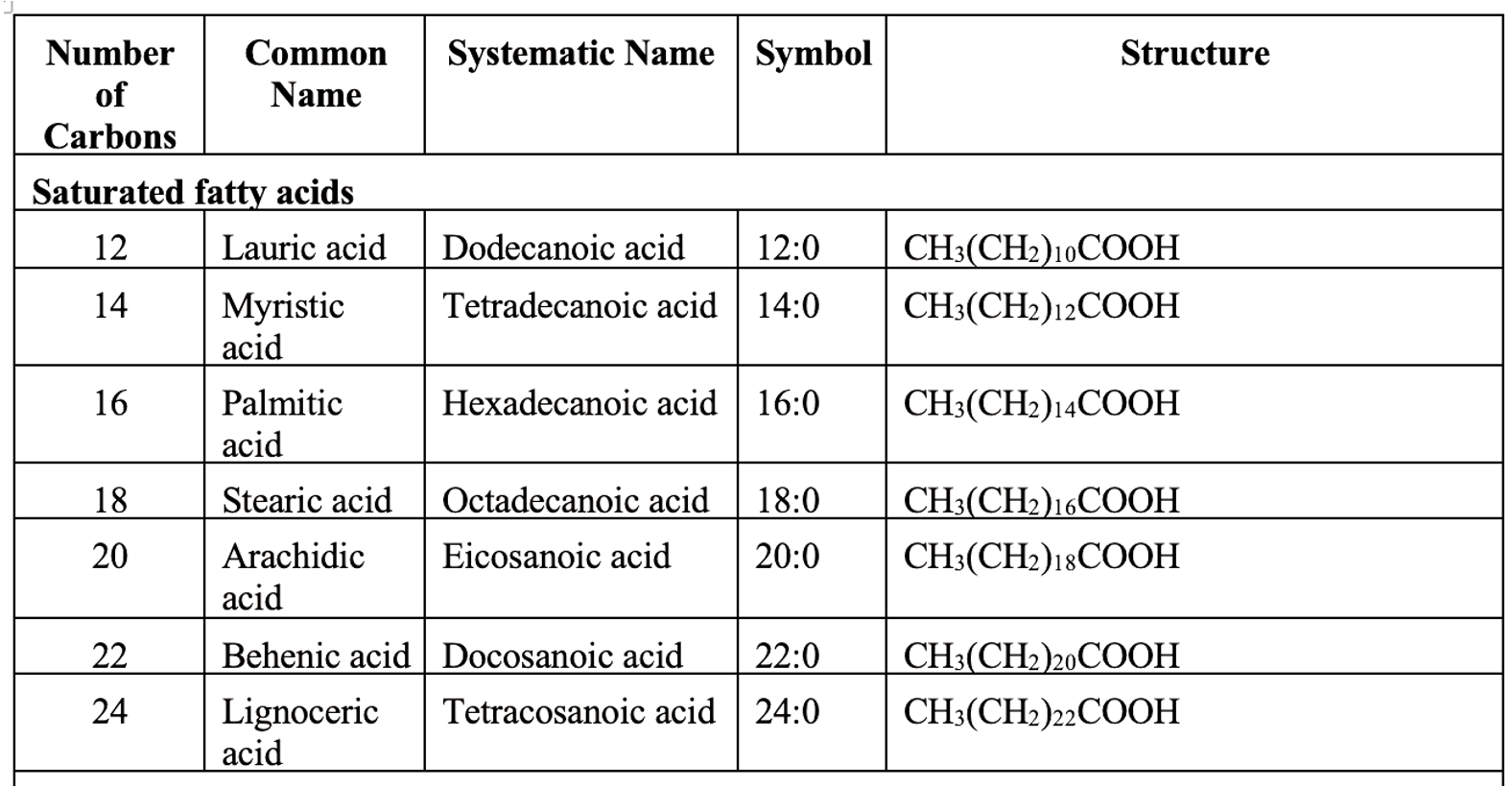
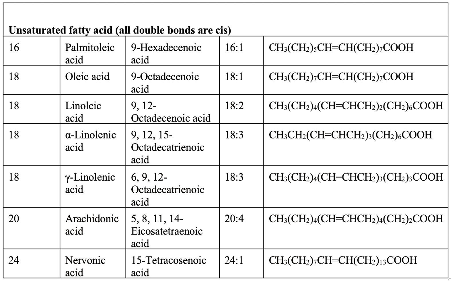
Fats or Triacylglycerols Are Fatty Acid Esters of Glycerol:
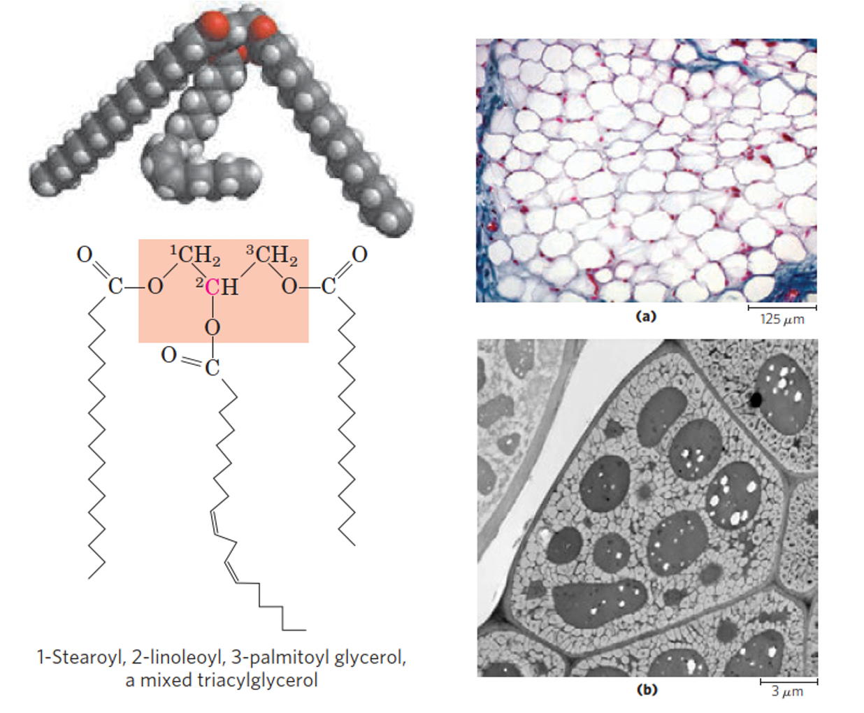
Waxes Serve as Energy Stores and Water Repellents:
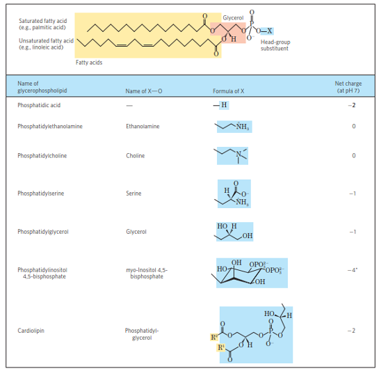

Non-Saponifiable Lipids :
Terpenes:
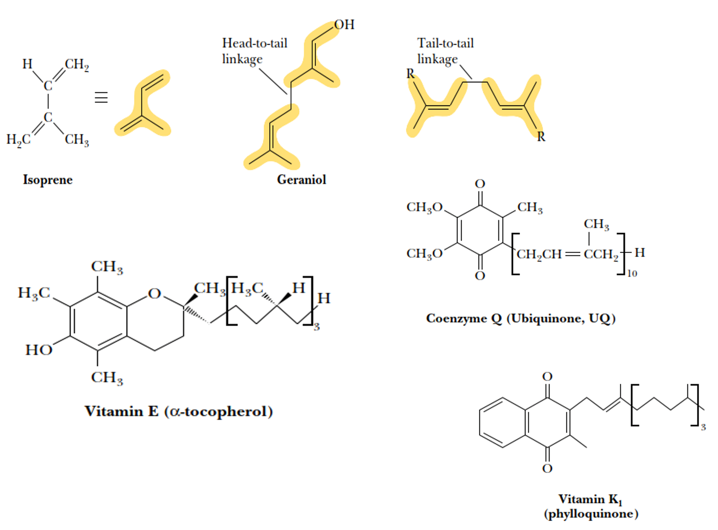
Glycolipids:
- Glycolipids are a type of complex lipids comprising carbohydrate, fatty acids, sphingolipids or a glycerol group. The term glycolipids mainly describe any compound containing one or more monosaccharide residues bound by a glycosidic linkage. These molecules are widely distributed in tissue, brain and also in nerve cells.
Types of Glycolipids:
- Glycolipids are structural lipids, which are generally found on the extracellular face of the eukaryotic cellular membrane. The different types of glycolipids are:
Cerebrosides:
- These types of glycolipids or glycosphingolipids are found primarily in the brain and in the peripheral nervous tissue. It acts as an insulator and is mainly involved in providing a protective coating to the nerve cells.
Gangliosides:
- These types of glycolipids or glycosphingolipids are neutral compounds and are composed of sialic acid residues. They are the main components of the animal’s cell membrane and are abundantly found in the plasma membrane of the neurons.
The main functions of Glycolipid are: :
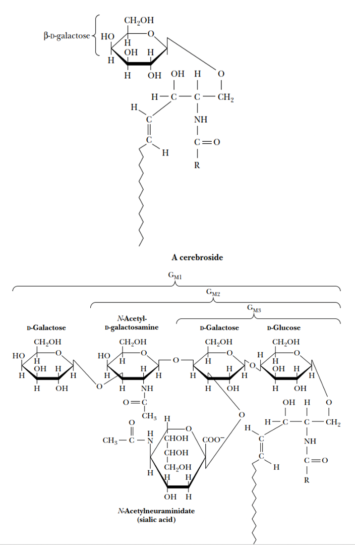
- The Prostaglandins (PG) are a group of physiologically active lipid compounds called eicosanoids having diverse hormone-like effects in animals. Prostaglandins have been found in almost every tissue in humans and other animals. They are derived enzymatically from the fatty acid arachidonic acid. Every prostaglandin contains 20 carbon atoms, including a 5-carbon ring. They are a subclass of eicosanoids and of the prostanoid class of fatty acid derivatives.
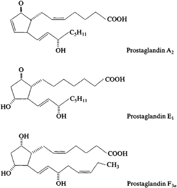
Proteins
- Beta, proteins are polypeptide chains. Poly ka matlab ‘many’ and peptide ka matlab hai yhan par peptide bond se. Peptide bond ek aisi amide linkage hai jo ek amino acid ko dusre amino acid se jodti hai. Toh amino acids hi wo cheej hai jo ek ke sath ek jud ke polypeptide ya protein bnati hai.
- Ek amino acid me ek centre me ek Carbon atom hai, ussey juda hua ek hydrogen atom, ek -COOH (carboxyl) group and ek -NH2 (amine) group ke sath sath ek variable R group hota hai.
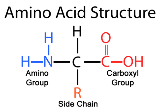
- Yeah basic structure hai kisi bhi amino acid ka. Humare pass keval 20 amino acids hai jinme keval R group different hota hai like kisi me R group simple -H se lekr, -CH3 ya even aromatic ring systems bhi ho sakte hai.
- Inko hum substituted methanes bhi bolte hain.
- Types of amino acids :
- Acidic : glutamic acid, aspartic acid
- Basic amino acids : lysine, arginine, histidine
- Neutral amino acids : glycine, alanine and valine
- Sulphur containing amino acids : methionine and cysteine.
- Alcoholic amino acids : threonine and serine.
- Aromatic amino acids : tyrosine, phenylalanine and tryptophan.
- Jo amino acids hume protein bnane ke liye diet se chahiye hote hai unko hum essential amino acids kehte hain jaise ki : phenylalanine, valine, tryptophan, threonine, isoleucine, methionine, histidine, arginine, lysine and leucine. In 10 essential amino acids me se histidine and arginine adults ke liye non-essential hai.
- Primary structure : proteins me amino acids ki keval ek linear chain jisme intra chain hydrogen bonds na ho usko primary structure bolte hai.
- Primary structure me new interaction jaise ki hydrogen bonds develop hote hai ek hi chain ke different amino acids ke beech me jo ki more condensed structure bnate hai jaise ki α helix and beta-pleated sheet structure. Again, aage chal kar different types ke α helices and beta pleated sheets hoti hai jo hume iss stage pe nahi padhne. Alpha helix ka example hai α keratin and β sheet ka example hai β-keratin and fibroin proteins.
- Further interactions like disulphide linkages, ionic linkages and van der wall interaction ek tertiary functional form bnate hai jisko tertiary structure bola jata hai. Yeah globular ya fibrous ho sakte hai.
- ek se jyada polypeptide chain ek dusre ke sath interact karke ek multimeric form bnati hai jisko tertiary structure kehte hai jaise ki adult hemoglobin molecule jisme 2 α and 2 β chain hain.
- Conjugate proteins wo proteins hote hai jinme proteins ke sath koi dusra molecule juda ho jaise ki glycoproteins me glucose/sugar + proteins hote hai, nucleoproteins me nucleic acids + proteins hote hain.
Nucleic Acids
- These are polynucleotides. A nucleotide has three chemically distinct components. One is a heterocyclic compound, the second is a monosaccharide and the third a phosphoric acid or phosphate.
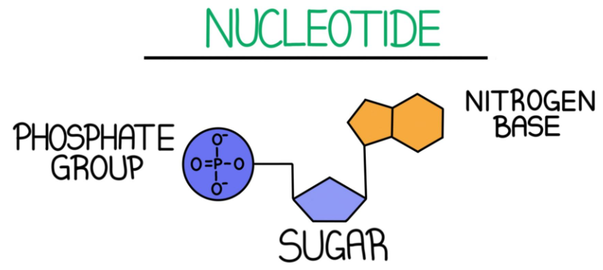
- The heterocyclic compounds in nucleic acids are the nitrogenous bases named adenine, guanine, uracil, cytosine, and thymine. Adenine and Guanine are substituted purines while the rest are substituted pyrimidines.
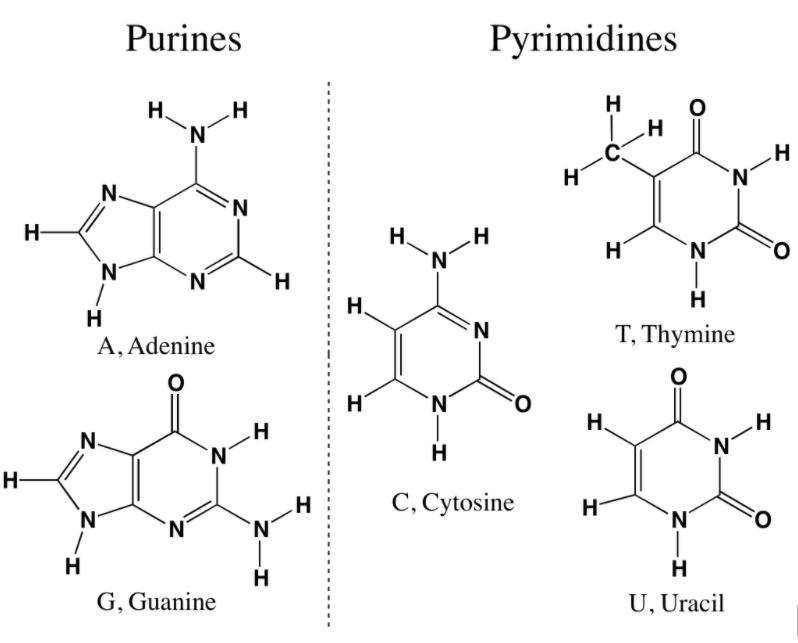
- The sugar found in polynucleotides is either ribose (a monosaccharide pentose) or 2’ deoxyribose.
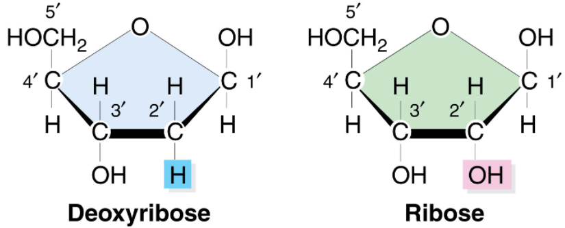
- A nucleic acid containing deoxyribose is called deoxyribonucleic acid (DNA) while that which contains ribose is called ribonucleic acid (RNA).
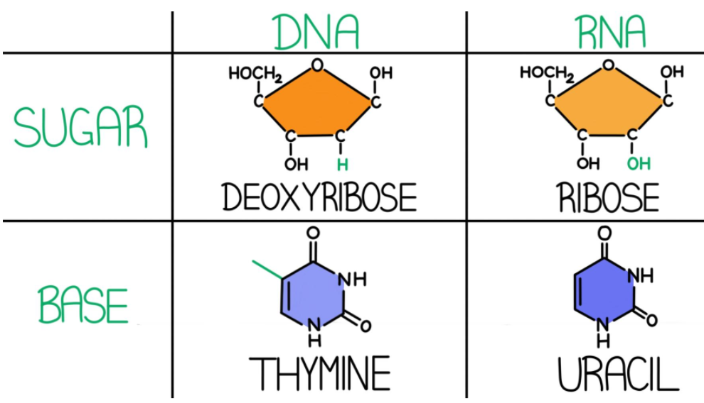
Nucleic Acids
- These are polynucleotides. A nucleotide has three chemically distinct components. One is a heterocyclic compound, the second is a monosaccharide and the third a phosphoric acid or phosphate.

- The heterocyclic compounds in nucleic acids are the nitrogenous bases named adenine, guanine, uracil, cytosine, and thymine. Adenine and Guanine are substituted purines while the rest are substituted pyrimidines.

- The sugar found in polynucleotides is either ribose (a monosaccharide pentose) or 2’ deoxyribose.

- A nucleic acid containing deoxyribose is called deoxyribonucleic acid (DNA) while that which contains ribose is called ribonucleic acid (RNA).

Nucleic Acid
- In 1865, Mendel showed that “Merkmalen” (now “genes”) transmitted genetic information, and in the first part of the twentieth century, their patterns of transmission were studied extensively. Although these classical genetic studies provided little insight into the molecular nature of genes, they did demonstrate that the genetic material must perform three essential functions:
- The genotypic function, replication. The genetic material must store genetic information and accurately transmit that information from parents to offspring, generation after generation.
- The phenotypic function, gene expression. The genetic material must control the development of the phenotype of the organism. That is, the genetic material must dictate the growth of the organism from the single-celled zygote to the mature adult.
- The evolutionary function, mutation. The genetic material must undergo changes to produce variations that allow organisms to adapt to modifications in the environment so that evolution can occur.
Other early genetic studies established a precise correlation between the patterns of transmission of genes and the behavior of chromosomes during sexual reproduction, providing strong evidence that genes are usually located on chromosomes.
Proof That Genetic Information Is Stored in DNA:
- Griffith's experiment, reported in 1928 by Frederick Griffith, was the first experiment suggesting that bacteria are capable of transferring genetic information through a process known as transformation. Griffith's findings were followed by research in the late 1930s and early 40s that isolated DNA as the material that communicated this genetic information.
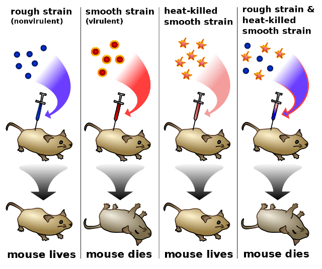
In 1944 Oswald Avery, Colin MacLeod, and Maclyn McCarty’s demonstrated that the “transforming principle” in S. pneumoniae is DNA. Avery and colleagues showed that DNA is the only component of the Type IIIS cells required to transform Type IIR cells to Type IIIS. The most definitive experiments in Avery, MacLeod, and McCarty’s proof that DNA was the transforming principle involved the use of enzymes that degrade DNA, RNA, or protein.
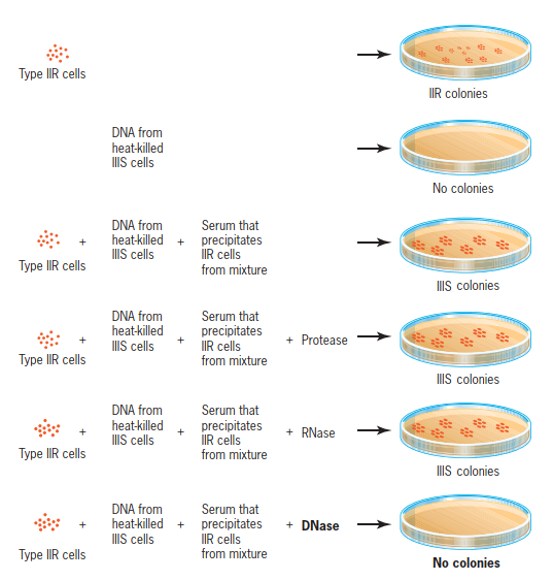
In separate experiments, highly purified DNA from Type IIIS cells was treated with the enzymes (1) deoxyribonuclease (DNase), which degrades DNA, (2) ribonuclease (RNase), which degrades RNA, or (3) proteases, which degrade proteins; the DNA was then tested for its ability to transform Type IIR cells to Type IIIS. Only DNase treatment had any effect on the transforming activity of the DNA preparation—it eliminated all transforming activity.
PROOF THAT RNA STORES THE GENETIC INFORMATION IN SOME VIRUSES:
- As more and more viruses were identified and studied, it became apparent that many of them contain RNA and proteins, but no DNA. In all cases studied to date, it is clear that these RNA viruses—like all other organisms—store their genetic information in nucleic acids rather than in proteins, although in these viruses the nucleic acid is RNA. One of the first experiments that established RNA as the genetic material in RNA viruses was the so-called reconstitution experiment of Heinz Fraenkel-Conrat and coworkers, published in 1957. Their simple, but definitive, experiment was done with tobacco mosaic virus (TMV), a small virus composed of a single molecule of RNA encapsulated in a protein coat. Different strains of TMV can be identified on the basis of differences in the chemical composition of their protein coats.
Nucleic acids, the major components of Miescher’s nuclein, are macromolecules composed of repeating subunits called nucleotides. Each nucleotide is composed of (1) a phosphate group, (2) a five-carbon sugar, or pentose, and (3) a cyclic nitrogen-containing compound called a base. In DNA, the sugar is 2-deoxyribose (thus the name deoxyribonucleic acid); in RNA, the sugar is ribose (thus ribonucleic acid). Four different bases commonly are found in DNA: adenine (A), guanine (G), thymine (T), and cytosine (C). RNA also usually contains adenine, guanine, and cytosine but has a different base, uracil (U), in place of thymine. Adenine and guanine are double-ring bases called purines; cytosine, thymine, and uracil are single-ring bases called pyrimidines. Both DNA and RNA, therefore, contain four different subunits, or nucleotides: two purine nucleotides and two pyrimidine nucleotides. In polynucleotides such as DNA and RNA, these subunits are joined together in long chains. RNA usually exists as a single-stranded polymer that is composed of a long sequence of nucleotides. DNA has one additional—and very important—level of organization: it is usually a double-stranded molecule.
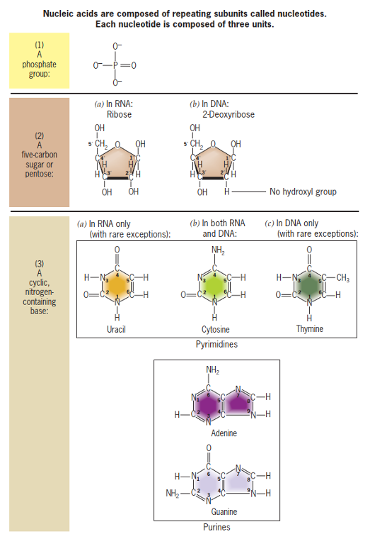
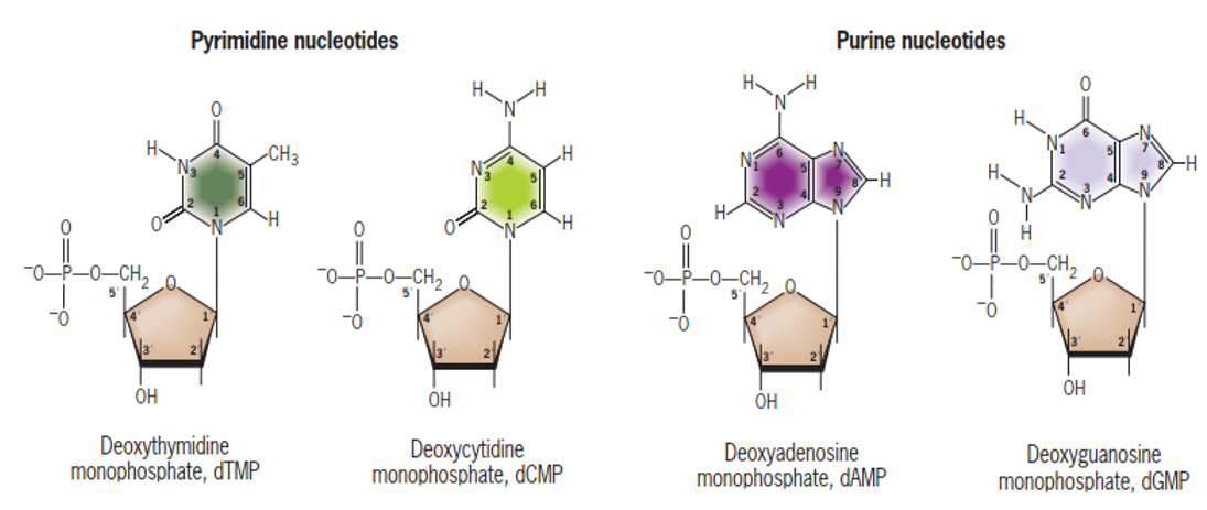
DNA STRUCTURE: THE DOUBLE HELIX :
- One of the most exciting breakthroughs in the history of biology occurred in 1953 when James Watson and Francis Crick deduced the correct structure of DNA. Their double-helix model of the DNA molecule immediately suggested an elegant mechanism for the transmission of genetic information Watson and Crick’s double-helix structure was based on two major kinds of evidence:
- When Erwin Chargaff and colleagues analyzed the composition of DNA from many different organisms, they found that the concentration of thymine was always equal to the concentration of adenine and the concentration of cytosine was always equal to the concentration of guanine.
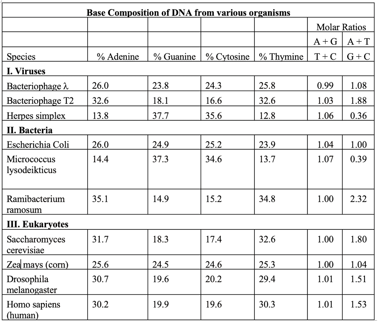
- When X rays are focused through fibers of purified molecules, the rays are deflected by the atoms of the molecules in specific patterns, called diffraction patterns, which provide information about the organization of the components of the molecules. These X-ray diffraction patterns can be recorded on X-ray-sensitive film just as patterns of light can be recorded with a camera and light-sensitive film. Watson and Crick used X-ray diffraction data on DNA structure provided by Maurice Wilkins, Rosalind Franklin, and their coworkers. These data indicated that DNA was a highly ordered, two-stranded structure with repeating substructures spaced every 0.34 nanometer along the axis of the molecule.
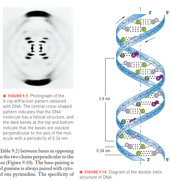 On the basis of Chargaff’s chemical data, Wilkins’ and Franklin’s X-ray diffraction data, and inferences from model building, Watson and Crick proposed that DNA exists as a right-handed double helix in which the two poly-nucleotide chains are coiled about one another in a spiral. Watson, Crick, and Wilkins shared the 1962 Nobel Prize in Physiology or Medicine for their work on the double-helix model. Unfortunately, Franklin died prematurely (age 37) in 1958, and Nobel Prizes cannot be awarded posthumously. Each of the two polynucleotide chains in a double helix consists of a sequence of nucleotides linked together by phosphodiester bonds, joining adjacent deoxyribose moieties.
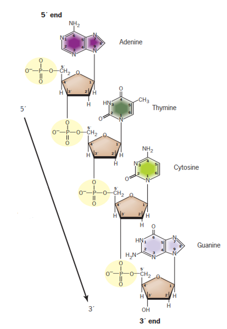
The two polynucleotide strands are held together in their helical configuration by hydrogen bonding between bases in opposing strands; the resulting base pairs are stacked between the two chains perpendicular to the axis of the molecule like the steps of a spiral staircase.
The base-pairing is specific: adenine is always paired with thymine, and guanine is always paired with cytosine. Thus, all base pairs consist of one purine and one pyrimidine. The specificity of base-pairing results from the hydrogen-bonding capacities of the bases in their normal configurations. In their common structural configurations, adenine and thymine form two hydrogen bonds, and guanine and cytosine form three hydrogen bonds.
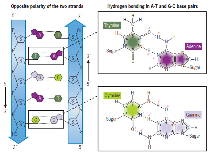 Hydrogen bonding is not possible between cytosine and adenine or thymine and guanine when they exist in their common structural states. Once the sequence of bases in one strand of a DNA double helix is known, the sequence of bases in the other strand is also known because of the specific base-pairing.
 DNA double helix are thus said to be complementary. This property, the complementarity of the two strands of the double helix, makes DNA uniquely suited to store and transmit genetic information from generation to generation.

The base pairs in DNA are stacked about 0.34 nm apart, with 10 base pairs per turn (360°) of the double helix. The sugar-phosphate backbones of the two complementary strands are antiparallel.
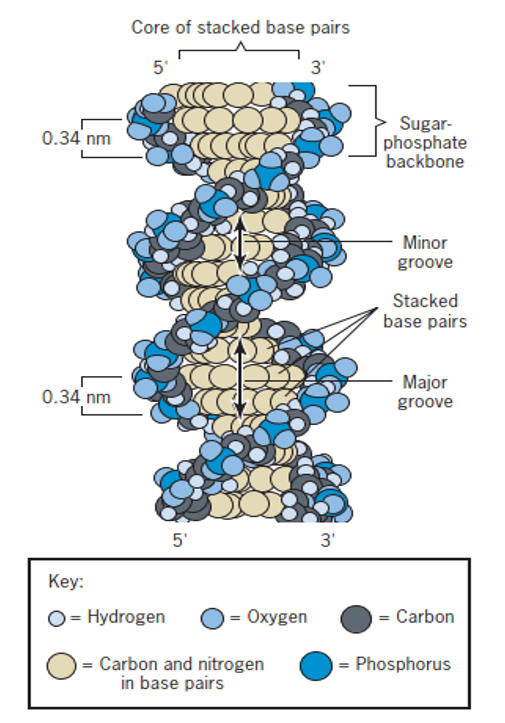
DNA STRUCTURE: ALTERNATE FORMS OF THE DOUBLE HELIX :
- The Watson–Crick double-helix structure just described is called B-DNA. B-DNA is the conformation that DNA takes under physiological conditions (in aqueous solutions containing low concentrations of salts). The vast majority of the DNA molecules present in the aqueous protoplasms of living cells exist in the B conformation.
The structures of DNA molecules change as a function of their environment. The exact conformation of a given DNA molecule or segment of a DNA molecule will depend on the nature of the molecules with which it is interacting. In fact, intracellular B-DNA appears to have an average of 10.4 nucleotide pairs per turn, rather than precisely 10. In high concentrations of salts or in a partially dehydrated state, DNA exists as A-DNA, which is a right-handed helix like B-DNA, but with 11 nucleotide pairs per turn. A-DNA is a shorter, thicker double helix with a diameter of 2.3 nm. However, the A-DNA conformation is important because DNA-RNA heteroduplexes (double helices containing a DNA strand base-paired with a complementary RNA strand) or RNA-RNA duplexes exist in a very similar structure in vivo.

Certain DNA sequences have been shown to exist in a left-handed, double-helical form called Z-DNA (Z for the zigzagged path of the sugar-phosphate backbones of the structure). Z-DNA was discovered by X-ray diffraction analysis of crystals formed by DNA oligomers containing alternating G:C and C:G base pairs. Z-DNA occurs in double helices that are G:C-rich and contain alternating purine and pyrimidine residues. In addition to its unique left-handed helical structure, Z-DNA differs from the A and B conformations in having 12 base pairs per turn, a diameter of 1.8 nm, and a single deep groove.
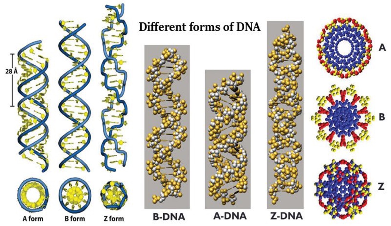
Proteins
- Proteins are polypeptides. They are linear chains of amino acids linked by peptide bonds.

Each protein is a polymer of amino acids. As there are 20 types of amino acids (e.g., alanine, cysteine, proline, tryptophan, lysine, etc.), a protein is a heteropolymer and not a homopolymer. A homopolymer has only one type of monomer repeating ‘n’ number of times.

Amino acids can be essential or non-essential. The latter are those which our body can make, while we get essential amino acids through our diet/food.

Proteins carry out many functions in living organisms, some transport nutrients across cell membrane, some fight infectious organisms, some are hormones, some are enzymes.

Structure of Proteins
Primary Structure : Simple linear chain of amino acids joined together by peptide bonds.
Secondary Structure : New interactions like hydrogen bonds between different groups arise in the primary structure giving rise to a folded structure which is a bit more complex than the previous linear structure known as secondary structure. Types of secondary structures are ‘alpha’-helix, β-sheet and collagen helix.

Common features of α-helical structure :

Tertiary Structure : this is the 3D structure of a protein which forms due to interactions (like hydrophobic or van der waals force and/or electrostatic interactions) between α-helix and β-pleated sheets. It is of 2 types : globular and fibrous.

Quaternary Structure : some proteins are multimeric so the interactions between different polypeptide chains of a protein give rise to a quaternary structure. Ex : hemoglobin molecule.

Nature Of Bonds Linking Monomers In A Polymer
- Proteins are formed of either a single polypeptide or from multiple polypeptide chains as seen in the quaternary structure of a protein in the previous section. Amino acids in polypeptide chains are linked to one another by a special bond named as amide bond or amide linkage. Amino acids have an amino and a carboxyl group so, the carboxyl group of one amino acid and amino group of another amino acid undergo dehydration to form an amide bond while releasing a molecule of water.
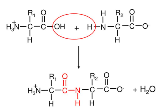
- In a polysaccharide the individual monosaccharides are linked by a glycosidic bond. This bond is also formed by dehydration. This bond is formed between two carbon atoms of two adjacent monosaccharides.
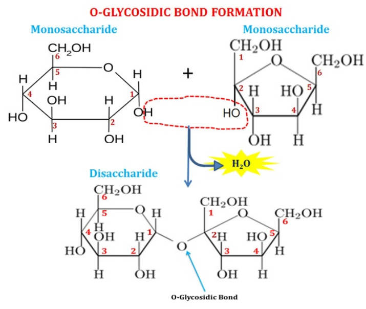
O and N-glycosidic Bonds :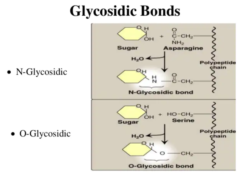
- In a nucleic acid a phosphate moiety links the 3’-carbon of one sugar of one nucleotide to the 5’-carbon of the sugar of the succeeding nucleotide. The bond between the phosphate and hydroxyl group of sugar is an ester bond. As there is one such ester bond on either side, it is called phosphodiester bond.
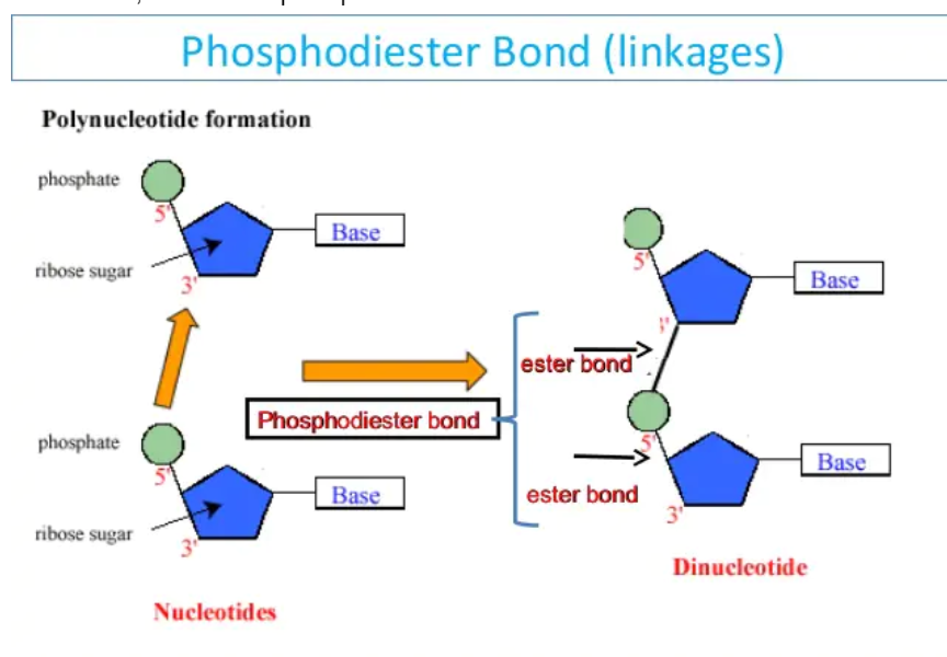
- The two strands of polynucleotides are antiparallel, i.e., run in the opposite direction. The backbone is formed by the sugar-phosphate-sugar chain. The nitrogen bases are projected more or less perpendicular to this backbone but face inside. A and G of one strand compulsorily base pairs with T and C, respectively, on the other strand. There are two hydrogen bonds between A and T and three hydrogen bonds between G and C. Each strand appears like a helical staircase. Each step of ascent is represented by a pair of bases. At each step of ascent, the strand turns 36°. One full turn of the helical strand would involve ten steps or ten base pairs. Attempt drawing a line diagram. The pitch would be 34Å. The rise per base pair would be 3.4Å. This form of DNA with the above mentioned salient features is called B-DNA. In higher classes, you will be told that there are more than a dozen forms of DNA named after English alphabets with unique structural features.
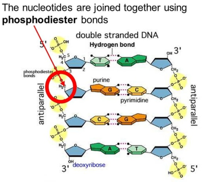
Nature Of Bonds Linking Monomers In A Polymer
- Proteins are formed of either a single polypeptide or from multiple polypeptide chains as seen in the quaternary structure of a protein in the previous section. Amino acids in polypeptide chains are linked to one another by a special bond named as amide bond or amide linkage. Amino acids have an amino and a carboxyl group so, the carboxyl group of one amino acid and amino group of another amino acid undergo dehydration to form an amide bond while releasing a molecule of water.

- In a polysaccharide the individual monosaccharides are linked by a glycosidic bond. This bond is also formed by dehydration. This bond is formed between two carbon atoms of two adjacent monosaccharides.

O and N-glycosidic Bonds :
- In a nucleic acid a phosphate moiety links the 3’-carbon of one sugar of one nucleotide to the 5’-carbon of the sugar of the succeeding nucleotide. The bond between the phosphate and hydroxyl group of sugar is an ester bond. As there is one such ester bond on either side, it is called phosphodiester bond.

- The two strands of polynucleotides are antiparallel, i.e., run in the opposite direction. The backbone is formed by the sugar-phosphate-sugar chain. The nitrogen bases are projected more or less perpendicular to this backbone but face inside. A and G of one strand compulsorily base pairs with T and C, respectively, on the other strand. There are two hydrogen bonds between A and T and three hydrogen bonds between G and C. Each strand appears like a helical staircase. Each step of ascent is represented by a pair of bases. At each step of ascent, the strand turns 36°. One full turn of the helical strand would involve ten steps or ten base pairs. Attempt drawing a line diagram. The pitch would be 34Å. The rise per base pair would be 3.4Å. This form of DNA with the above mentioned salient features is called B-DNA. In higher classes, you will be told that there are more than a dozen forms of DNA named after English alphabets with unique structural features.

Summary
- Although there is a bewildering diversity of living organisms, their chemical composition and metabolic reactions appear to be remarkably similar. The elemental composition of living tissues and non-living matter appear also to be similar when analysed qualitatively. However, a closer examination reveals that the relative abundance of carbon, hydrogen and oxygen is higher in living systems when compared to inanimate matter. The most abundant chemical in living organisms is water. There are thousands of small molecular weight (<1000 Da) biomolecules. Amino acids, monosaccharide and disaccharide sugars, fatty acids, glycerol, nucleotides, nucleosides and nitrogen bases are some of the organic compounds seen in living organisms. There are 20 types of amino acids and 5 types of nucleotides. Fats and oils are glycerides in which fatty acids are esterified to glycerol. Phospholipids contain, in addition, a phosphorylated nitrogenous compound. Only three types of macromolecules, i.e., proteins, nucleic acids and polysaccharides are found in living systems. Lipids, because of their association with membranes separate in the macromolecular fraction.Biomacromolecules are polymers. They are made of building blocks which are different. Proteins are heteropolymers made of amino acids. Nucleic acids (RNA and DNA) are composed of nucleotides. Biomacromolecules have a hierarchy of structures – primary, secondary, tertiary and quaternary. Nucleic acids serve as genetic material. Polysaccharides are components of cell wall in plants, fungi and also of the exoskeleton of arthropods. They also are storage forms of energy (e.g., starch and glycogen). Proteins serve a variety of cellular functions. Many of them are enzymes, some are antibodies, some are receptors, some are hormones and some others are structural proteins. Collagen is the most abundant protein in animal world and Ribulose bisphosphate Carboxylase-Oxygenase (RuBisCO) is the most abundant protein in the whole of the biosphere. Enzymes are proteins which catalyse biochemical reactions in the cells. Ribozymes are nucleic acids with catalytic power. Proteinaceous enzymes exhibit substrate specificity, require optimum temperature and pH for maximal activity. They are denatured at high temperatures. Enzymes lower activation energy of reactions and enhance greatly the rate of the reactions. Nucleic acids carry hereditary information and are passed on from parental generation to progeny.
Carbohydrates
- Beta, carbohydrates ko define karte hain as ‘hydrates of carbon’, jinka general formula hai Cn(H2O)n and n ki value ki range hai from 3-7 carbon atoms. Inme aldehyde ya fir ketone me se koi bhi ek functional group ho sakta hai.
- Generally, carbohydrates ko further 3 categories me divide kiya gya hai : monosaccharides, oligosaccharides and polysaccharides.
- Monosaccharides : single unit of sugar jaise ki keval ek glucose ka molecule jisko further agar degrade kardenge toh wo carbohydrate nahi reh jayega. Toh yeah sabse choti unit hai. Neeche table me types given hai :
Types of Monosaccharides
No. of Carbon atoms Aldehyde Ketones 3 : Trioses Glyceraldehyde Dihydroxyacetone 4 : Tetroses Erythrose Erythrulose 5 : Pentoses Deoxyribose, Ribose Ribulose 6 : Hexoses Glucose, Galactose, Mannose Fructose 7 : Heptoses L-glycero-D-manno-heptose Sedoheptulose - Glucose : dextrorotatory, also known as grape sugar, corn sugar, blood sugar and dextrose. Pyran ring structure involving 6 members.
- Reducing & Non reducing Sugar : kisi bhi sugar molecule ko reducing hone ke liye, ya toh uske pass ek free aldehyde group ho ya fir wo isomerism se ek produce kare. Agar yeah condition meet hoti hai toh reducing sugar hai otherwise usko non reducing kehte hai. Reducing sugars positive fehling's solution test, positive benedict’s reagent test deti hai jabki non reducing nahi. Reducing sugars : all monosaccharides, lactose and maltose. Non reducing sugars : sucrose and most polysaccharides.
- Derived sugars : inko baki chemical se reaction ke dwara bnaya jata hai jaise ki glucose + amino group = glucosamine sugar.
- Oligosaccharides : jab 2-9 monosaccharides units milkr ek condensed structure bnti hai toh usko oligosaccharides kehte hai and 9 se jyada subunits hone pe usko hum polysaccharides keh dete hai. Ex : Maltose = α-glucose + α-glucose. Lactose = glucose + galactose. Sucrose = glucose + fructose. These are disaccharides.
- polysaccharides, bahut sare monosaccharides ke condensation se banta hai. Glycogen me 30,000 glucose units hoti hai. Inulin me 30-35 fructose units hoti hai, cellulose me 6000 β-glucose units hoti hai.
Dynamic State Of Body Constituents – Concept Of Metabolism
- The sum total of all the chemical reactions occurring in a living organism are known as metabolic reactions and this state is known as metabolism. These reaction fall under 2 general categories : anabolism and catabolism. Ex : A few examples for such metabolic transformations are: removal of CO2 from amino acids making an amino acid into an amine, removal of amino group in a nucleotide base; hydrolysis of a glycosidic bond in a disaccharide, etc.

- Anabolism means formation of new compounds from raw materials like in photosynthesis, carbohydrates are synthesized using water and carbon dioxide. Catabolism on the other hand means breakdown. Example : Glycolysis, a process in which we breakdown a glucose molecule to obtain energy (ATP) from it.
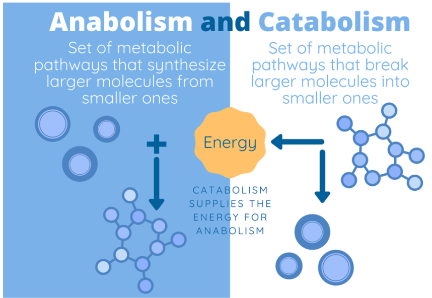
- There is no uncatalysed metabolic conversion in living systems. Even CO2 dissolving in water, a physical process, is a catalysed reaction in living systems. The catalysts which hasten the rate of a given metabolic conversation are also proteins. These proteins with catalytic power are named enzymes.
- the living state is a non-equilibrium steady-state to be able to perform work; the living process is a constant effort to prevent falling into equilibrium. This is achieved by energy input. Metabolism provides a mechanism for the production of energy. Hence the living state and metabolism are synonymous. Without metabolism there cannot be a living state.
Dynamic State Of Body Constituents – Concept Of Metabolism
- The sum total of all the chemical reactions occurring in a living organism are known as metabolic reactions and this state is known as metabolism. These reaction fall under 2 general categories : anabolism and catabolism. Ex : A few examples for such metabolic transformations are: removal of CO2 from amino acids making an amino acid into an amine, removal of amino group in a nucleotide base; hydrolysis of a glycosidic bond in a disaccharide, etc.

- Anabolism means formation of new compounds from raw materials like in photosynthesis, carbohydrates are synthesized using water and carbon dioxide. Catabolism on the other hand means breakdown. Example : Glycolysis, a process in which we breakdown a glucose molecule to obtain energy (ATP) from it.

- There is no uncatalysed metabolic conversion in living systems. Even CO2 dissolving in water, a physical process, is a catalysed reaction in living systems. The catalysts which hasten the rate of a given metabolic conversation are also proteins. These proteins with catalytic power are named enzymes.
- the living state is a non-equilibrium steady-state to be able to perform work; the living process is a constant effort to prevent falling into equilibrium. This is achieved by energy input. Metabolism provides a mechanism for the production of energy. Hence the living state and metabolism are synonymous. Without metabolism there cannot be a living state.
Polysaccharides
- Polysaccharides are long chains of sugars. They are threads (literally a cotton thread) containing different monosaccharides (mono=single, saccharide=sugar unit) as building blocks.
- For example, cellulose is a polymeric polysaccharide consisting of only one type of monosaccharide i.e., glucose. Cellulose is a homopolymer.

- Starch is a variant of this but present as a store house of energy in plant tissues.

- Animals have another variant called glycogen.

- Inulin is a polymer of fructose.

- In a polysaccharide chain (say glycogen), the right end is called the reducing end and the left end is called the non-reducing end.

- Starch forms helical secondary structures. In fact, starch can hold I2 molecules in the helical portion. The starch-I2 is blue in colour. Cellulose does not contain complex helices and hence cannot hold I2.

- There are more complex polysaccharides in nature. They have as building blocks, amino-sugars and chemically modified sugars (e.g., glucosamine, N-acetyl galactosamine, etc.).

- Exoskeletons of arthropods, for example, have a complex polysaccharide called chitin.

ENZYMES
- Enzymes are mostly proteins , but sometimes they are nucleic acids which work like enzymes as well , hence these are called Ribozymes.
- All enzymes catalyse biochemical reactions in living cells and therefore they are also called BIOCATALYSTS Just like any other proteins, Enzymes also have various structures like secondary and tertiary , where in tertiary structure , the backbone of this chain folds itself up and forms a site where the substrate attaches itself with the enzyme , this particular site is called the “Active site ”.
- Enzymes are called substrate specific , this is because the active site attaches to a certain substrate only , hence we see that enzymes are substrate specific . These compounds through their active sites catalyse the chemical reactions at a much higher rate .
-
Enzymes are biological catalysts or organic catalysts , but they differ from inorganic catalysts.
- There are certain organisms which are found in very high temperatures called thermophiles , enzymes isolated from these thermophilic organisms are thermally very stable and can function at very high temperatures also (80- 90 degrees celsius ) .
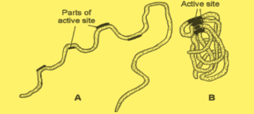
Chemical nature of Enzymes
- A large number of enzymes require an additional non-protein group for their efficient activity. Thus there are two types of enzymes.
- Simple Enzyme: Enzymes which are purely made up of proteins only are called simple enzymes e.g., pepsin, trypsin, amylase, urease.
- Conjugate Enzyme: These enzymes are made up of two parts viz. protein part called apoenzyme and non-protein part called cofactor. The complete conjugate enzyme is called Holoenzyme, e.g. dehydrogenase, hexokinase.
- Cofactors are of following types :
- Prosthetic groups : These are organic compounds and are distinguished from other cofactors in that they are tightly bound to the apoenzyme. For example, in peroxidase and catalase, which catalyze the breakdown of hydrogen peroxide to water and oxygen, haem is the prosthetic group and it is a part of the active site of the enzyme.
- Co-enzymes : These are also organic compounds but their association with the apoenzyme is only transient, usually occurring during the course of catalysis. Furthermore, co-enzymes serve as co-factors in a number of different enzyme catalyzed reactions. The essential chemical components of many coenzymes are vitamins, e.g., coenzyme nicotinamide adenine dinucleotide (NAD) and NADP contain the vitamin niacin.
- Metal ions : A number of enzymes require metal ions for their activity which form coordination bonds with side chains at the active site and at the same time form one or more coordination bonds with the substrate, e.g., zinc is a cofactor for the proteolytic enzyme carboxypeptidase
How do Enzymes bring about such High Rates of Chemical Conversions?
- It is now understood that the substrate 'S' has to bind the enzyme at its 'active site' within a given cleft or pocket. The substrate has to diffuse towards the 'active site'. There is thus, an obligatory formation of an 'ES' complex. E stands for enzyme. This complex formation is a transient phenomenon.
- During the state where substrate is bound to the enzyme active site, a new structure of the substrate called transition state structure is formed.Very soon, after the expected bond breaking/making is completed, the product is released from the active site. In other words, the structure of substrate gets transformed into the structure of product(s). The pathway of this transformation must go through the so-called transition state structure.
- There could be many more 'altered structural states' between the stable substrate and the product. Implicit in this statement is the fact that all other intermediate structural states are unstable.
- Stability is something related to energy status of the molecule or the structure. Hence, when we look at this pictorially through a graph it looks like something as in the given figure. The y-axis represents the potential energy content. The x-axis represents the progression of the structural transformation or states through the 'transition state'.
- It is important to note two things. The energy level difference between S and P. If 'P' is at a lower level than 'S', the reaction is an exothermic reaction. One need not supply energy (by heating) in order to form the product. However, whether it is an exothermic or spontaneous reaction or an endothermic or energy requiring reaction, the 'S' has to go through a much higher energy state or transition state. The difference in average energy content of 'S' from that of this transition state is called 'activation energy'. Enzymes eventually bring down this energy barrier making the transition of 'S' to 'P' more easy.
- Activation energy is the minimum amount of energy required to start the reaction. Activation energy increases the kinetic energy of the system and brings about forceful collisions between the reactants. Enzymes lower the activation energy required for a reaction.
- The catalytic cycle of an enzyme action can be described in the following steps:
- First, the substrate binds to the active site of the enzyme, fitting into the active site.
- The binding of the substrate induces the enzyme to alter its shape, fitting more tightly around the substrate.
- The active site of the enzyme, now in close proximity of the substrate breaks the chemical bonds of the substrate and the new enzyme- product complex is formed.
- The enzyme releases the products of the reaction and the free enzyme is ready to bind to another molecule of the substrate and run through the catalytic cycle once again.
Mode of enzyme action
- Two hypothesis have been put forward to explain the mode of enzyme action :
- Lock and Key hypothesis :
- This hypothesis was given by Emil Fischer (1894). According to this hypothesis, both enzyme and substrate molecules have specific geometrical shapes. It is similar to the system of lock and key, which have, special geometrical shapes in the region of their activity.
- Just as a lock can be opened by its specific key, a substrate molecule can be acted upon by a particular enzyme.
- This also explains the specificity of enzyme action. After coming in contact with the active site of the enzyme, the substrate molecules or reactants form a complex called enzyme substrate complex. In the enzyme substrate complex, the molecules of the substrate undergo chemical change and form products.
- This theory explains how a small concentration of enzyme can act upon a large amount of the substrate. It also explains how the enzyme remains unaffected at the end of chemical reaction.
- This explains how the enzyme remains unaffected at the end of chemical reaction.
- Induced fit hypothesis :
- This hypothesis was proposed by Koshland (1960). According to this hypothesis the active site of the enzyme does not initially exist in a shape that is complementary to the substrate but is induced to assume the complementary shape as the substrate becomes bound to the enzyme. There are two ends:
(a) Buttressing group is meant for supporting the substrate.
(b) Catalytic group is meant for catalysing the reaction.
- This hypothesis was proposed by Koshland (1960). According to this hypothesis the active site of the enzyme does not initially exist in a shape that is complementary to the substrate but is induced to assume the complementary shape as the substrate becomes bound to the enzyme. There are two ends:
Factors affecting enzyme activity
- Optimum pH
- Each enzyme shows its highest activity at a specific pH. This is called the optimum pH.
- Activity declines both above and below the optimum pH. Most intracellular enzymes function best around neutral pH. Some digestive enzymes have their optimum in the acidic or alkaline range.
- For example, the protein digesting enzyme pepsin found in stomach, has an optimum pH of 2.0. Another protein-digesting enzyme, trypsin, found in the duodenum, functions best in an alkaline pH of 8.5.
- Optimum Temperature
- Enzymes generally function in a narrow range of temperature which usually corresponds to the body temperature of the organism. Each enzyme shows its highest activity at a particular temperature called optimum temperature.
- Activity declines both above and below the optimum temperature. Low temperature preserves the enzyme in a temporarily inactive state. Food may be preserved for a long time in a frozen state because neither microbial enzymes nor enzymes in the food can act at low temperatures to cause its spoilage.
- High temperature destroys enzyme activity, because proteins are denatured by heat. For this reason, only a few cells can tolerate temperatures above 45º C. Some heat resistant microorganisms living in hot springs at temperatures close to 100º C, possess heat-resistant enzymes.
- Effect of substrate concentration
- With the increase in substrate concentration (S), the velocity V of the enzymatic reaction rises at first. The reaction ultimately reaches a maximum velocity(Vmax), which is not exceeded by any further rise in substrate concentration. This happens because enzyme molecules are fewer than the substrate molecules. Any more increase in susbtrate concentration will saturate all the enzyme molecules. No enzyme s left free to bind with additional molecules of the substrate.
- Michaelis Menten constant (Km): It is a constant which indicates the substrate concentration at which the chemical reaction catalysed by an enzyme attains half its maximum velocity. Michaelis Menten constant, Km, generally lies between 10–1 to 10 –6 M. A high Km indicates low affinity while a low Km, shows strong affinity. If an enzyme acts on more than one substrate it shows different Km values for them.
- Enzyme Concentration: If an excess of substrate is present, doubling the enzyme concentration usually doubles the rate of reaction.
- Concentration of end products: As end products accumulate, the rate of reaction decreases
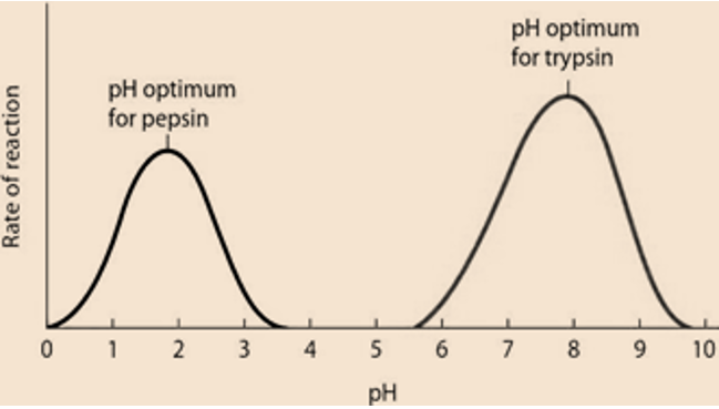
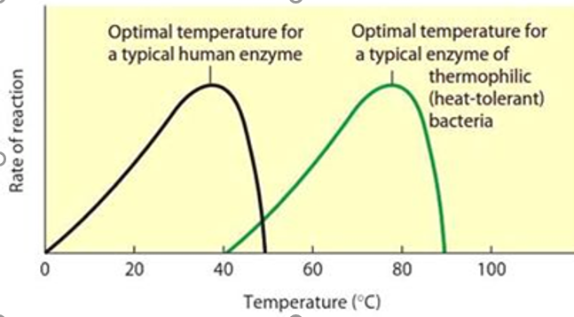
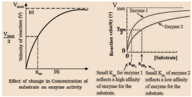
Nomenclature and Classification of enzymes
Naming the enzymes is called nomenclature. There are three ways of naming enzymes :- Duclaux (1883) provided a system for naming enzymes by adding suffix '- ase' either at the end of the substrate of which enzyme acts or after the type of reaction catalysed by enzyme. By adding suffix '- ase' after the substrate upon which they act e.g., sucrase, lipase, nuclease (which act upon sucrose, lipids, nucleic acids, respectively).
- According to the function : catalysed by enzymes e.g., dehydrogenase (remove hydrogen), carboxylase (addition of CO2) , decarboxylase (removal of CO2), etc.
- Double naming of enzyme : In this, the enzyme is given a name by using two words, the first word is after substrate and the second word is according to function performed by the enzyme e.g., succinic dehydrogenase. It removes hydrogen from succinic acid.
- According to International Union of Biochemists (IUB), the enzymes are classified into 6 classes or categories on the basis of type of reactions they catalyse.
- The oxidoreductases (class 1) are enzymes which catalyse oxido - reduction between two substrates S and S'. e.g., In these reactions one compound is oxidised and the other is reduced. Example - Dehydrogenases, oxidases, peroxidases, reductases, catalases.
- The transferases (class 2) catalyze the transfer of other groups from one molecule to another. Enzymes catalysing a transfer of a group, G (other than hydrogen) between a pair of substrate S and S' e.g., Example - Transaminases (transfer amino groups), Kinases (catalyse the phosphorylation of substrate by transferring phosphate group usually from ATP), Polymerases (DNA & RNA polymerase) involved in nucleic acid synthesis.
- The hydrolases (class 3) hydrolases cause cleavage of bond using water. Enzymes catalysing hydrolysis of ester, ether, peptide, glycosidic, C-C, C-halide or P-N bonds.Examples - Protease, trypsin, chymotrypsin, sucrose, lactase etc. DNA helicase breaks down the hydrogen bonds between the DNA strands of a DNA molecule. Nucleases such as DNase & RNase hydrolyse DNA and RNA respectively.
- Lyases (class 4, often also referred to as"synthases") are enzymes that catalyse removal of groups from substrates by mechanisms other than hydrolysis leaving double bonds. Cleavage of bond does not require water. Examples - In glycolysis, the lyase called aldolase catalyzes the readily reversible splitting of fructose 1,6-bisphosphate (F-1,6-BP),into the products glyceraldehyde 3-phosphate (GAP) and dihydroxyacetone phosphate (DHAP). This is an example of a lyase that helps to cleave carbon-carbon bonds.
- The isomerases (class 5) move groups within a molecule, without changing the gross composition of the substrate. Includes all enzymes catalysing inter-conversion of optical, geometric or positional isomers. Examples - Phosphohexose isomerase, Triose isomerase, Topoisomerase. Triose isomerase catalyses the inter- conversion of glyceraldehyde 3-phosphate (GAP) and dihydroxyacetone phosphate (DHAP).
- The ligation reactions catalyzed by ligases ("synthetases," class 6) are energy-dependent and are therefore always coupled to the hydrolysis of nucleoside triphosphates Examples - RUBP carboxylase, PEP carboxylase, Amino-acyl tRNA synthetase, DNA ligase and RNA ligase.
International Classification of Enzymes
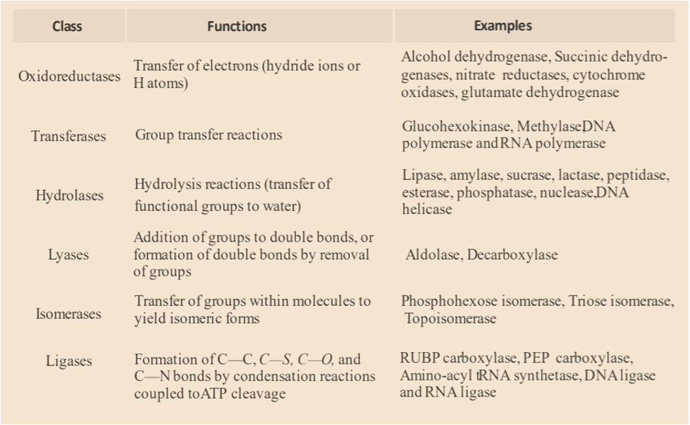
ENZYMES
- Enzymes are mostly proteins , but sometimes they are nucleic acids which work like enzymes as well , hence these are called Ribozymes.
- All enzymes catalyse biochemical reactions in living cells and therefore they are also called BIOCATALYSTS Just like any other proteins, Enzymes also have various structures like secondary and tertiary , where in tertiary structure , the backbone of this chain folds itself up and forms a site where the substrate attaches itself with the enzyme , this particular site is called the “Active site ”.
- Enzymes are called substrate specific , this is because the active site attaches to a certain substrate only , hence we see that enzymes are substrate specific . These compounds through their active sites catalyse the chemical reactions at a much higher rate .
-
Enzymes are biological catalysts or organic catalysts , but they differ from inorganic catalysts.
- There are certain organisms which are found in very high temperatures called thermophiles , enzymes isolated from these thermophilic organisms are thermally very stable and can function at very high temperatures also (80- 90 degrees celsius ) .

Chemical nature of Enzymes
- A large number of enzymes require an additional non-protein group for their efficient activity. Thus there are two types of enzymes.
- Simple Enzyme: Enzymes which are purely made up of proteins only are called simple enzymes e.g., pepsin, trypsin, amylase, urease.
- Conjugate Enzyme: These enzymes are made up of two parts viz. protein part called apoenzyme and non-protein part called cofactor. The complete conjugate enzyme is called Holoenzyme, e.g. dehydrogenase, hexokinase.
- Cofactors are of following types :
- Prosthetic groups : These are organic compounds and are distinguished from other cofactors in that they are tightly bound to the apoenzyme. For example, in peroxidase and catalase, which catalyze the breakdown of hydrogen peroxide to water and oxygen, haem is the prosthetic group and it is a part of the active site of the enzyme.
- Co-enzymes : These are also organic compounds but their association with the apoenzyme is only transient, usually occurring during the course of catalysis. Furthermore, co-enzymes serve as co-factors in a number of different enzyme catalyzed reactions. The essential chemical components of many coenzymes are vitamins, e.g., coenzyme nicotinamide adenine dinucleotide (NAD) and NADP contain the vitamin niacin.
- Metal ions : A number of enzymes require metal ions for their activity which form coordination bonds with side chains at the active site and at the same time form one or more coordination bonds with the substrate, e.g., zinc is a cofactor for the proteolytic enzyme carboxypeptidase
How do Enzymes bring about such High Rates of Chemical Conversions?
- It is now understood that the substrate 'S' has to bind the enzyme at its 'active site' within a given cleft or pocket. The substrate has to diffuse towards the 'active site'. There is thus, an obligatory formation of an 'ES' complex. E stands for enzyme. This complex formation is a transient phenomenon.
- During the state where substrate is bound to the enzyme active site, a new structure of the substrate called transition state structure is formed.Very soon, after the expected bond breaking/making is completed, the product is released from the active site. In other words, the structure of substrate gets transformed into the structure of product(s). The pathway of this transformation must go through the so-called transition state structure.
- There could be many more 'altered structural states' between the stable substrate and the product. Implicit in this statement is the fact that all other intermediate structural states are unstable.
- Stability is something related to energy status of the molecule or the structure. Hence, when we look at this pictorially through a graph it looks like something as in the given figure. The y-axis represents the potential energy content. The x-axis represents the progression of the structural transformation or states through the 'transition state'.
- It is important to note two things. The energy level difference between S and P. If 'P' is at a lower level than 'S', the reaction is an exothermic reaction. One need not supply energy (by heating) in order to form the product. However, whether it is an exothermic or spontaneous reaction or an endothermic or energy requiring reaction, the 'S' has to go through a much higher energy state or transition state. The difference in average energy content of 'S' from that of this transition state is called 'activation energy'. Enzymes eventually bring down this energy barrier making the transition of 'S' to 'P' more easy.
- Activation energy is the minimum amount of energy required to start the reaction. Activation energy increases the kinetic energy of the system and brings about forceful collisions between the reactants. Enzymes lower the activation energy required for a reaction.
- The catalytic cycle of an enzyme action can be described in the following steps:
- First, the substrate binds to the active site of the enzyme, fitting into the active site.
- The binding of the substrate induces the enzyme to alter its shape, fitting more tightly around the substrate.
- The active site of the enzyme, now in close proximity of the substrate breaks the chemical bonds of the substrate and the new enzyme- product complex is formed.
- The enzyme releases the products of the reaction and the free enzyme is ready to bind to another molecule of the substrate and run through the catalytic cycle once again.
Mode of enzyme action
- Two hypothesis have been put forward to explain the mode of enzyme action :
- Lock and Key hypothesis :
- This hypothesis was given by Emil Fischer (1894). According to this hypothesis, both enzyme and substrate molecules have specific geometrical shapes. It is similar to the system of lock and key, which have, special geometrical shapes in the region of their activity.
- Just as a lock can be opened by its specific key, a substrate molecule can be acted upon by a particular enzyme.
- This also explains the specificity of enzyme action. After coming in contact with the active site of the enzyme, the substrate molecules or reactants form a complex called enzyme substrate complex. In the enzyme substrate complex, the molecules of the substrate undergo chemical change and form products.
- This theory explains how a small concentration of enzyme can act upon a large amount of the substrate. It also explains how the enzyme remains unaffected at the end of chemical reaction.
- This explains how the enzyme remains unaffected at the end of chemical reaction.
- Induced fit hypothesis :
- This hypothesis was proposed by Koshland (1960). According to this hypothesis the active site of the enzyme does not initially exist in a shape that is complementary to the substrate but is induced to assume the complementary shape as the substrate becomes bound to the enzyme. There are two ends:
(a) Buttressing group is meant for supporting the substrate.
(b) Catalytic group is meant for catalysing the reaction.
- This hypothesis was proposed by Koshland (1960). According to this hypothesis the active site of the enzyme does not initially exist in a shape that is complementary to the substrate but is induced to assume the complementary shape as the substrate becomes bound to the enzyme. There are two ends:
Factors affecting enzyme activity
- Optimum pH
- Each enzyme shows its highest activity at a specific pH. This is called the optimum pH.
- Activity declines both above and below the optimum pH. Most intracellular enzymes function best around neutral pH. Some digestive enzymes have their optimum in the acidic or alkaline range.
- For example, the protein digesting enzyme pepsin found in stomach, has an optimum pH of 2.0. Another protein-digesting enzyme, trypsin, found in the duodenum, functions best in an alkaline pH of 8.5.
- Optimum Temperature
- Enzymes generally function in a narrow range of temperature which usually corresponds to the body temperature of the organism. Each enzyme shows its highest activity at a particular temperature called optimum temperature.
- Activity declines both above and below the optimum temperature. Low temperature preserves the enzyme in a temporarily inactive state. Food may be preserved for a long time in a frozen state because neither microbial enzymes nor enzymes in the food can act at low temperatures to cause its spoilage.
- High temperature destroys enzyme activity, because proteins are denatured by heat. For this reason, only a few cells can tolerate temperatures above 45º C. Some heat resistant microorganisms living in hot springs at temperatures close to 100º C, possess heat-resistant enzymes.
- Effect of substrate concentration
- With the increase in substrate concentration (S), the velocity V of the enzymatic reaction rises at first. The reaction ultimately reaches a maximum velocity(Vmax), which is not exceeded by any further rise in substrate concentration. This happens because enzyme molecules are fewer than the substrate molecules. Any more increase in susbtrate concentration will saturate all the enzyme molecules. No enzyme s left free to bind with additional molecules of the substrate.
- Michaelis Menten constant (Km): It is a constant which indicates the substrate concentration at which the chemical reaction catalysed by an enzyme attains half its maximum velocity. Michaelis Menten constant, Km, generally lies between 10–1 to 10 –6 M. A high Km indicates low affinity while a low Km, shows strong affinity. If an enzyme acts on more than one substrate it shows different Km values for them.
- Enzyme Concentration: If an excess of substrate is present, doubling the enzyme concentration usually doubles the rate of reaction.
- Concentration of end products: As end products accumulate, the rate of reaction decreases


Nomenclature and Classification of enzymes
Naming the enzymes is called nomenclature. There are three ways of naming enzymes :- Duclaux (1883) provided a system for naming enzymes by adding suffix '- ase' either at the end of the substrate of which enzyme acts or after the type of reaction catalysed by enzyme. By adding suffix '- ase' after the substrate upon which they act e.g., sucrase, lipase, nuclease (which act upon sucrose, lipids, nucleic acids, respectively).
- According to the function : catalysed by enzymes e.g., dehydrogenase (remove hydrogen), carboxylase (addition of CO2) , decarboxylase (removal of CO2), etc.
- Double naming of enzyme : In this, the enzyme is given a name by using two words, the first word is after substrate and the second word is according to function performed by the enzyme e.g., succinic dehydrogenase. It removes hydrogen from succinic acid.
- According to International Union of Biochemists (IUB), the enzymes are classified into 6 classes or categories on the basis of type of reactions they catalyse.
- The oxidoreductases (class 1) are enzymes which catalyse oxido - reduction between two substrates S and S'. e.g., In these reactions one compound is oxidised and the other is reduced. Example - Dehydrogenases, oxidases, peroxidases, reductases, catalases.
- The transferases (class 2) catalyze the transfer of other groups from one molecule to another. Enzymes catalysing a transfer of a group, G (other than hydrogen) between a pair of substrate S and S' e.g., Example - Transaminases (transfer amino groups), Kinases (catalyse the phosphorylation of substrate by transferring phosphate group usually from ATP), Polymerases (DNA & RNA polymerase) involved in nucleic acid synthesis.
- The hydrolases (class 3) hydrolases cause cleavage of bond using water. Enzymes catalysing hydrolysis of ester, ether, peptide, glycosidic, C-C, C-halide or P-N bonds.Examples - Protease, trypsin, chymotrypsin, sucrose, lactase etc. DNA helicase breaks down the hydrogen bonds between the DNA strands of a DNA molecule. Nucleases such as DNase & RNase hydrolyse DNA and RNA respectively.
- Lyases (class 4, often also referred to as"synthases") are enzymes that catalyse removal of groups from substrates by mechanisms other than hydrolysis leaving double bonds. Cleavage of bond does not require water. Examples - In glycolysis, the lyase called aldolase catalyzes the readily reversible splitting of fructose 1,6-bisphosphate (F-1,6-BP),into the products glyceraldehyde 3-phosphate (GAP) and dihydroxyacetone phosphate (DHAP). This is an example of a lyase that helps to cleave carbon-carbon bonds.
- The isomerases (class 5) move groups within a molecule, without changing the gross composition of the substrate. Includes all enzymes catalysing inter-conversion of optical, geometric or positional isomers. Examples - Phosphohexose isomerase, Triose isomerase, Topoisomerase. Triose isomerase catalyses the inter- conversion of glyceraldehyde 3-phosphate (GAP) and dihydroxyacetone phosphate (DHAP).
- The ligation reactions catalyzed by ligases ("synthetases," class 6) are energy-dependent and are therefore always coupled to the hydrolysis of nucleoside triphosphates Examples - RUBP carboxylase, PEP carboxylase, Amino-acyl tRNA synthetase, DNA ligase and RNA ligase.
International Classification of Enzymes

Nucleic Acids
- Nucleus me jab studies ho rhi thi wo scientists ne paaya ki nucleus ke ander jo substance hai wo nature me acidic hai toh uska naam nucleic acid rakh dia gya. Ab hum iss category me DNA and RNA ko include karte hai.
- Yeah nucleotides ki ek chain yani ki polynucleotides hote hai.
- ek nucleotide ke 3 part hote hai : pentose sugar (DNA me deoxyribose sugar and RNA me ribose sugar) + nitrogenous bases + phosphate group.
- nitrogenous bases 2 types ke hote hai : purine and pyrimidine. Purines me 9 members ki ring hoti hai. Example hai : adenine and guanine. Pyrimidine me keval 6 member ki ring system hoti hai. Example hai : thymine, uracil and cytosine.
- Sugar, nitrogenous base se join karegi via N-glycosidic bonds. Phosphate and sugar ke beech me ester bond hota hai. 2 nucleotides aapas me phosphodiester bonds se jude honge.
- DNA ke bhi kafi types hai, like A-DNA, B-DNA, C-DNA & Z-DNA. Details inki nahi chahiye.
- Ek cell me 3 types ke RNA hai : r-RNA, t-RNA and m-RNA.
- Nucleic acids ki further details and inke functions class 12 me discuss kiye jayenge.
Watson and Crick ne DNA ka double helical structure dia tha jisme helix ke 360o turn me 10 base pairs aate thy toh 2 base pairs ke beech me 36o ka rotation hua. 2 base pair ke beech me 3.4 angstrom distance hai and pitch hui 34 Ao (angstrom) ki.
Summary
- Although there is a bewildering diversity of living organisms, their chemical composition and metabolic reactions appear to be remarkably similar. The elemental composition of living tissues and non-living matter appear also to be similar when analysed qualitatively. However, a closer examination reveals that the relative abundance of carbon, hydrogen and oxygen is higher in living systems when compared to inanimate matter. The most abundant chemical in living organisms is water. There are thousands of small molecular weight (<1000 Da) biomolecules. Amino acids, monosaccharide and disaccharide sugars, fatty acids, glycerol, nucleotides, nucleosides and nitrogen bases are some of the organic compounds seen in living organisms. There are 20 types of amino acids and 5 types of nucleotides. Fats and oils are glycerides in which fatty acids are esterified to glycerol. Phospholipids contain, in addition, a phosphorylated nitrogenous compound. Only three types of macromolecules, i.e., proteins, nucleic acids and polysaccharides are found in living systems. Lipids, because of their association with membranes separate in the macromolecular fraction.Biomacromolecules are polymers. They are made of building blocks which are different. Proteins are heteropolymers made of amino acids. Nucleic acids (RNA and DNA) are composed of nucleotides. Biomacromolecules have a hierarchy of structures – primary, secondary, tertiary and quaternary. Nucleic acids serve as genetic material. Polysaccharides are components of cell wall in plants, fungi and also of the exoskeleton of arthropods. They also are storage forms of energy (e.g., starch and glycogen). Proteins serve a variety of cellular functions. Many of them are enzymes, some are antibodies, some are receptors, some are hormones and some others are structural proteins. Collagen is the most abundant protein in animal world and Ribulose bisphosphate Carboxylase-Oxygenase (RuBisCO) is the most abundant protein in the whole of the biosphere. Enzymes are proteins which catalyse biochemical reactions in the cells. Ribozymes are nucleic acids with catalytic power. Proteinaceous enzymes exhibit substrate specificity, require optimum temperature and pH for maximal activity. They are denatured at high temperatures. Enzymes lower activation energy of reactions and enhance greatly the rate of the reactions. Nucleic acids carry hereditary information and are passed on from parental generation to progeny.
Nucleic Acids
- These are polynucleotides. A nucleotide has three chemically distinct components. One is a heterocyclic compound, the second is a monosaccharide and the third a phosphoric acid or phosphate.

- The heterocyclic compounds in nucleic acids are the nitrogenous bases named adenine, guanine, uracil, cytosine, and thymine. Adenine and Guanine are substituted purines while the rest are substituted pyrimidines.

- The sugar found in polynucleotides is either ribose (a monosaccharide pentose) or 2’ deoxyribose.

- A nucleic acid containing deoxyribose is called deoxyribonucleic acid (DNA) while that which contains ribose is called ribonucleic acid (RNA).

Nature Of Bonds Linking Monomers In A Polymer
- Jo polymers padhe hai namely : carbs, proteins and lipids unme har ek me, ek monomer dusre se ek chemical bond se juda hota hai jiska har case me special naam hai. For example, polysaccharides me jo monomeric monosaccharides ki unit hoti hai wo ek dusre se glycosidic bond se judi hoti hai. Even pentose sugar and nitrogenous base me jo linkage hoti hai wo bhi glycosidic linkage hai but N-glycosidic linkage kyunki vhaan pe bod pentose sugar ke 1st carbon and nitrogen base ke Nitrogen (N) atom ke beech me hota hai lekin ek polysaccharide me 2 monosaccharides ke beech ka glycosidic bond O (oxygen) atom k through hai toh usko hum O-glycosidic linkage kehte hai.
- Next hai proteins jinme amino acids ek dusre se peptide bond ya amide bond se jude hote hai jo ek amino acid ke carboxyl group and dusre amino acid ke amino group ke beech me condensation (dehydration) se banta hai.
- Nucleotide me 3 components hote hai : pentose sugar, nitrogen bases (purine and pyrimidine) and phosphate bond. Pentose sugar and phosphate group ke beech me ester bond hota hai. 2 nucleotides ke beech me 2 ester bond hai toh hum in general keh dete hai ki 2 nucleotides apas me ek phosphodiester bond se jude hote hai.
Nature Of Bonds Linking Monomers In A Polymer
- Proteins are formed of either a single polypeptide or from multiple polypeptide chains as seen in the quaternary structure of a protein in the previous section. Amino acids in polypeptide chains are linked to one another by a special bond named as amide bond or amide linkage. Amino acids have an amino and a carboxyl group so, the carboxyl group of one amino acid and amino group of another amino acid undergo dehydration to form an amide bond while releasing a molecule of water.

- In a polysaccharide the individual monosaccharides are linked by a glycosidic bond. This bond is also formed by dehydration. This bond is formed between two carbon atoms of two adjacent monosaccharides.

O and N-glycosidic Bonds :
- In a nucleic acid a phosphate moiety links the 3’-carbon of one sugar of one nucleotide to the 5’-carbon of the sugar of the succeeding nucleotide. The bond between the phosphate and hydroxyl group of sugar is an ester bond. As there is one such ester bond on either side, it is called phosphodiester bond.

- The two strands of polynucleotides are antiparallel, i.e., run in the opposite direction. The backbone is formed by the sugar-phosphate-sugar chain. The nitrogen bases are projected more or less perpendicular to this backbone but face inside. A and G of one strand compulsorily base pairs with T and C, respectively, on the other strand. There are two hydrogen bonds between A and T and three hydrogen bonds between G and C. Each strand appears like a helical staircase. Each step of ascent is represented by a pair of bases. At each step of ascent, the strand turns 36°. One full turn of the helical strand would involve ten steps or ten base pairs. Attempt drawing a line diagram. The pitch would be 34Å. The rise per base pair would be 3.4Å. This form of DNA with the above mentioned salient features is called B-DNA. In higher classes, you will be told that there are more than a dozen forms of DNA named after English alphabets with unique structural features.

Dynamic State Of Body Constituents – Concept Of Metabolism
- The sum total of all the chemical reactions occurring in a living organism are known as metabolic reactions and this state is known as metabolism. These reaction fall under 2 general categories : anabolism and catabolism. Ex : A few examples for such metabolic transformations are: removal of CO2 from amino acids making an amino acid into an amine, removal of amino group in a nucleotide base; hydrolysis of a glycosidic bond in a disaccharide, etc.

- Anabolism means formation of new compounds from raw materials like in photosynthesis, carbohydrates are synthesized using water and carbon dioxide. Catabolism on the other hand means breakdown. Example : Glycolysis, a process in which we breakdown a glucose molecule to obtain energy (ATP) from it.

- There is no uncatalysed metabolic conversion in living systems. Even CO2 dissolving in water, a physical process, is a catalysed reaction in living systems. The catalysts which hasten the rate of a given metabolic conversation are also proteins. These proteins with catalytic power are named enzymes.
- the living state is a non-equilibrium steady-state to be able to perform work; the living process is a constant effort to prevent falling into equilibrium. This is achieved by energy input. Metabolism provides a mechanism for the production of energy. Hence the living state and metabolism are synonymous. Without metabolism there cannot be a living state.
Enzymes
- Beta iss section main hum enzymes padh rahe hain or humne abi padha ki enzymes proteins hote h zada tar , par kuch enzymes nucleic acid ke bhi hote h is liye hum une “RIBOZYMES” kehte hain . Enzymes ke pass ek alag se folding structure hota hai jisse hum active site kehte h jo ap picture mein dekh sakte ho.
ENZYMES humare ek substrate yani ki jo initially use hone wali cheez h , uske liye kaafi specific hain , matlab , hum keh skte hai ki har enzymes ki active site pe alag alag substrate honge .
ab kuch enzymes humare bhot garam jagah bhi milte h , aise enzymes kaafi tez garmi ko seh skte h jo kareeb 80-90 degree celsius tak ho skti hai
Ab ,beta bhot sare aise enzymes hain jinmein non protein groups ki zarurat hoti hai taaki vo ache se kaam kar sakein. Aur iske do types hote hain : A) Simple Enzymes : Enzymes jo sirf protein ke bane hote hain unhe hum simple enzymes kehte hain jaise , pepsin , trypsin etc .
B)Conjugate Enzymes: aise enzymes jimei extra part hota h apart from protein jisse hum cofactor kehte hain . yeh sab abi hum next segment mei padhenge saaath hi hum yeh bhi padhenge ki enzymes itna high rate kaise maintain karte hain or konse factors isse associated hain . Ab beta , kyuki hum sab factors aane wale hain topic mei study karenge hi detail mei toh yeh jaan na important hai ki unke theories kya h ,yeh important hain ki hum enzymatic actions padh sakein , ismei humare 2 types ke theories hain , lock and key and induced fit . Induced fir theory humari widely accepted theory hai. Saath hi aane wale portion mein hum enzymes ka nomenclature bhi padhenge jo kaafi important hai exams ke perspective se .
ENZYMES
- Enzymes are mostly proteins , but sometimes they are nucleic acids which work like enzymes as well , hence these are called Ribozymes.
- All enzymes catalyse biochemical reactions in living cells and therefore they are also called BIOCATALYSTSJust like any other proteins, Enzymes also have various structures like secondary and tertiary , where in tertiary structure , the backbone of this chain folds itself up and forms a site where the substrate attaches itself with the enzyme , this particular site is called the “Active site ”.
- Enzymes are called substrate specific , this is because the active site attaches to a certain substrate only , hence we see that enzymes are substrate specific . These compounds through their active sites catalyse the chemical reactions at a much higher rate .
- Enzymes are biological catalysts or organic catalysts , but they differ from inorganic catalysts.
- There are certain organisms which are found in very high temperatures called thermophiles , enzymes isolated from these thermophilic organisms are thermally very stable and can function at very high temperatures also (80- 90 degrees celsius ) .

Chemical nature of Enzymes
- A large number of enzymes require an additional non-protein group for their efficient activity. Thus there are two types of enzymes.
- Simple Enzyme: Enzymes which are purely made up of proteins only are called simple enzymes e.g., pepsin, trypsin, amylase, urease.
- Conjugate Enzyme: These enzymes are made up of two parts viz. protein part called apoenzyme and non-protein part called cofactor. The complete conjugate enzyme is called Holoenzyme, e.g. dehydrogenase, hexokinase.
- Cofactors are of following types :
- Prosthetic groups : These are organic compounds and are distinguished from other cofactors in that they are tightly bound to the apoenzyme. For example, in peroxidase and catalase, which catalyze the breakdown of hydrogen peroxide to water and oxygen, haem is the prosthetic group and it is a part of the active site of the enzyme.
- Co-enzymes : These are also organic compounds but their association with the apoenzyme is only transient, usually occurring during the course of catalysis. Furthermore, co-enzymes serve as co-factors in a number of different enzyme catalyzed reactions. The essential chemical components of many coenzymes are vitamins, e.g., coenzyme nicotinamide adenine dinucleotide (NAD) and NADP contain the vitamin niacin.
- Metal ions : A number of enzymes require metal ions for their activity which form coordination bonds with side chains at the active site and at the same time form one or more coordination bonds with the substrate, e.g., zinc is a cofactor for the proteolytic enzyme carboxypeptidase
How do Enzymes bring about such High Rates of Chemical Conversions?
- It is now understood that the substrate 'S' has to bind the enzyme at its 'active site' within a given cleft or pocket. The substrate has to diffuse towards the 'active site'. There is thus, an obligatory formation of an 'ES' complex. E stands for enzyme. This complex formation is a transient phenomenon.
- During the state where substrate is bound to the enzyme active site, a new structure of the substrate called transition state structure is formed.Very soon, after the expected bond breaking/making is completed, the product is released from the active site. In other words, the structure of substrate gets transformed into the structure of product(s). The pathway of this transformation must go through the so-called transition state structure.
- There could be many more 'altered structural states' between the stable substrate and the product. Implicit in this statement is the fact that all other intermediate structural states are unstable.
- Stability is something related to energy status of the molecule or the structure. Hence, when we look at this pictorially through a graph it looks like something as in the given figure. The y-axis represents the potential energy content. The x-axis represents the progression of the structural transformation or states through the 'transition state'.
- It is important to note two things. The energy level difference between S and P. If 'P' is at a lower level than 'S', the reaction is an exothermic reaction. One need not supply energy (by heating) in order to form the product. However, whether it is an exothermic or spontaneous reaction or an endothermic or energy requiring reaction, the 'S' has to go through a much higher energy state or transition state. The difference in average energy content of 'S' from that of this transition state is called 'activation energy'. Enzymes eventually bring down this energy barrier making the transition of 'S' to 'P' more easy.
- Activation energy is the minimum amount of energy required to start the reaction. Activation energy increases the kinetic energy of the system and brings about forceful collisions between the reactants. Enzymes lower the activation energy required for a reaction.
- The catalytic cycle of an enzyme action can be described in the following steps:
- First, the substrate binds to the active site of the enzyme, fitting into the active site.
- The binding of the substrate induces the enzyme to alter its shape, fitting more tightly around the substrate.
- The active site of the enzyme, now in close proximity of the substrate breaks the chemical bonds of the substrate and the new enzyme- product complex is formed.
- The enzyme releases the products of the reaction and the free enzyme is ready to bind to another molecule of the substrate and run through the catalytic cycle once again.
Mode of enzyme action
- Two hypothesis have been put forward to explain the mode of enzyme action :
- Lock and Key hypothesis :
- This hypothesis was given by Emil Fischer (1894). According to this hypothesis, both enzyme and substrate molecules have specific geometrical shapes. It is similar to the system of lock and key, which have, special geometrical shapes in the region of their activity.
- Just as a lock can be opened by its specific key, a substrate molecule can be acted upon by a particular enzyme.
- This also explains the specificity of enzyme action. After coming in contact with the active site of the enzyme, the substrate molecules or reactants form a complex called enzyme substrate complex. In the enzyme substrate complex, the molecules of the substrate undergo chemical change and form products.
- This theory explains how a small concentration of enzyme can act upon a large amount of the substrate. It also explains how the enzyme remains unaffected at the end of chemical reaction.
- This explains how the enzyme remains unaffected at the end of chemical reaction.
- Induced fit hypothesis :
- This hypothesis was proposed by Koshland (1960). According to this hypothesis the active site of the enzyme does not initially exist in a shape that is complementary to the substrate but is induced to assume the complementary shape as the substrate becomes bound to the enzyme. There are two ends:
(a) Buttressing group is meant for supporting the substrate.
(b) Catalytic group is meant for catalysing the reaction.
- This hypothesis was proposed by Koshland (1960). According to this hypothesis the active site of the enzyme does not initially exist in a shape that is complementary to the substrate but is induced to assume the complementary shape as the substrate becomes bound to the enzyme. There are two ends:
Factors affecting enzyme activity
- Optimum pH
- Each enzyme shows its highest activity at a specific pH. This is called the optimum pH.
- Activity declines both above and below the optimum pH. Most intracellular enzymes function best around neutral pH. Some digestive enzymes have their optimum in the acidic or alkaline range.
- For example, the protein digesting enzyme pepsin found in stomach, has an optimum pH of 2.0. Another protein-digesting enzyme, trypsin, found in the duodenum, functions best in an alkaline pH of 8.5.
- Optimum Temperature
- Enzymes generally function in a narrow range of temperature which usually corresponds to the body temperature of the organism. Each enzyme shows its highest activity at a particular temperature called optimum temperature.
- Activity declines both above and below the optimum temperature. Low temperature preserves the enzyme in a temporarily inactive state. Food may be preserved for a long time in a frozen state because neither microbial enzymes nor enzymes in the food can act at low temperatures to cause its spoilage.
- High temperature destroys enzyme activity, because proteins are denatured by heat. For this reason, only a few cells can tolerate temperatures above 45º C. Some heat resistant microorganisms living in hot springs at temperatures close to 100º C, possess heat-resistant enzymes.
- Effect of substrate concentration
- With the increase in substrate concentration (S), the velocity V of the enzymatic reaction rises at first. The reaction ultimately reaches a maximum velocity(Vmax), which is not exceeded by any further rise in substrate concentration. This happens because enzyme molecules are fewer than the substrate molecules. Any more increase in susbtrate concentration will saturate all the enzyme molecules. No enzyme s left free to bind with additional molecules of the substrate.
- Michaelis Menten constant (Km): It is a constant which indicates the substrate concentration at which the chemical reaction catalysed by an enzyme attains half its maximum velocity. Michaelis Menten constant, Km, generally lies between 10–1 to 10 –6 M. A high Km indicates low affinity while a low Km, shows strong affinity. If an enzyme acts on more than one substrate it shows different Km values for them.
- Enzyme Concentration: If an excess of substrate is present, doubling the enzyme concentration usually doubles the rate of reaction.
- Concentration of end products: As end products accumulate, the rate of reaction decreases


Nomenclature and Classification of enzymes
Naming the enzymes is called nomenclature. There are three ways of naming enzymes :- Duclaux (1883) provided a system for naming enzymes by adding suffix '- ase' either at the end of the substrate of which enzyme acts or after the type of reaction catalysed by enzyme. By adding suffix '- ase' after the substrate upon which they act e.g., sucrase, lipase, nuclease (which act upon sucrose, lipids, nucleic acids, respectively).
- According to the function : catalysed by enzymes e.g., dehydrogenase (remove hydrogen), carboxylase (addition of CO2) , decarboxylase (removal of CO2), etc.
- Double naming of enzyme : In this, the enzyme is given a name by using two words, the first word is after substrate and the second word is according to function performed by the enzyme e.g., succinic dehydrogenase. It removes hydrogen from succinic acid.
- According to International Union of Biochemists (IUB), the enzymes are classified into 6 classes or categories on the basis of type of reactions they catalyse.
- The oxidoreductases (class 1) are enzymes which catalyse oxido - reduction between two substrates S and S'. e.g., In these reactions one compound is oxidised and the other is reduced. Example - Dehydrogenases, oxidases, peroxidases, reductases, catalases.
- The transferases (class 2) catalyze the transfer of other groups from one molecule to another. Enzymes catalysing a transfer of a group, G (other than hydrogen) between a pair of substrate S and S' e.g., Example - Transaminases (transfer amino groups), Kinases (catalyse the phosphorylation of substrate by transferring phosphate group usually from ATP), Polymerases (DNA & RNA polymerase) involved in nucleic acid synthesis.
- The hydrolases (class 3) hydrolases cause cleavage of bond using water. Enzymes catalysing hydrolysis of ester, ether, peptide, glycosidic, C-C, C-halide or P-N bonds.Examples - Protease, trypsin, chymotrypsin, sucrose, lactase etc. DNA helicase breaks down the hydrogen bonds between the DNA strands of a DNA molecule. Nucleases such as DNase & RNase hydrolyse DNA and RNA respectively.
- Lyases (class 4, often also referred to as"synthases") are enzymes that catalyse removal of groups from substrates by mechanisms other than hydrolysis leaving double bonds. Cleavage of bond does not require water. Examples - In glycolysis, the lyase called aldolase catalyzes the readily reversible splitting of fructose 1,6-bisphosphate (F-1,6-BP),into the products glyceraldehyde 3-phosphate (GAP) and dihydroxyacetone phosphate (DHAP). This is an example of a lyase that helps to cleave carbon-carbon bonds.
- The isomerases (class 5) move groups within a molecule, without changing the gross composition of the substrate. Includes all enzymes catalysing inter-conversion of optical, geometric or positional isomers. Examples - Phosphohexose isomerase, Triose isomerase, Topoisomerase. Triose isomerase catalyses the inter- conversion of glyceraldehyde 3-phosphate (GAP) and dihydroxyacetone phosphate (DHAP).
- The ligation reactions catalyzed by ligases ("synthetases," class 6) are energy-dependent and are therefore always coupled to the hydrolysis of nucleoside triphosphates Examples - RUBP carboxylase, PEP carboxylase, Amino-acyl tRNA synthetase, DNA ligase and RNA ligase.
International Classification of Enzymes

Summary
- Although there is a bewildering diversity of living organisms, their chemical composition and metabolic reactions appear to be remarkably similar. The elemental composition of living tissues and non-living matter appear also to be similar when analysed qualitatively. However, a closer examination reveals that the relative abundance of carbon, hydrogen and oxygen is higher in living systems when compared to inanimate matter. The most abundant chemical in living organisms is water. There are thousands of small molecular weight (<1000 Da) biomolecules. Amino acids, monosaccharide and disaccharide sugars, fatty acids, glycerol, nucleotides, nucleosides and nitrogen bases are some of the organic compounds seen in living organisms. There are 20 types of amino acids and 5 types of nucleotides. Fats and oils are glycerides in which fatty acids are esterified to glycerol. Phospholipids contain, in addition, a phosphorylated nitrogenous compound. Only three types of macromolecules, i.e., proteins, nucleic acids and polysaccharides are found in living systems. Lipids, because of their association with membranes separate in the macromolecular fraction.Biomacromolecules are polymers. They are made of building blocks which are different. Proteins are heteropolymers made of amino acids. Nucleic acids (RNA and DNA) are composed of nucleotides. Biomacromolecules have a hierarchy of structures – primary, secondary, tertiary and quaternary. Nucleic acids serve as genetic material. Polysaccharides are components of cell wall in plants, fungi and also of the exoskeleton of arthropods. They also are storage forms of energy (e.g., starch and glycogen). Proteins serve a variety of cellular functions. Many of them are enzymes, some are antibodies, some are receptors, some are hormones and some others are structural proteins. Collagen is the most abundant protein in animal world and Ribulose bisphosphate Carboxylase-Oxygenase (RuBisCO) is the most abundant protein in the whole of the biosphere. Enzymes are proteins which catalyse biochemical reactions in the cells. Ribozymes are nucleic acids with catalytic power. Proteinaceous enzymes exhibit substrate specificity, require optimum temperature and pH for maximal activity. They are denatured at high temperatures. Enzymes lower activation energy of reactions and enhance greatly the rate of the reactions. Nucleic acids carry hereditary information and are passed on from parental generation to progeny.

