BODY FLUIDS AND CIRCULATION for Class 11 BIOLOGY
INTRODUCTION
- All living cells have to be provided with nutrients, Oxygen and other essential substances, waste or harmful substances produced, have to be removed continuously for healthy functioning of tissues. It is therefore essential to have efficient mechanisms for the movement of these substances to the cells and from the cells.
Simple organisms like sponges and coelenterates circulate water from their surroundings through their body cavities to facilitate the cells to exchange these substances. More complex organisms use special fluids within their bodies to transport such materials.
Human Circulatory system, also called the blood vascular system, consists of a muscular chambered heart, a network of closed branching blood vessels and the blood, the fluid which is circulated.
CHAPTER WEIGHTAGE
This chapter is important because it has a whole range of new topics jinse questions aate hi aate hai. generally lymp pe short note ya cardiac cycle ya heart ka strucutre ya ECG jaise topics kafi jyada repeated hote hai final exams me.
INTRODUCTION
- All living cells have to be provided with nutrients, Oxygen and other essential substances, waste or harmful substances produced, have to be removed continuously for healthy functioning of tissues. It is therefore essential to have efficient mechanisms for the movement of these substances to the cells and from the cells.
Simple organisms like sponges and coelenterates circulate water from their surroundings through their body cavities to facilitate the cells to exchange these substances. More complex organisms use special fluids within their bodies to transport such materials.
Human Circulatory system, also called the blood vascular system, consists of a muscular chambered heart, a network of closed branching blood vessels and the blood, the fluid which is circulated.
CHAPTER WEIGHTAGE
This chapter is important because it has a weightage of 2-3 Questions.
BLOOD
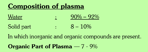
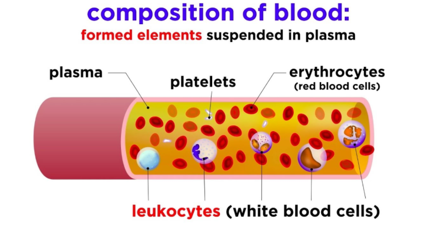
- Haematology is the study of blood. Blood is a special connective tissue consisting of fibre-free fluid matrix plasma, and formed elements. The amount of blood in a healthy human being is 5.0-5.5 litres.
- Plasma: It is a straw coloured, slightly alkaline(pH 7.4) viscous fluid constituting nearly 55% of the blood. Plasma mein proteins, lipids, salts, wastes, nutrients, hormones, water etc hota hai.
- Proteins : Albumins - help in osmotic balance. Their deficiency reduces the water content of blood and increases that of tissue fluid, causing oedema. Globulins - primarily are involved in defense mechanisms of the body and transport of substances.Fibrinogens - are needed for clotting or coagulation of blood. Prothrombin- another clotting protein also present in blood. Prothrombin is synthesized in the liver with the help of Vitamin K. Fibrinogens, globulins and albumins are the major proteins.
- Plasma without clotting factors is called serum.
- Nutrients : Plasma includes glucose, amino acids and lipids etc. are always in transit in the body.
- Ions - it contains small amounts of ions like sodium, calcium, magnesium, chloride etc.
- Gases - Nitrogen, oxygen, carbon dioxide occur in a dissolved state.
- Wastes - Wastes like urea, uric acid, etc are present in plasma.
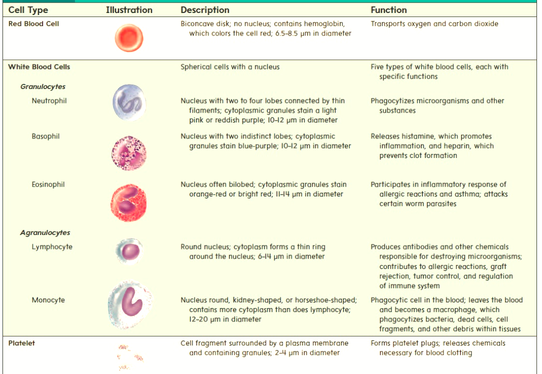
- Blood cells: These are of three types:
- Erythrocytes : Also called RBCs.
- Are the most abundant of all the cells in blood.
- A healthy adult man has, on an average, 5 millions to 5.5 millions of RBCs mm–3 of blood.
- RBCs are formed in the red bone marrow in adults.
- RBCs are devoid of nuclei in most mammals and are biconcave in shape.
- They have a red coloured, iron containing complex protein called haemoglobin.
- A healthy individual has 12-16 gms of haemoglobin in every 100 ml of blood.
- RBCs have an average life span of 120 days after which they are destroyed in the spleen (graveyard of RBCs).
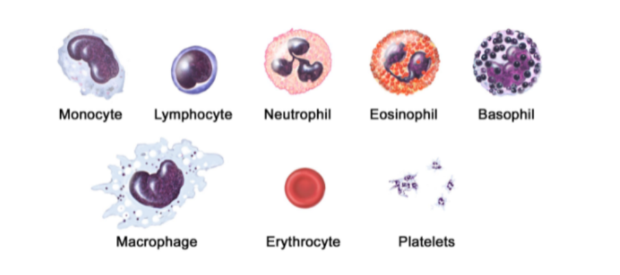
- Leucocytes: Also called WBCs.
- They are colourless due to the lack of haemoglobin. Inme nucleus present hota hai.
- Aur inka numbers average 6000-8000 mm–3 of blood.
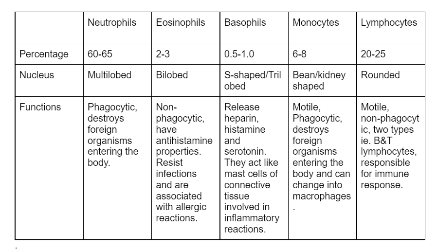
- Two main categories of WBCs are – granulocytes and agranulocytes.
- Granulocytes contain granules in their cytoplasm and have leucocytes which possess lobulated nuclei. They are also called polymorphonuclear leukocytes. Eg - neutrophils, eosinophils and basophils.
- Agranulocytes are leukocytes which do not possess granules as such. Eg lymphocytes and monocytes.
- Increase in WBCs count is called leukocytosis whereas decrease in WBCs count is called leukopenia.
- Abnormal increase of WBCs also occurs in malignancies(cancer) like leukemia.
- Blood platelets(thrombocytes) :
- These are non nucleated colorless cell fragments rather than true cells.
- Platelets are formed in bone marrow from large nucleated cells called megakaryocytes.
- Their size is generally 2-4 micrometre and their number is 1,50,000-3,50,000/mm3.
- They have a lifespan of about a week.
- Reduction in their number can lead to clotting disorder which will lead to excessive loss of blood from the body.
- Increase in platelet number is called thrombocytosis whereas decrease in platelet number is called thrombocytopenia.
- Dengue fever is characterised by an increase in platelet count.
- Erythrocytes : Also called RBCs.
- Blood is considered as a connective tissue because it circulates throughout the body and connects all parts transporting materials and performs a number of functions.

BLOOD
- Haematology is the study of blood. Blood is a special connective tissue consisting of fibre-free fluid matrix plasma, and formed elements. The amount of blood in a healthy human being is 5.0-5.5 litres.
- Plasma: It is a straw coloured, slightly alkaline(pH 7.4) viscous fluid constituting nearly 55% of the blood. Plasma mein proteins, lipids, salts, wastes, nutrients, hormones, water etc hota hai.
- Proteins : Albumins - help in osmotic balance. Their deficiency reduces the water content of blood and increases that of tissue fluid, causing oedema. Globulins - primarily are involved in defense mechanisms of the body and transport of substances. Fibrinogens - are needed for clotting or coagulation of blood. Prothrombin- another clotting protein also present in blood. Prothrombin is synthesized in the liver with the help of Vitamin K. Fibrinogens, globulins and albumins are the major proteins.
- Plasma without clotting factors is called serum.
- Nutrients : Plasma includes glucose, amino acids and lipids etc. are always in transit in the body.
- Ions - it contains small amounts of ions like sodium, calcium, magnesium, chloride etc.
- Gases - Nitrogen, oxygen, carbon dioxide occur in a dissolved state.
- Wastes - Wastes like urea, uric acid, etc are present in plasma.
- Blood cells: These are of three types:

- Erythrocytes : Also called RBCs.
- Are the most abundant of all the cells in blood.
- A healthy adult man has, on an average, 5 millions to 5.5 millions of RBCs mm–3 of blood.
- RBCs are formed in the red bone marrow in adults.
- RBCs are devoid of nuclei in most mammals and are biconcave in shape.
- They have a red coloured, iron containing complex protein called haemoglobin.
- A healthy individual has 12-16 gms of haemoglobin in every 100 ml of blood.
- RBCs have an average life span of 120 days after which they are destroyed in the spleen (graveyard of RBCs).
- Leucocytes: Also called WBCs.
- They are colourless due to the lack of haemoglobin. Inme nucleus present hota hai.
- Aur inka numbers average 6000-8000 mm–3 of blood.
- Two main categories of WBCs are – granulocytes and agranulocytes.
- Granulocytes contain granules in their cytoplasm and have leucocytes which possess lobulated nuclei. They are also called polymorphonuclear leukocytes. Eg - neutrophils, eosinophils and basophils.
- Agranulocytes are leukocytes which do not possess granules as such. Eg lymphocytes and monocytes.
- Increase in WBCs count is called leukocytosis whereas decrease in WBCs count is called leukopenia.
- Abnormal increase of WBCs also occurs in malignancies(cancer) like leukemia.
- Blood platelets(thrombocytes) :
- These are non nucleated colorless cell fragments rather than true cells.
- Platelets are formed in bone marrow from large nucleated cells called megakaryocytes.
- Their size is generally 2-4 micrometre and their number is 1,50,000-3,50,000/mm3.
- They have a lifespan of about a week.
- Reduction in their number can lead to clotting disorder which will lead to excessive loss of blood from the body.
- Increase in platelet number is called thrombocytosis whereas decrease in platelet number is called thrombocytopenia.
- Dengue fever is characterised by an increase in platelet count.

- Erythrocytes : Also called RBCs.
- Blood is considered as a connective tissue because it circulates throughout the body and connects all parts transporting materials and performs a number of functions.


Blood & Lymph
- Blood ek special connective tissue hai jo different organs ko apas me connect karta hai in the sense ki blood puri body me jitne bhi tissues and cells hai, un sabke beech me connectivity maintain karke rakhta hai. Blood ke 2 main parts hai : liquid component (55%) and cellular component jisko formed elements (45%) bhi kaha jata hai.liquid component ko hi hum plasma kehte hai jisme 90-92% water hota hai and baki ki blood proteins like albumins, glucose, amino acids, lipids, ions etc hote hai. Formed elements me aapke different types ke blood cells aate hai jaise ki RBCs, WBCs and platelets (yeah actual me cell fragments hote hai). RBCs red isiliye hote hai kyunki inme red pigment hota hai which is haemoglobin and yhi Hb oxygen se bond karke oxygen carry karne me help karta hai and yeah sab hum breathing and exchange of gases wale chapter me padh chuke hai. Right? WBCs basically immune cells hai yani ki yeah hume immunity provide karte hai and infections vgarah se ladne me help karte hai. Different types ke WBCs hote hai jinko hum majorly granulocytes and agranulocytes me divide kar dete hai. Another thing is, kafi WBCs phagocytic cells hote hai but not all WBCs. Neutrophils, eosinophils and basophils different granulocytes hai jinme different chemicals se filled granules hote hai. Yeah cells jab bhi pathogen ko recognise karte hai toh in chemicals ko release krte hai jo ki uss pathogen ko maar denge & yeah cells baki inflammatory molecules and signalling molecules bhi secrete karte hai. Baki cells bhi foreign particles ko engulf karte hai ya dusre methods se unko marte hai lekin unme aise granules nahi hote toh hum unko agranulocytes bol dete hai jaise ki macrophagic cells, B & T lymphocytes hote hai. B & T cells generally antibodies banane ke function ke sath associated hai jo ki particularly B cells bnate hai and kuch T cells keval B cell ki help karte hai antibody bnane me toh unko hum Helper T cells kehte hai and these helper cells are actually very crucial for B cells. Macrophagic cells are basically foreign particles and pathogen ko engulf karte hai yani ke yeah phagocytic cell hai. WBCs ko hi hum immune cells keh dete hai and inko immunology me alag se padha jata hai because these are so vast in their function, morphology that they require a separate study of their own. Platelets ko hi thrombocytes kehte hai jo ki actual me cell fragments hote hai. Inka blood clotting me role hai and platelets ki kami me blood clot nahi kar pata jo ki mild hemorrhages ko bhi fatal bna deta hai.
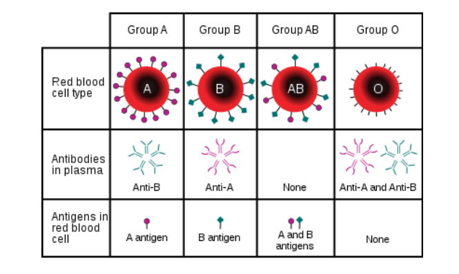
Now, coming back to RBCs, inke surface me kuch glycoprotein molecules hote hai jo ki cell-cell interaction and recognition me involved hote hai and yeah actually me immune cells ko stimulate kar sakte hai. Inko hum antigens kehte hai. Antigen ek kafi broad term hai toh don’t focus much on it now. Now har individual ki body me ek type ka antigen hote hai and uss antigen ko chor kar baki antigens ke against wo individual antibodies bna skata hai and bnata bhi hai. Humne in antigens ko naam dedia ki kisi pe antigen A hai and kisi me antigen B hai and kisi me antigen AB dono hai and kisi kisi ke RBCs pe koi antigen nahi. Toh jiski body me A antigen hai uska blood group A hoga, jiske bas keval B antigen hai usko B blood group bolenge and jiske pass AB hai usko AB blood group and jiske pas koi bhi blood group nahi hai usko hum O blood group bolte hai. Toh yeah basically ABO blood grouping hai jo ki bahut hi basic hai. Jiska blood A hai usme B ke against antibodies hogi toh agar A blood group wale person ko humne B blood dedia toh uska immune system B antigen wale RBCs pe attack karega and unke against antibodies bnane shuru karega toh destroy those foreign RBCs and yeah reaction fatal hota hai. So hospitals me hamesha blood typing hoti hai jinme aapka blood group check kia jata hai. Now these ABO blood grouping bahut hi basic hai. Iske alawa ek suna hoga ki koi kehta hai ki me B+ hu ya B- jabki koi A+ hota hai ya O+ ya O- toh what does this + and - mean?
→ Jo abhi 2 A and B antigen padhe unke alawa bhi antigens hote toh kafi hai but another common one is Rh antigen. Yeah sabme nhi hota. Jisme hota hai usko + bolte hai and jisme nhi hota wo Rh -ve hota hai. For example kisi person me agar dono B and Rh antigen hai toh iss blood group ko hum B+ blood kehte hai. Ek dikkat Rh blood group ke karan tab aati hai jab mom Rh-ve ho and foetus Rh+ve ho. Pehli pregnancy me dikkat nhi aati. Lekin jab pehli delivery hori hoti hai toh placenta rupture hoti and and mother’s immune system is exposed to Rh antigen jo ki mom ki body ke liye ek foreign antigen and wo uske against antibodies bnana shuru karenge. Yeah sab abhi tak koi dikkat nahi hai kyunki baby born hogya so no issue. Lekin issue second pregnancy me create hota hai. Mom ki body me jo antibodies hai wo placenta cross kar sakti hai and wo foetus ke RBCs ke jakr chipak jati hai and foetal RBCs ko destroy kar deti hai. Ab iske karan baby ko anemia hogya. Kabhi kabhi itna severe anemia ho jata hai ki foetus could be a stillbirth. RBCs destruction ke karan jo pigments release hue hai wo accumulate hona shuru karte hai and baby ko jaundice bhi hojata hai anemia ke sath. Issi condition ko erythroblastosis foetalis kehte hai jisko doctors anti-Rh-antibodies (Rhogam vaccine) mom me administer karke prevent kar dete hai.
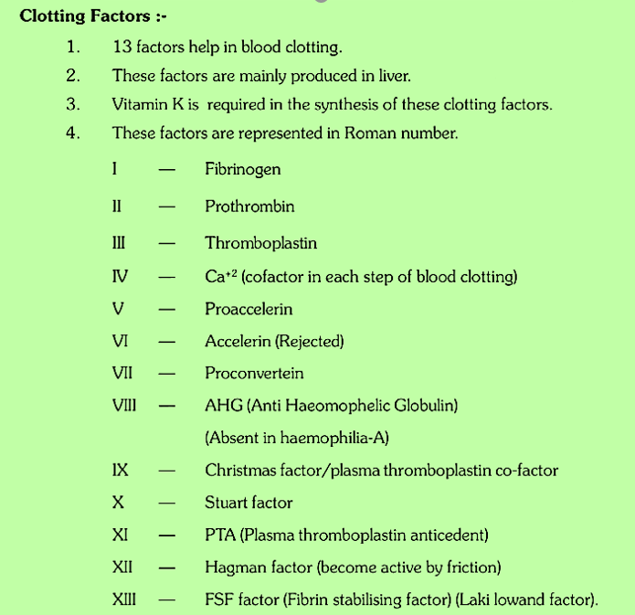
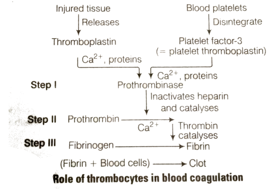
Blood Coagulation : Jab bhi chot lagti hai toh toh jo damaged tissue hota hai wo kuch coagulation promoting substance release karta hai jo ki prothrombinase naam ke enzyme ko activate kar dete hai. Yeah enzyme activate hone ke baad, prothrombin ko thrombin me convert karta hai. Thrombin basically fibrinogen ko fibrin me convert karta hai. Fibrin insoluble protein mesh hai jo ko blood clot ka ek main part hai. Issi fibrin ke sath platelets and baki blood cells milkar ek har solid structure form karte hai jo ki blood clot kehlata hai. Yeah blood clot excessive bleeding ko prevent karta hai and tissue ko heal karne me help karta hai. Exact blood clot ki formation ke pathways me hum different clotting factors chahie hote hai jo ki 12 factors and Ca
Lymph ko tissue fluid kehte hai. Imagine your blood as chai with tea leaves and full/sabut elaichi vgarah in it. The liquid part is basically the plasma jisme cheeni ghuli hai chai ke case me and different proteins, ions, sugars, aminoacids, gases, wastes, hormones etc ghuley hue hai blood ke case me. Blood arteries se capillaries ke through tissue tak jata hai. Ab yahan par samjho ki capillaries actually kafi thin hai and inke endothelial cells ke beech me spaces hote hai just like the chalni/strainer we use at home. Toh us chalni ki tar ko capillary endothelial cells maan lo. Jab chai ko strain karte hai toh basically kya hota hai? Tea leaves, adrak, elaichi vgarah sab chalni me reh jata hai and chai strain hokar neeche aa jati hai. Right? Same way me jab capillaries ke through blood pressure me flow karta hai toh kuch plasma bahar aa jati hai lekin blood cells nhi kyunki just like adrak and elaichi, blood cells ka size bada and wo wo capillary se bahar nhi nikal sakte. Ab yeah fluid jo bahar nikli hai issi ko hum tissue fluid kehte hai jisko lymph bhi bol dete hai. Chemical composition me yeah lagbhag plasma ke comparable hai lekin totally 100% identical nahi hai kyunki isme large molecules like large proteins vgarah nhi hote. Issi lymph ya tissue fluid ke through aas pass ke cells apni nutrient requirements ko pura karte hai and substances ka aadan-pradaan hota hai. Lekin aise agar yeah body ke har tissue me lymph nikalta rahe toh eventually blood ki volume kam ho jayegi and shareer ke sabhi hisso me sujan aajyegi kyunki lymph wapis blood me nhi mil pa rha toh we have lymphatic ducts jo ki iss lymph ko collect karke heart tak bhej deti hai and vhaan par yeah lymph fir se blood ke sath mil jata hai.
BLOOD GROUPS
- Blood of human beings differ in certain aspects but appear similar. Various types of grouping of blood have been done. Two such groupings are ABO and Rh.
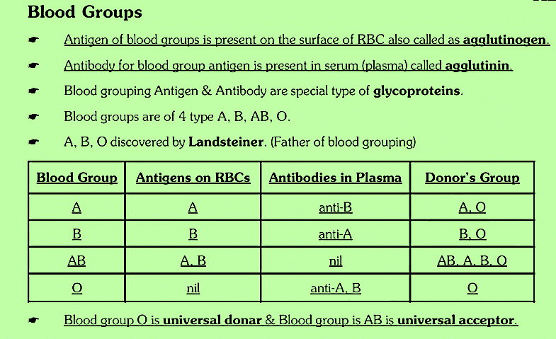
- It is based on the presence or absence of two surface antigens on the RBCs namely A and B. The plasma of different individuals contains two natural antibodies.
- Distribution of antigens and antibodies in different blood groups is given:
- During blood transfusion, any type of blood cannot be used but the blood of the donor should be carefully matched with that of the recipient to avoid clumping.
- “O” blood group can be donated to persons with any other blood group and hence O group individuals are universal donors Persons with “AB” group are universal recipients.
- Another antigen, the Rh antigen similar to that present in Rhesus monkeys, is also observed on the surface of RBCs of the majority (nearly 80%) of humans. Such individuals are Rh +ve while those without this antigen are Rh -ve.
- When Rh +ve blood is transfused to any Rh -ve individual, it will cause the formation of specific antibodies against Rh antigen.
- A special case of Rh incompatibility is observed between Rh -ve blood of a pregnant mother with Rh +ve blood of the foetus. Rh antigens of the foetus do not get exposed to Rh -ve blood of the mother in the first pregnancy as these two are separated by placenta.
- During delivery of the first child, there is a possibility of exposure of maternal blood to small amounts of the Rh +ve blood from foetus. The mother starts preparing antibodies against Rh antigens in her blood.
- In case of subsequent pregnancies, the antibodies from the mother’s blood can leak into the blood of the foetus(Rh +ve) and destroy foetal RBCs. This is called erythroblastosis fetalis/ Haemolytic disease of the newborn.
- This can be avoided by administering anti-Rh antibodies to the mother immediately after the delivery of the first child.
- Blood exhibits coagulation or clotting in response to an injury or trauma. This is a mechanism to prevent excessive loss of blood from the body.
- A dark reddish brown scum formed at the site of a cut or an injury over a period of time. It is a clot or coagulum jo mainly thread jese network fibrins se banta hai jisme mare hue aur kharab cells jakade jate hai.
- Fibrins are formed by the conversion of inactive fibrinogens in the plasma by the enzyme thrombin.
- Thrombins, in turn, are formed from another inactive substance present in the plasma membrane called prothrombin.
- An enzyme complex, thrombokinase, is required for the above reaction. This complex is formed by a series of linked enzymatic reactions(cascade process) involving a number of factors present in plasma in an inactive state.
- An injury or a trauma stimulates the platelets in the blood to release certain factors which activate the mechanism of coagulation. Certain factors released by the tissues at the site of injury also can initiate coagulation.
- A pale yellow fluid oozing from the clot is called serum.
- Heparin prevents blood clotting inside blood vessels.
ABO grouping
Rh Grouping :
COAGULATION OF BLOOD
BLOOD GROUPS
- Blood of human beings differ in certain aspects but appear similar. Various types of grouping of blood have been done. Two such groupings are ABO and Rh.
- It is based on the presence or absence of two surface antigens on the RBCs namely A and B. The plasma of different individuals contains two natural antibodies.
- Distribution of antigens and antibodies in different blood groups is given:

- During blood transfusion, any type of blood cannot be used but the blood of the donor should be carefully matched with that of the recipient to avoid clumping.
- “O” blood group can be donated to persons with any other blood group and hence O group individuals are universal donors Persons with “AB” group are universal recipients.
- Another antigen, the Rh antigen similar to that present in Rhesus monkeys, is also observed on the surface of RBCs of the majority (nearly 80%) of humans. Such individuals are Rh +ve while those without this antigen are Rh -ve.
- When Rh +ve blood is transfused to any Rh -ve individual, it will cause the formation of specific antibodies against Rh antigen.
- A special case of Rh incompatibility is observed between Rh -ve blood of a pregnant mother with Rh +ve blood of the foetus. Rh antigens of the foetus do not get exposed to Rh -ve blood of the mother in the first pregnancy as these two are separated by placenta.
- During delivery of the first child, there is a possibility of exposure of maternal blood to small amounts of the Rh +ve blood from foetus. The mother starts preparing antibodies against Rh antigens in her blood.
- In case of subsequent pregnancies, the antibodies from the mother’s blood can leak into the blood of the foetus(Rh +ve) and destroy foetal RBCs. This is called erythroblastosis fetalis/ Haemolytic disease of the newborn.
- This can be avoided by administering anti-Rh antibodies to the mother immediately after the delivery of the first child.
- Blood exhibits coagulation or clotting in response to an injury or trauma. This is a mechanism to prevent excessive loss of blood from the body.
- A dark reddish brown scum formed at the site of a cut or an injury over a period of time. It is a clot or coagulum jo mainly thread jese network fibrins se banta hai jisme mare hue aur kharab cells jakade jate hai.
- Fibrins are formed by the conversion of inactive fibrinogens in the plasma by the enzyme thrombin.
- Thrombins, in turn, are formed from another inactive substance present in the plasma membrane called prothrombin.
- An enzyme complex, thrombokinase, is required for the above reaction. This complex is formed by a series of linked enzymatic reactions(cascade process) involving a number of factors present in plasma in an inactive state.
- An injury or a trauma stimulates the platelets in the blood to release certain factors which activate the mechanism of coagulation. Certain factors released by the tissues at the site of injury also can initiate coagulation.
- A pale yellow fluid oozing from the clot is called serum.
- Heparin prevents blood clotting inside blood vessels.
ABO grouping
Rh Grouping :
COAGULATION OF BLOOD


HUMAN CIRCULATORY SYSTEM
- It is a reddish, roughly conical, highly muscular and mesodermal hollow organ with the size of a clenched fist (band muthi).
- It lies behind the sternum in the mediastinum space of thoracic cavity in between two lungs, hlka sa left side ki trf muda hua hota hai.
- Heart is enclosed in a tough, double walled membranous bag, the pericardium - outer parietal pericardium and inner visceral pericardium. Between these layers occur a narrow pericardial cavity(space) filled with pericardial fluid. Which provide frictionless movement and protection from shock and mechanical injury.
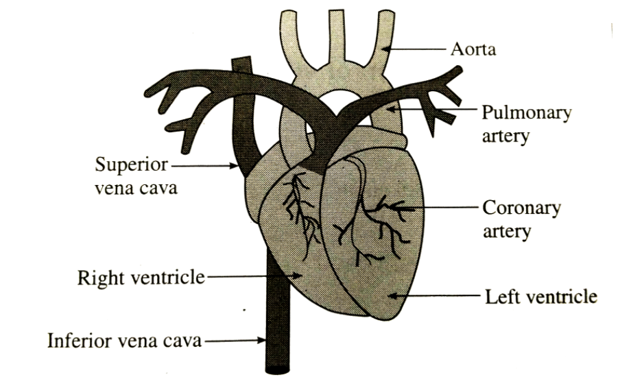
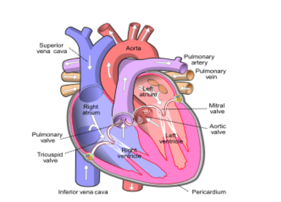
External Structure Of Heart: - The left and right atria are separated externally by a shallow ventral interatrial groove. The atria are separated externally from the ventricles by an oblique groove called atrio-ventricular sulcus.(coronary sulcus). There is an interventricular groove between the two ventricles, jisme coronary blood vessels hoti h.
- In the human heart, there are four chambers, two relatively small upper chambers called atria(collecting chambers) and two larger lower chambers called ventricles(distributing chambers).
- A thin, muscular wall called the interatrial septum separates the right and the left atria, whereas a thick wall, the interventricular septum, separates the left and the right ventricles.
- The atrium and the ventricles of the same side are also separated by a thick fibrous tissue called the atrio-ventricular septum. Each of these septa are provided with an opening through which the two chambers of the same side are connected.
- The opening between the right atrium and the right ventricle is guarded by a valve formed of three muscular flaps or cusps, the tricuspid valve, whereas a bicuspid or mitral valve gerd the opening between the left atrium and the left ventricle.
- The valves in the heart allow the flow of blood only in one direction i.e. from the atria to the ventricles and from the ventricles to the pulmonary artery or aorta. These valves prevent any backward flow.
- The walls of the ventricles are internally raised into a number of thick, muscular, column shaped projections called columnanae carnage or trabecular carnage; and a few large muscular elevations called papillary muscles
- Papillary muscles act as anchors for chordae tendineae which are strong, inelastic thread like tendons in the heart.
- Opening of superior vena cava and of pulmonary veins are without valves.
- Eustachian valve is present at the opening of inferior vena cava, Thebesian or coronary valve is present at the opening of coronary sinus and semilunar valve are present at the openings of pulmonary aorta and systemic aorta.
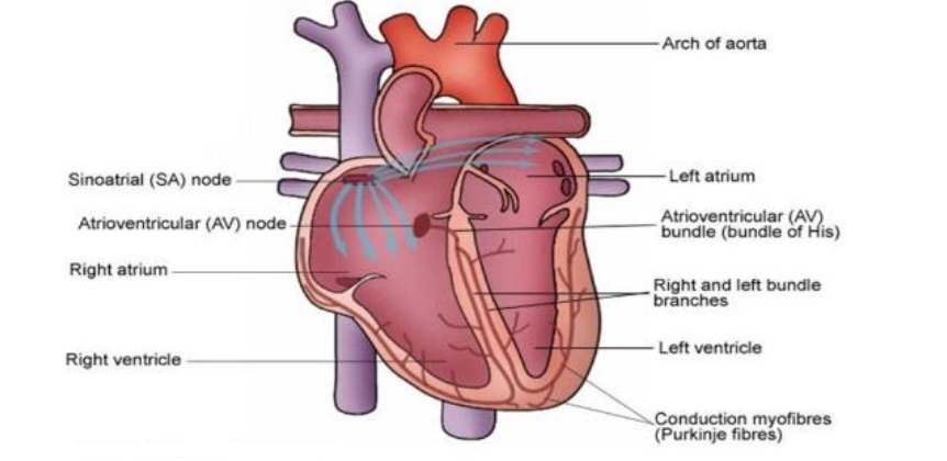
- Human heart is made of cardiac muscle. The walls of the ventricles are much thicker than that of the atria. A specialised cardiac musculature called the nodal tissue is also distributed in the heart. A patch of this tissue is present in the upper right corner of the right atrium called the sino-atrial node(SAN). Another mass of this tissue is seen in the lower left corner of the right atrium close to the atrio-ventricular septum called the atrio-ventricular node(AVN).
- A bundle of nodal fibres, atrioventricular bundle (AV bundle) continues from the AVN which passes through the atrio-ventricular septa to emerge on the top of the interventricular septum and immediately divides into a right and left bundle.
- These branches give rise to minute fibres throughout the ventricular musculature of the respective sides and are called purkinje fibres. These fibers along with right and left bundles are known as bundle of HIS.
- The nodal musculature has the ability to generate action potentials without any external stimuli, i.e. it is autoexcitable.
- Number of action potentials that could be generated per minute vary at different parts of the nodal system.
- SAN can generate a maximum number of action potentials (70-75/min) which is responsible for initiating and maintaining the rhythmic contractile activity of the heart i.e. it sets the pace of activities of the heart, therefore it is called the pacemaker.
- Normal average heart rate is 72 beats/min.
- SAN →AVN → AV bundle → Left and Right bundle branch → Purkinje fibres.
- Sequential event in the heart which is cyclically repeated is called the cardiac cycle and it consists of systole and diastole of both atria and ventricles.
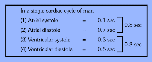
- As heart beats 72 times per minute, the duration of a cardiac cycle is about 0.8 seconds, hence many cardiac cycles are performed per minute.
- The cardiac cycle has three phases- Atrial systole, ventricular systole and joint diastole.
- Joint diastole : To begin with, all the four chambers of heart are in a relaxed state, i.e. they are in joint diastole. As the tricuspid and bicuspid valves are open, blood from the pulmonary veins and vena cava flows in to the left and the right ventricle respectively through the left and the right atria. The semilunar valves are closed at this stage.
- Atrial systole : SAN generates an action potential which stimulates both the atria to undergo a simultaneous contraction- atrial systole. This increases flow of blood into the ventricles by about 30% causing active filling of ventricles.
- Ventricular systole : Action potential is conducted to the ventricular side by AVN and AV bundle from where a bundle of HIS transmits it through entire ventricular musculature. This causes ventricular muscles to contract, (ventricular systole), atria undergoes relaxation(diastole), coinciding with ventricular systole. Ventricular systole increases ventricular pressure causing closure of tricuspid and bicuspid valves due to attempted backflow of blood into atria.
- As ventricular pressure increases further, semilunar valves guarding pulmonary artery(right side) and aorta(left side) are forced open, allowing the blood in the ventricles to flow through these vessels into the circulatory pathways.
- The ventricles now relax(ventricular diastole) and the ventricular pressure falls causing the closure of semilunar valves which prevents the backflow of blood into the ventricles. As the ventricular pressure declines further, the tricuspid and bicuspid valves are pushed open by the pressure in the atria exerted by the blood which was being emptied into them by the veins.
- The blood now once again moves freely to the ventricles. Soon the SAN generates a new action potential and the events described above are repeated in the above described sequence and the process continues.
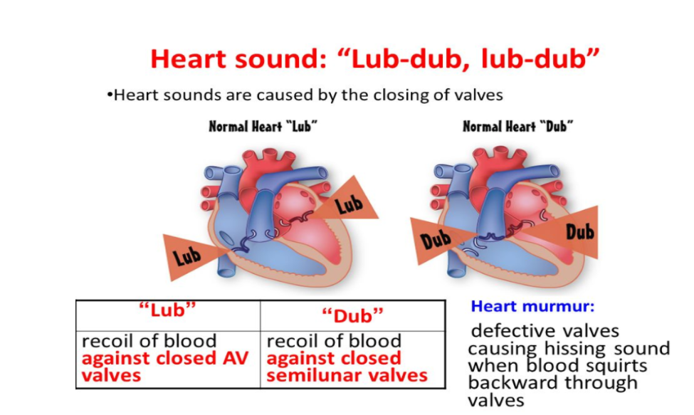
- These are generated due to closure of heart valves and heard with the help of a stethoscope.
- Defective/damaged valves lead to backflow of blood and cause an abnormal hissing sound(shhh) called murmur of the heart.
- Heart sounds are of great clinical significance.
- Volume of blood pumped by each ventricle in one minute is called cardiac output. It is also called minute volume.
- Cardiac output = stroke volume into heart rate.
- Stroke volume/beat volume is the volume of blood pumped by each ventricle in one beat/one cardiac cycle.
- Its amount is approximately 70ml.
- The body has the ability to alter the stroke volume as well as heart rate and thereby the cardiac output. For example, the cardiac output of an athlete will be much higher than that of an ordinary man.
- Adrenal medullary hormones can also increase cardiac output.
- Venous return : Amount of blood returning to the right atrium per minute is called venous return and is equal to 5.25 litres. Normally venous return and cardiac output are equal.
- The spontaneous and rhythmic contraction and relaxation of the heart to pump out and receive blood to and from the body is called heartbeat.
- Heart rate: It is heart beats per minute.
- Rhythmia - Normal heart rate
- Arrhythmia - Irregular heart rate
- Bradycardia - Decrease in heart rate
- Tachycardia - Increase in heart rate
- Smaller animals have a high metabolic rate and hence have higher heart rate.
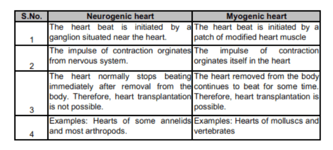
- Nervous control : Normal activities of the heart are regulated intrinsically, i.e., auto regulated by specialised muscles (nodal tissue), hence the heart is called myogenic.
→ A special neural centre in the medulla oblongata can moderate cardiac function through autonomic nervous system (ANS).Neural signals through the sympathetic nerves (part of ANS) can increase the rate of heart beat, the strength of ventricular contraction and thereby the cardiac output.
→ On the other hand, parasympathetic neural signals (another component of ANS) decrease the rate of heart beat, speed of conduction of action potential and thereby the cardiac output. - Hormonal control : Adrenal medullary hormones( Adrenaline and noradrenaline) can also increase the cardiac output, Nor-adrenaline under normal conditions and adrenaline at the time of emergency.
→ Thyroxine hormone also increases the heart rate by increasing energy production.
→ Acetylcholine decreases the heart rate. - ECG is a graphical representation of the electrical activity of the heart during a cardiac cycle. The machine (electro-cardiograph) is used to obtain an electrocardiogram (ECG).
- ECG k liye, a patient is connected to the machine with three electrical leads (one to each wrist(kalai) and to the left ankle) that continuously monitor the heart activity.
- Agr hume zyada detail m ECG chahiye toh, multiple leads are attached to the chest region.
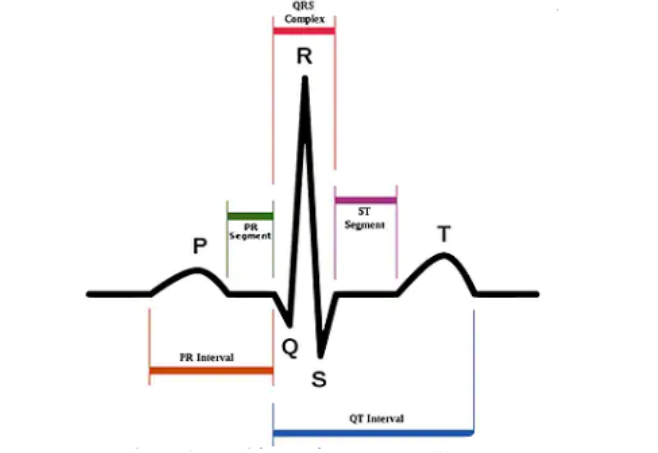
- Ek manak ECG mein, each peak is identified with a letter from P to T that corresponds to a specific electrical activity of the heart.
- The P-wave represents the electrical excitation (or depolarisation) of the atria, which leads to the contraction of both the atria.
- The QRS complex represents the depolarisation of the ventricles, which initiates the ventricular contraction. The contraction starts shortly after Q and marks the beginning of the systole.
- The T-wave represents the return of the ventricles from excited to normal state (repolarisation). The end of the T-wave marks the end of systole.
- By counting the number of QRS complexes that occur in a given time period, one can determine the heart beat rate of an individual.Since the ECGs obtained from different individuals have roughly the same shape for a given lead configuration, any deviation from this shape indicates a possible abnormality or disease. Hence, it is of a great clinical significance.
Internal Structure :
Types Of Heart On The Basis Of Origin Of Impulse:
Conducting System Of Heart
Mechanism Of Working Of Heart
Heart Sounds
Cardiac Output
Heartbeat
Regulation Of Heart Beat
ECG (Electrocardiogram)
HUMAN CIRCULATORY SYSTEM
- It is a reddish, roughly conical, highly muscular and mesodermal hollow organ with the size of a clenched fist (band muthi).
- It lies behind the sternum in the mediastinum space of thoracic cavity in between two lungs, hlka sa left side ki trf muda hua hota hai.
- Heart is enclosed in a tough, double walled membranous bag, the pericardium - outer parietal pericardium and inner visceral pericardium. Between these layers occur a narrow pericardial cavity(space) filled with pericardial fluid. Which provide frictionless movement and protection from shock and mechanical injury.
External Structure Of Heart: - The left and right atria are separated externally by a shallow ventral interatrial groove. The atria are separated externally from the ventricles by an oblique groove called atrio-ventricular sulcus.(coronary sulcus). There is an interventricular groove between the two ventricles, jisme coronary blood vessels hoti h.
- In the human heart, there are four chambers, two relatively small upper chambers called atria(collecting chambers) and two larger lower chambers called ventricles(distributing chambers).
- A thin, muscular wall called the interatrial septum separates the right and the left atria, whereas a thick wall, the interventricular septum, separates the left and the right ventricles.
- The atrium and the ventricles of the same side are also separated by a thick fibrous tissue called the atrio-ventricular septum. Each of these septa are provided with an opening through which the two chambers of the same side are connected.
- The opening between the right atrium and the right ventricle is guarded by a valve formed of three muscular flaps or cusps, the tricuspid valve, whereas a bicuspid or mitral valve gerd the opening between the left atrium and the left ventricle.
- The valves in the heart allow the flow of blood only in one direction i.e. from the atria to the ventricles and from the ventricles to the pulmonary artery or aorta. These valves prevent any backward flow.
- The walls of the ventricles are internally raised into a number of thick, muscular, column shaped projections called columnanae carnage or trabecular carnage; and a few large muscular elevations called papillary muscles
- Papillary muscles act as anchors for chordae tendineae which are strong, inelastic thread like tendons in the heart.
- Opening of superior vena cava and of pulmonary veins are without valves.
- Eustachian valve is present at the opening of inferior vena cava, Thebesian or coronary valve is present at the opening of coronary sinus and semilunar valve are present at the openings of pulmonary aorta and systemic aorta.
- Human heart is made of cardiac muscle. The walls of the ventricles are much thicker than that of the atria. A specialised cardiac musculature called the nodal tissue is also distributed in the heart. A patch of this tissue is present in the upper right corner of the right atrium called the sino-atrial node(SAN). Another mass of this tissue is seen in the lower left corner of the right atrium close to the atrio-ventricular septum called the atrio-ventricular node(AVN).
- A bundle of nodal fibres, atrioventricular bundle (AV bundle) continues from the AVN which passes through the atrio-ventricular septa to emerge on the top of the interventricular septum and immediately divides into a right and left bundle.
- These branches give rise to minute fibres throughout the ventricular musculature of the respective sides and are called purkinje fibres. These fibers along with right and left bundles are known as bundle of HIS.
- The nodal musculature has the ability to generate action potentials without any external stimuli, i.e. it is autoexcitable.
- Number of action potentials that could be generated per minute vary at different parts of the nodal system.
- SAN can generate a maximum number of action potentials (70-75/min) which is responsible for initiating and maintaining the rhythmic contractile activity of the heart i.e. it sets the pace of activities of the heart, therefore it is called the pacemaker.
- Normal average heart rate is 72 beats/min.
- SAN →AVN → AV bundle → Left and Right bundle branch → Purkinje fibres.
- Sequential event in the heart which is cyclically repeated is called the cardiac cycle and it consists of systole and diastole of both atria and ventricles.
- As heart beats 72 times per minute, the duration of a cardiac cycle is about 0.8 seconds, hence many cardiac cycles are performed per minute.
- The cardiac cycle has three phases- Atrial systole, ventricular systole and joint diastole.
- Joint diastole : To begin with, all the four chambers of heart are in a relaxed state, i.e. they are in joint diastole. As the tricuspid and bicuspid valves are open, blood from the pulmonary veins and vena cava flows in to the left and the right ventricle respectively through the left and the right atria. The semilunar valves are closed at this stage.
- Atrial systole : SAN generates an action potential which stimulates both the atria to undergo a simultaneous contraction- atrial systole. This increases flow of blood into the ventricles by about 30% causing active filling of ventricles.
- Ventricular systole : Action potential is conducted to the ventricular side by AVN and AV bundle from where a bundle of HIS transmits it through entire ventricular musculature. This causes ventricular muscles to contract, (ventricular systole), atria undergoes relaxation(diastole), coinciding with ventricular systole. Ventricular systole increases ventricular pressure causing closure of tricuspid and bicuspid valves due to attempted backflow of blood into atria.
- As ventricular pressure increases further, semilunar valves guarding pulmonary artery(right side) and aorta(left side) are forced open, allowing the blood in the ventricles to flow through these vessels into the circulatory pathways.
- The ventricles now relax(ventricular diastole) and the ventricular pressure falls causing the closure of semilunar valves which prevents the backflow of blood into the ventricles. As the ventricular pressure declines further, the tricuspid and bicuspid valves are pushed open by the pressure in the atria exerted by the blood which was being emptied into them by the veins.
- The blood now once again moves freely to the ventricles. Soon the SAN generates a new action potential and the events described above are repeated in the above described sequence and the process continues.
- These are generated due to closure of heart valves and heard with the help of a stethoscope.
- Defective/damaged valves lead to backflow of blood and cause an abnormal hissing sound(shhh) called murmur of the heart.
- Heart sounds are of great clinical significance.
- Volume of blood pumped by each ventricle in one minute is called cardiac output. It is also called minute volume.
- Cardiac output = stroke volume into heart rate.
- Stroke volume/beat volume is the volume of blood pumped by each ventricle in one beat/one cardiac cycle.
- Its amount is approximately 70ml.
- The body has the ability to alter the stroke volume as well as heart rate and thereby the cardiac output. For example, the cardiac output of an athlete will be much higher than that of an ordinary man.
- Adrenal medullary hormones can also increase cardiac output.
- Venous return : Amount of blood returning to the right atrium per minute is called venous return and is equal to 5.25 litres. Normally venous return and cardiac output are equal.

- The spontaneous and rhythmic contraction and relaxation of the heart to pump out and receive blood to and from the body is called heartbeat.
- Heart rate: It is heart beats per minute.
- Rhythmia - Normal heart rate
- Arrhythmia - Irregular heart rate
- Bradycardia - Decrease in heart rate
- Tachycardia - Increase in heart rate
- Smaller animals have a high metabolic rate and hence have higher heart rate.
- Nervous control : Normal activities of the heart are regulated intrinsically, i.e., auto regulated by specialised muscles (nodal tissue), hence the heart is called myogenic.
→ A special neural centre in the medulla oblongata can moderate cardiac function through autonomic nervous system (ANS).Neural signals through the sympathetic nerves (part of ANS) can increase the rate of heart beat, the strength of ventricular contraction and thereby the cardiac output.
→ On the other hand, parasympathetic neural signals (another component of ANS) decrease the rate of heart beat, speed of conduction of action potential and thereby the cardiac output. - Hormonal control : Adrenal medullary hormones( Adrenaline and noradrenaline) can also increase the cardiac output, Nor-adrenaline under normal conditions and adrenaline at the time of emergency.
→ Thyroxine hormone also increases the heart rate by increasing energy production.
→ Acetylcholine decreases the heart rate. - ECG is a graphical representation of the electrical activity of the heart during a cardiac cycle. The machine (electro-cardiograph) is used to obtain an electrocardiogram (ECG).
- ECG k liye, a patient is connected to the machine with three electrical leads (one to each wrist(kalai) and to the left ankle) that continuously monitor the heart activity.
- Agr hume zyada detail m ECG chahiye toh, multiple leads are attached to the chest region.
- Ek manak ECG mein, each peak is identified with a letter from P to T that corresponds to a specific electrical activity of the heart.
- The P-wave represents the electrical excitation (or depolarisation) of the atria, which leads to the contraction of both the atria.
- The QRS complex represents the depolarisation of the ventricles, which initiates the ventricular contraction. The contraction starts shortly after Q and marks the beginning of the systole.
- The T-wave represents the return of the ventricles from excited to normal state (repolarisation). The end of the T-wave marks the end of systole.
- By counting the number of QRS complexes that occur in a given time period, one can determine the heart beat rate of an individual.Since the ECGs obtained from different individuals have roughly the same shape for a given lead configuration, any deviation from this shape indicates a possible abnormality or disease. Hence, it is of a great clinical significance.

Internal Structure :

Types Of Heart On The Basis Of Origin Of Impulse:

Conducting System Of Heart

Mechanism Of Working Of Heart
Heart Sounds

Cardiac Output
Heartbeat
Regulation Of Heart Beat
ECG (Electrocardiogram)

BLOOD VESSELS
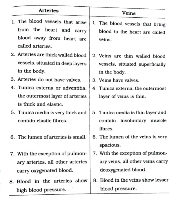
- The blood flows strictly by a fixed route through blood vessels. Wall of blood vessels is made up of three layers:
- Tunica externa(outer layer) - It is made up of some elastic fibres and collagen fibres.
- Tunica media(middle layer) - It has smooth muscles in addition to fibres.
- Tunica interna (inner layer) - There is outer layer of elastic fibres and an inner layer of endothelium.
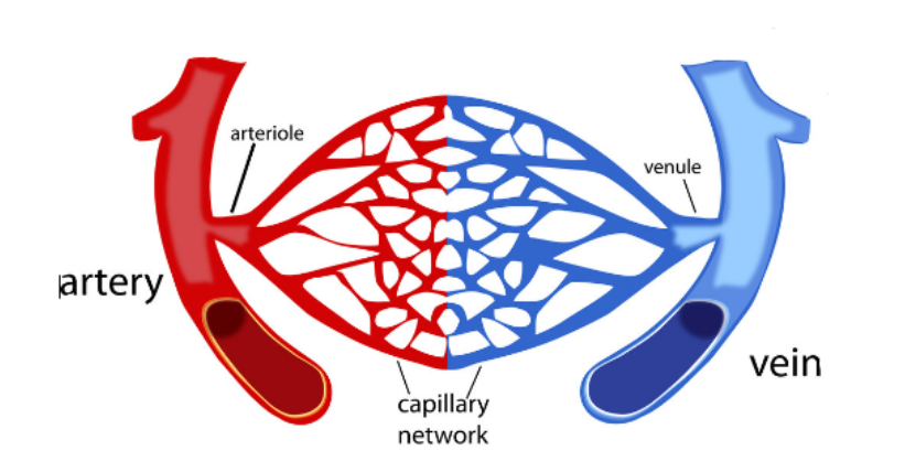
- Types of blood vessels: The blood vessels are the part of the circulatory system that transports blood throughout the body. There are three major types of blood vessels: arteries, capillaries and veins.
- Capillaries: These enable the actual exchange of water and chemicals between the blood and the tissues.
→ These are made up of Tunica interna and they connect the arteries to the veins. These are the smallest blood vessels. - The pathway of blood flow : Heart → Arteries → Arterioles → Capillaries → Venule → Veins → Heart
- Coronary circulation : System of blood vessels is present in out body exclusively for the circulation of blood to and from the cardiac musculature.
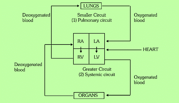
- Pulmonary circulation : The blood pumped by the right ventricle enters the pulmonary artery, whereas the left ventricle pumps blood into the aorta. The deoxygenated blood pumped into the pulmonary artery is passed on to the lungs from where the oxygenated blood is carried by the pulmonary veins into the left atrium. This pathway constitutes the pulmonary circulation.
- Systemic circulation : The oxygenated blood entering the aorta is carried by a network of arteries, arterioles and capillaries to the tissues from where the deoxygenated blood is collected by a system of venules, veins and vena cava and emptied into the right atrium. This is the systemic circulation.The systemic circulation provides nutrients, O2 and other essential substances to the tissues and takes CO2 and other harmful substances away for elimination.
- Portal circulation : A unique vascular connection exists between the digestive tract and liver called hepatic portal system. The hepatic portal vein carries blood from intestine to the liver before it is delivered to the systemic circulation.
- A special coronary system of blood vessels is present in our body exclusively for the circulation of blood to and from the cardiac musculature.
- This is a network of capillaries, vessels, ducts carrying fluid called lymph. Lymph is a colourless fluid containing specialised lymphocytes which are responsible for the immune responses of the body.It is a mobile connective tissue formed by the filtration of blood.
- As the blood passes through the capillaries in tissues, some water along with many small water soluble substances move out into the spaces between the cells of tissues leaving the larger proteins and most of the formed elements in the blood vessels. This fluid released out is called the interstitial fluid or tissue fluid. It has the same mineral distribution as that in plasma.
- Lymph is also an important carrier for nutrients, hormones, etc. Fats are absorbed through lymph in the lacteals present in the intestinal villi.
- Normally the rate of lymph formation is equal to the rate of its return to the bloodstream.
Types Of Blood Circulation
Lymphatic System
Human Circulatory System
- Pehle ek baar simple overview dekh lete hai ki hota kya hai humans and baki mammals ke circulatory system. Circulating fluid yani ki blood ka main kaam hai gaseous exchange me help karna and baki ke substances like food, waste and hormones ka transport but for the time being apna focus hoga oxygen ki delivery pe. Heart ki left side oxygenated blood pump karta hai puri body ko and heart ki right side deoxygenated blood ko lungs tak pump karta hai. Jo bhi tissues se used blood aara hai jisme carbon dioxide ki conc. Jyada hai wo purey shareer se different vein lekr aati hai and vena cava (superior and inferior) me daal deti hai. Vena cava se blod right auricle/atrium me jata hai jo right ventricle tak blood bhejdega. Right ventricle fir iss blood of pulmonary arteries ke through lungs tak pump karta hai. Lungs me CO2 bahar nikal jati hai and O2 RBCs ke Hb se bind hojati hai. This oxygen rich blood is oxygenated blood. Fir yeah blood lungs se pulmonary veins ke through heart ki left auricle me aata hai and vhan se left ventricle me. Left ventricle se aorta ke through puri body me pump kiya jata hai. Toh dekho same blood ko 2 baar circulate kiya gya hai : ek baar toh usko lungs tak bheja oxygenation ke liye - this is known as pulmonary circulation and dusri baar jab wahi oxygenated blood heart me aaya toh usko aorta ke through puri body tak pump kia gya - this is known as systemic circulation; toh yeah 2 baar circulation hi double circulation kehlata hai.
Ab jab hume pta hai ki okay roughly aisa aisa hota hai circulation ke time pe toh ab hum heart ka structure thodi se detail me study karenge - keval utna jitna CBSE syllabus me required hai. Heart me 4 chambers hote hai : 2 right and left atria jo ki upper chambers hai and inter atrial septum ki help se ek dusre se separated hai. Then lower side me 2 - right and left ventricles hote hai jo ki in general atria ke comparison me thick hote hai and in dono me se bhi left ventricle thickest hota hai. Dono ventricles ek dusre se ventricular septum ki help se separated hote hai. Yeah separation basically oxygenated and deoxygenated blood ko alag alag rakhta hai and mix nahi honey deta. Heart cardiac muscles se bna hua hai. Iske charo taraf ek protective layer hoti hai jisko pericardial sac kehte hai and yeah double layered hota hai. Dono layers ke beech me pericardial fluid hoti hai jo heart ko mechanical damage se protect karti hai, buoyancy provide karti hai and neighbor organs se friction kam karti hai. Right atria me upper right corner ki side sinoatrial node hoti hai and lower left corner par atrioventricular node hoti hai. SA node heart ka pacemaker hai kyunki yeah electrical pulses generate karta hai jo heart ko contract karti hai. Heart ki contraction directly brain ya nervous system se nahi hoti yani ki hum heart neurogenic nahi balki myogenic hai. Atria or ventricle ke beech me atrioventricular valves hoti hai jo blood ka backflow prevent karti hai. Right atria and right ventricle ke beech me tricuspid valve hoti hai and left atria and left ventricle ke beech me bicuspid/mitral valve hoti hai. Pulmonary artery me aorta me bhi semilunar valves hoti hoti. Sabhi valves ka kaam hai ki unidirectional blood flow ko maintain kare and backflow ko prevent kare.
→ SA node impulse generate karti hai and atria contract karte hai toh AV valves khul jati hai and jitna blood atria me uss waqt hoga wo neeche ventricles me chla jata hai and AV close hojati hai taki jab ventricles contract kare toh blood wapis atria me na jaye and uss waqt semilunar valves khul jati hai toh blood aorta and pulmonary trunk me jata hai. Another thing is left ventricle ki thickness sabse jyada hai kyunki usko puri body ko blood pump karna hai toh it has to contract with greater force jiske liye jyada muscle fibres chahiye hote hai. AV node se bundle of his nerve impulse ko carry karta hai and purkinje fibres tak bhejta hai. Purkinje fibres ventricles me embedded hote hai and jab excitation pulse ko conduct karte hai taki ventricles contract kar paye. Heart beats 70 times per minute. Infants ki heart rate normally iss sey higher hoti hai and trained athletes me normal resting heart rate could go as low as 40 beats per minute which is okay and normal for them but not for rest of us. Normal se kam beats per minute, bradycardia kehlata hai and normal se jyada beats basically jab 100bets/min ke aas pass ya ussey jyada ho toh wo tachycardia kehlata hai. Dono hi prolonged time tak dangerous hote hai.
CIRCULATORY PATHWAYS
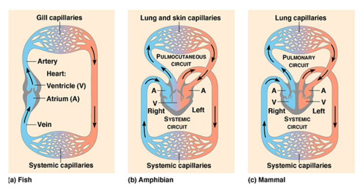
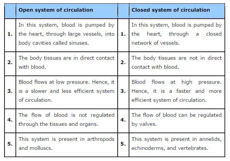
- The circulatory patterns are open or closed types.
- Closed circulation is of two types:
- Single circulation -In fish the heart pumps out deoxygenated blood which is oxygenated by the gills and supplied to the body parts from where deoxygenated blood is returned to the heart.
- Double circulation -
- Incomplete double circulation - In amphibians and reptiles, the left atrium receives oxygenated blood from the gills/lungs/skin and the right atrium gets the deoxygenated blood from other body parts. However, they get mixed up in the single ventricle which pumps out mixed blood.
- Complete double - In such circulation two separate circulatory pathways i.e. pulmonary and systemic are present.. In birds and mammals, oxygenated and deoxygenated blood received by the left and right atria respectively passes on to the ventricles of the same sides. The ventricles pump it out without any mixing up, i.e., two separate circulatory pathways are present in these organisms, hence, these animals have double circulation.

BLOOD VESSELS
- The blood flows strictly by a fixed route through blood vessels. Wall of blood vessels is made up of three layers:
- Tunica externa(outer layer) - It is made up of some elastic fibres and collagen fibres.
- Tunica media(middle layer) - It has smooth muscles in addition to fibres.
- Tunica interna (inner layer) - There is outer layer of elastic fibres and an inner layer of endothelium.
- Types of blood vessels: The blood vessels are the part of the circulatory system that transports blood throughout the body. There are three major types of blood vessels: arteries, capillaries and veins.
- Capillaries: These enable the actual exchange of water and chemicals between the blood and the tissues.
→ These are made up of Tunica interna and they connect the arteries to the veins. These are the smallest blood vessels. - The pathway of blood flow : Heart → Arteries → Arterioles → Capillaries → Venule → Veins → Heart
- Coronary circulation : System of blood vessels is present in out body exclusively for the circulation of blood to and from the cardiac musculature.
- Pulmonary circulation : The blood pumped by the right ventricle enters the pulmonary artery, whereas the left ventricle pumps blood into the aorta. The deoxygenated blood pumped into the pulmonary artery is passed on to the lungs from where the oxygenated blood is carried by the pulmonary veins into the left atrium. This pathway constitutes the pulmonary circulation.
- Systemic circulation : The oxygenated blood entering the aorta is carried by a network of arteries, arterioles and capillaries to the tissues from where the deoxygenated blood is collected by a system of venules, veins and vena cava and emptied into the right atrium. This is the systemic circulation.The systemic circulation provides nutrients, O2 and other essential substances to the tissues and takes CO2 and other harmful substances away for elimination.
- Portal circulation : A unique vascular connection exists between the digestive tract and liver called hepatic portal system. The hepatic portal vein carries blood from intestine to the liver before it is delivered to the systemic circulation.
- A special coronary system of blood vessels is present in our body exclusively for the circulation of blood to and from the cardiac musculature.
- This is a network of capillaries, vessels, ducts carrying fluid called lymph.Lymph is a colourless fluid containing specialised lymphocytes which are responsible for the immune responses of the body.It is a mobile connective tissue formed by the filtration of blood.
- As the blood passes through the capillaries in tissues, some water along with many small water soluble substances move out into the spaces between the cells of tissues leaving the larger proteins and most of the formed elements in the blood vessels. This fluid released out is called the interstitial fluid or tissue fluid. It has the same mineral distribution as that in plasma.
- Lymph is also an important carrier for nutrients, hormones, etc. Fats are absorbed through lymph in the lacteals present in the intestinal villi.
- Normally the rate of lymph formation is equal to the rate of its return to the bloodstream.


Types Of Blood Circulation
Lymphatic System
DISORDERS OF CIRCULATORY SYSTEM
- High Blood Pressure (Hypertension): Hypertension is the term for blood pressure that is higher than normal (120/80). In this measurement 120 mm Hg (millimetres of mercury pressure) is the systolic, or pumping, pressure and 80 mm Hg is the diastolic, or resting, pressure. If repeated checks of blood pressure of an individual is 140/90 (140 over 90) or higher, it shows hypertension. High blood pressure leads to heart diseases and also affects vital organs like the brain and kidney.
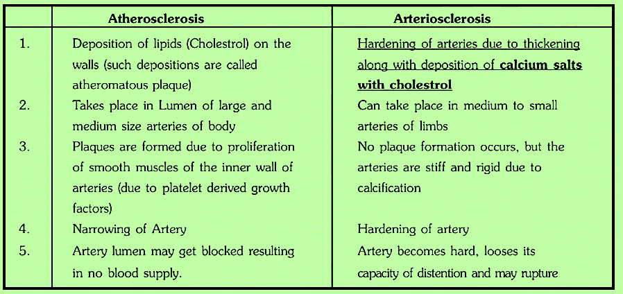
- Coronary Artery Disease (CAD): Coronary Artery Disease, often referred to as atherosclerosis, affects the vessels that supply blood to the heart muscle. It is caused by deposits of calcium, fat, cholesterol and fibrous tissues, which makes the lumen of arteries narrower.
- Angina: It is also called ‘angina pectoris’. A symptom of acute chest pain appears when not enough oxygen is reaching the heart muscle. Angina can occur in men and women of any age but it is more common among the middle-aged and elderly. It occurs due to conditions that affect the blood flow.
- Heart Failure: Heart failure means the state of heart when it is not pumping blood effectively enough to meet the needs of the body. It is sometimes called congestive heart failure because congestion of the lungs is one of the main symptoms of this disease.
- Cardiac arrest : when the heart stops beating. The electrocardiograph machine will make the sound….pip...pip...pip…..peeeeeeeeeeeeeeeeeeeee.
- Heart attack : When the heart muscle is suddenly damaged by an inadequate blood supply.
Cardiac Cycle & ECG
- Heart har beat ke sath contract and relax karta hai and yeah sab 0.8 seconds me ho jata hai. Issi ko hum cardiac cycle kehte hai. 3 phase hote hai cardiac cycle me : atrial systole, ventricular systole and joint diastole. Diastole ka matlab and systole ka matlab contract. Shuru karte hai joint diastole me jab av valves open hoti hai toh vena cava se aane wala blood directly right ventricle me jayega and pulmonary vein se aane wala oxygenated blood left atria se seedhe left ventricle me jata hai kyunki av valves open hai. This leads to 70% filling of ventricles. Then atria contract karna shuru karte hai and bacha hua blood ventricles me aajata hai. Yeah tha atrial systole jab atria contract karte hai jo ki nearly 0.1 second ke liye hota hai. Then AV valves close hoti hai and ventricles contract karna shuru karte hai. This is the begining of ventricular systole which lasts for about 0.3 seconds. Iss phase ki shuruat me semilunar valves close hoti hai toh jaise jaise ventricles contract karna shuru karte hai, av valves closed hai toh ventricles me se initally contraction ke time par blood nhi nikal pata jiske karan ventricles me pressure rise up hona shuru karta hai. Ek point ke baad ventricles ka pressure jaise hi pulmonary trunk and aorta me jo pressure hai usko cross karega toh semilunar valves open ho jayegi and ventricles se blood pulmonary artery and aorta me flow karna shuru karta hai the ventricles eject the blood. Sara 100% blood eject nhi kiya jata. Then jab blood lagbhag pump ho gya and keval thoda sa reh gya toh pressure ventricles ka fall karna shuru kar deta hai and pulmonary valves close. Yeah ventricular systole complete hogya. Ab ventricles relax karna shuru karte hai and unme pressure kam hota jata hai. Ab jaise hi ventricular pressure falls below the atrial pressure, av valves open hoti hai and ventricles fill karna shuru kar dete hai. Atria ab relax karte hai and sath me ventricles bhi relax kar rhe hai toh yeah joint diastole hai. Atrial systole lasts approximately 100 ms and ends prior to ventricular systole, as the atrial muscle returns to diastole. Fir jaldi se SA node another impulse generate karke new cardiac cycle initiate karti hai. Per beat me heart 70mL volume eject karta hai and this volume is known as stroke volume.
raar Heart sounds : Lub - due to closure of av valves. Yeah long duration ki hoti hai generally 0.16 sec and it is louder and low pitched. Second heart sound Dub hoti hai jo ki semilunar valves ke close hone pe sunaie deti hai and it is of shorter duration : 0.1 sec. High pitched and less louder sound hai yeah. Toh jo aapko dil ki dhadkan ya jo Dhak Dhak sound aati hai wo actually me yeah heart sounds hai and inke alawa bhi or heart sounds hoti hai. Plus agar valves me koi gadbad hai toh doctor ko abnormal heart sounds sunaie dengi jo actually distinguish ki ja sakti hai to get an idea ki most probably yhaan pe dikkat and then doctor advises further tests to know the actual disease.
- Electrocardiograph ek machine hai jo heart ki electrical activity ko measure karke usko graph paper pe print karke electrocardiogram (ECG) ki form me deti hai. Jaise ki uper diagram me dekha ki ek normal ECG me 3 cheezein hai : P wave, QRS complex and T wave. These refer to different stages of heart activity. Basically jab SA node fir karti hai toh atria depolarise hote hai and it leads to atrial systole jo ki ECG me P wave ki form me capture kar li jati hai. Then we all know ki next ventricles depolarise karenge toh ventricles contract karenge toh wo cheez ECG me QRS complex ki form me aati hai. Ab P wave ka amplitude QRS ke comparison me chota hota hai kyunki atria small size ke hai jabki ventricles ka mass jyada toh electrical activity ventricles me obviously jyada hogi toh lambi QRS complex aati hai. T wave represent karti hai ventricular repolarisation ko. Ab normally as such ECG me atrial repolarisation ke liye koi different wave nhi hoti kyunki jab atria repolarise karte hai toh wo small electrical activity hai jo ki machine cath karegi but simultaneously ventricles repolarise kar rahe hote hai and unme electrical activity jyada hai toh T wave ka jo amplitude aata hai wo bda hota hai and it actually represents ventricular repolarisation. Normal parameters me P duration < 0.12 sec and P amplitude < 2.5 mm. QRS duration ≤ 0.10 sec. QT Interval ≤ 0.40 sec. Doctor inhi intervals, amplitudes and in waves ki polarity ko check karte hai kyunki different diseases me ECG me different distinguishing features hote hai. For example kisi person ke ECG me agar P wave ka amplitude bda hai toh atrial size badha hua ho sakta hai, PR interval gadbad hai P wave kahi bifurcated toh nhi hai : yeah sab atria related abnormalities ko suggest karte hai. Ventricle related diseases and conduction pathway me koi gadbad hai toh QRS complex abnormal dikhega. Toh we won’t discuss those things here kyunki hamesha straightforward nhi hota and different diseases and ECG ko check karne ke liye heart ki puri anatomy and baki electrophysiology aani chahiye jo ki apne aap me alag course hai pura so we will not dwell into those aspects.
SUMMARY
- Vertebrates circulate blood, a fluid connective tissue, in their body, to transport essential substances to the cells and to carry waste substances from there. Another fluid, lymph (tissue fluid) is also used for the transport of certain substances. Blood comprises a fluid matrix, plasma and formed elements.
- Red blood cells (RBCs, erythrocytes), white blood cells (WBCs, leucocytes) and platelets (thrombocytes) constitute the formed elements. Blood of humans are grouped into A, B, AB and O systems based on the presence or absence of two surface antigens, A, B on the RBCs. Another blood grouping is also done based on the presence or absence of another antigen called Rhesus factor (Rh) on the surface of RBCs. The spaces between cells in the tissues contain a fluid derived from blood called tissue fluid. This fluid called lymph is almost similar to blood except for the protein content and the formed elements.
- All vertebrates and a few invertebrates have a closed circulatory system. Our circulatory system consists of a muscular pumping organ, heart, a network of vessels and a fluid, blood. Heart has two atria and two ventricles.
- Cardiac musculature is auto-excitable. Sino-atrial node (SAN) generates the maximum number of action potentials per minute (70-75/min) and therefore, it sets the pace of the activities of the heart. Hence it is called the Pacemaker. The action potential causes the atria and then the ventricles to undergo contraction (systole) followed by their relaxation (diastole). The systole forces the blood to move from the atria to the ventricles and to the pulmonary artery and the aorta.
- The cardiac cycle is formed by sequential events in the heart which is cyclically repeated and is called the cardiac cycle. A healthy person shows 72 such cycles per minute. About 70 mL of blood is pumped out by each ventricle during a cardiac cycle and it is called the stroke or beat volume.
- Volume of blood pumped out by each ventricle of Coronary Artery Disease (CAD): Coronary Artery Disease, often referred to as atherosclerosis, affects the vessels that supply blood to the heart muscle. It is caused by deposits of calcium, fat, cholesterol and fibrous tissues, which makes the lumen of arteries narrower. Angina: It is also called ‘angina pectoris’. A symptom of acute chest pain appears when not enough oxygen is reaching the heart muscle. Angina can occur in men and women of any age but it is more common among the middle-aged and elderly. It occurs due to conditions that affect the blood flow. Heart Failure: Heart failure means the state of heart when it is not pumping blood effectively enough to meet the needs of the body. It is sometimes called congestive heart failure because congestion of the lungs is one of the main symptoms of this disease. Heart failure is not the same as cardiac arrest (when the heart stops beating) or a heart attack (when the heart muscle is suddenly damaged by an inadequate blood supply) heart per minute is called the cardiac output and it is equal to the product of stroke volume and heart rate (approx 5 litres).
- The electrical activity of the heart can be recorded from the body surface by using electrocardiograph and the recording is called electrocardiogram (ECG) which is of clinical importance.
- We have a complete double circulation, i.e., two circulatory pathways, namely, pulmonary and systemic, are present. The pulmonary circulation starts by the pumping of deoxygenated blood by the right ventricle which is carried to the lungs where it is oxygenated and returned to the left atrium.
- The systemic circulation starts with the pumping of oxygenated blood by the left ventricle to the aorta which is carried to all the body tissues and the deoxygenated blood from there is collected by the veins and returned to the right atrium. Though the heart is autoexcitable, its functions can be moderated by neural and hormonal mechanisms.
CIRCULATORY PATHWAYS
- The circulatory patterns are open or closed types.
- Closed circulation is of two types:
- Single circulation -In fish the heart pumps out deoxygenated blood which is oxygenated by the gills and supplied to the body parts from where deoxygenated blood is returned to the heart.
- Double circulation -
- Incomplete double circulation - In amphibians and reptiles, the left atrium receives oxygenated blood from the gills/lungs/skin and the right atrium gets the deoxygenated blood from other body parts. However, they get mixed up in the single ventricle which pumps out mixed blood.
- Complete double - In such circulation two separate circulatory pathways i.e. pulmonary and systemic are present.. In birds and mammals, oxygenated and deoxygenated blood received by the left and right atria respectively passes on to the ventricles of the same sides. The ventricles pump it out without any mixing up, i.e., two separate circulatory pathways are present in these organisms, hence, these animals have double circulation.


DISORDERS OF CIRCULATORY SYSTEM
- High Blood Pressure (Hypertension): Hypertension is the term for blood pressure that is higher than normal (120/80). In this measurement 120 mm Hg (millimetres of mercury pressure) is the systolic, or pumping, pressure and 80 mm Hg is the diastolic, or resting, pressure. If repeated checks of blood pressure of an individual is 140/90 (140 over 90) or higher, it shows hypertension. High blood pressure leads to heart diseases and also affects vital organs like the brain and kidney.
- Coronary Artery Disease (CAD): Coronary Artery Disease, often referred to as atherosclerosis, affects the vessels that supply blood to the heart muscle. It is caused by deposits of calcium, fat, cholesterol and fibrous tissues, which makes the lumen of arteries narrower.
- Angina: It is also called ‘angina pectoris’. A symptom of acute chest pain appears when not enough oxygen is reaching the heart muscle. Angina can occur in men and women of any age but it is more common among the middle-aged and elderly. It occurs due to conditions that affect the blood flow.
- Heart Failure: Heart failure means the state of heart when it is not pumping blood effectively enough to meet the needs of the body. It is sometimes called congestive heart failure because congestion of the lungs is one of the main symptoms of this disease.
- Cardiac arrest : when the heart stops beating. The electrocardiograph machine will make the sound….pip...pip...pip…..peeeeeeeeeeeeeeeeeeeee.
- Heart attack : When the heart muscle is suddenly damaged by an inadequate blood supply.

SUMMARY
- Vertebrates circulate blood, a fluid connective tissue, in their body, to transport essential substances to the cells and to carry waste substances from there. Another fluid, lymph (tissue fluid) is also used for the transport of certain substances. Blood comprises a fluid matrix, plasma and formed elements.
- Red blood cells (RBCs, erythrocytes), white blood cells (WBCs, leucocytes) and platelets (thrombocytes) constitute the formed elements. Blood of humans are grouped into A, B, AB and O systems based on the presence or absence of two surface antigens, A, B on the RBCs. Another blood grouping is also done based on the presence or absence of another antigen called Rhesus factor (Rh) on the surface of RBCs. The spaces between cells in the tissues contain a fluid derived from blood called tissue fluid. This fluid called lymph is almost similar to blood except for the protein content and the formed elements.
- All vertebrates and a few invertebrates have a closed circulatory system. Our circulatory system consists of a muscular pumping organ, heart, a network of vessels and a fluid, blood. Heart has two atria and two ventricles.
- Cardiac musculature is auto-excitable. Sino-atrial node (SAN) generates the maximum number of action potentials per minute (70-75/min) and therefore, it sets the pace of the activities of the heart. Hence it is called the Pacemaker. The action potential causes the atria and then the ventricles to undergo contraction (systole) followed by their relaxation (diastole). The systole forces the blood to move from the atria to the ventricles and to the pulmonary artery and the aorta.
- The cardiac cycle is formed by sequential events in the heart which is cyclically repeated and is called the cardiac cycle. A healthy person shows 72 such cycles per minute. About 70 mL of blood is pumped out by each ventricle during a cardiac cycle and it is called the stroke or beat volume.
- Volume of blood pumped out by each ventricle of Coronary Artery Disease (CAD): Coronary Artery Disease, often referred to as atherosclerosis, affects the vessels that supply blood to the heart muscle. It is caused by deposits of calcium, fat, cholesterol and fibrous tissues, which makes the lumen of arteries narrower. Angina: It is also called ‘angina pectoris’. A symptom of acute chest pain appears when not enough oxygen is reaching the heart muscle. Angina can occur in men and women of any age but it is more common among the middle-aged and elderly. It occurs due to conditions that affect the blood flow. Heart Failure: Heart failure means the state of heart when it is not pumping blood effectively enough to meet the needs of the body. It is sometimes called congestive heart failure because congestion of the lungs is one of the main symptoms of this disease. Heart failure is not the same as cardiac arrest (when the heart stops beating) or a heart attack (when the heart muscle is suddenly damaged by an inadequate blood supply) heart per minute is called the cardiac output and it is equal to the product of stroke volume and heart rate (approx 5 litres).
- The electrical activity of the heart can be recorded from the body surface by using electrocardiograph and the recording is called electrocardiogram (ECG) which is of clinical importance.
- We have a complete double circulation, i.e., two circulatory pathways, namely, pulmonary and systemic, are present. The pulmonary circulation starts by the pumping of deoxygenated blood by the right ventricle which is carried to the lungs where it is oxygenated and returned to the left atrium.
- The systemic circulation starts with the pumping of oxygenated blood by the left ventricle to the aorta which is carried to all the body tissues and the deoxygenated blood from there is collected by the veins and returned to the right atrium. Though the heart is autoexcitable, its functions can be moderated by neural and hormonal mechanisms.
