BREATHING AND EXCHANGE OF GASES for Class 11 BIOLOGY
INTRODUCTION
- As we know that oxygen is required by living organisms for the breakdown of food into simple molecules like glucose, amino acids, fatty acids, etc., to derive energy to perform various activities. During these catabolic reactions, Carbon Dioxide is also released which is harmful for organisms. Iss liye oxygen ki continuous supply aur carbon dioxide ka cells se bahar niklna is very important.This process of exchange of oxygen from the atmosphere with CO2 produced by the cells is called breathing, commonly known as respiration.In this chapter we will learn about breathing and respiration in organisms.
CHAPTER WEIGHTAGE
About 2-4 questions can be expected from this chapter, Important areas are exchange of gases , Volumes and capacities Mechanism, transport curves and transportation .
INTRODUCTION
- As we know that oxygen is required by living organisms for the breakdown of food into simple molecules like glucose, amino acids, fatty acids, etc., to derive energy to perform various activities. During these catabolic reactions, Carbon Dioxide is also released which is harmful for organisms. Iss liye oxygen ki continuous supply aur carbon dioxide ka cells se bahar niklna is very important.This process of exchange of oxygen from the atmosphere with CO2 produced by the cells is called breathing, commonly known as respiration.In this chapter we will learn about breathing and respiration in organisms.
CHAPTER WEIGHTAGE
About 2-4 questions can be expected from this chapter, Important areas are exchange of gases , Volumes and capacities Mechanism And transportation .
RESPIRATORY ORGANS
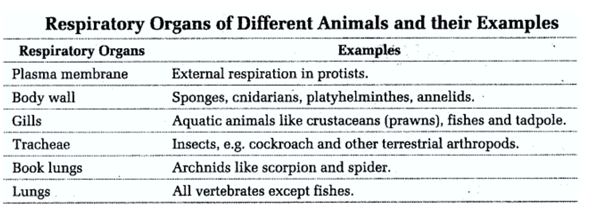
- Depending upon habitat and level of organisation, different animals have different organs for breathing/ respiration.
- Respiration involves the following steps :-
(i) Breathing or pulmonary ventilation by which atmospheric air is drawn in and CO2 rich alveolar air is released out.
(ii) Diffusion of gases (O2 and CO2) across alveolar membrane.
(iii)Transport of gases by blood.
(iv) Diffusion of O2and CO2 between blood and tissues.
(v) Utilisation of O2by the cell for catabolic reactions and resultant release of CO2.

Human Respiratory System
- Respiration ka matlab hota hai oxygen and carbon dioxide ka exchange and uss oxygen ko food ke oxidation ke liye use karna.
- Pulmonary Ventilation : ka matlab hai air ka lungs me inhalation saans lena) and lungs se exhalation (saans chorna).
- External Respiration ka matlab hai lungs me jo oxygen hai uska lungs me aane deoxygenated blood ki carbon dioxide se exchange karna. Simply bole toh toh oxygen inhaled air me thi wo ab lungs me flow karne wale me blood me diffuse hogyi.
- Internal Respiration ka matlab hai ki systemic capillaries me flow kar rhe oxygenated blood and demanding tisse ke beech me gaseous exchange. Yani ki iss final step me lungs se jo oxygen, oxygenated blood lekar puri body me ghum rha tha usko ab jaruratmand tissue lelega and apni CO2 ko uss blood me dump kar dega. This is what we mean when we say exchange of gases between capillary blood and tissue.
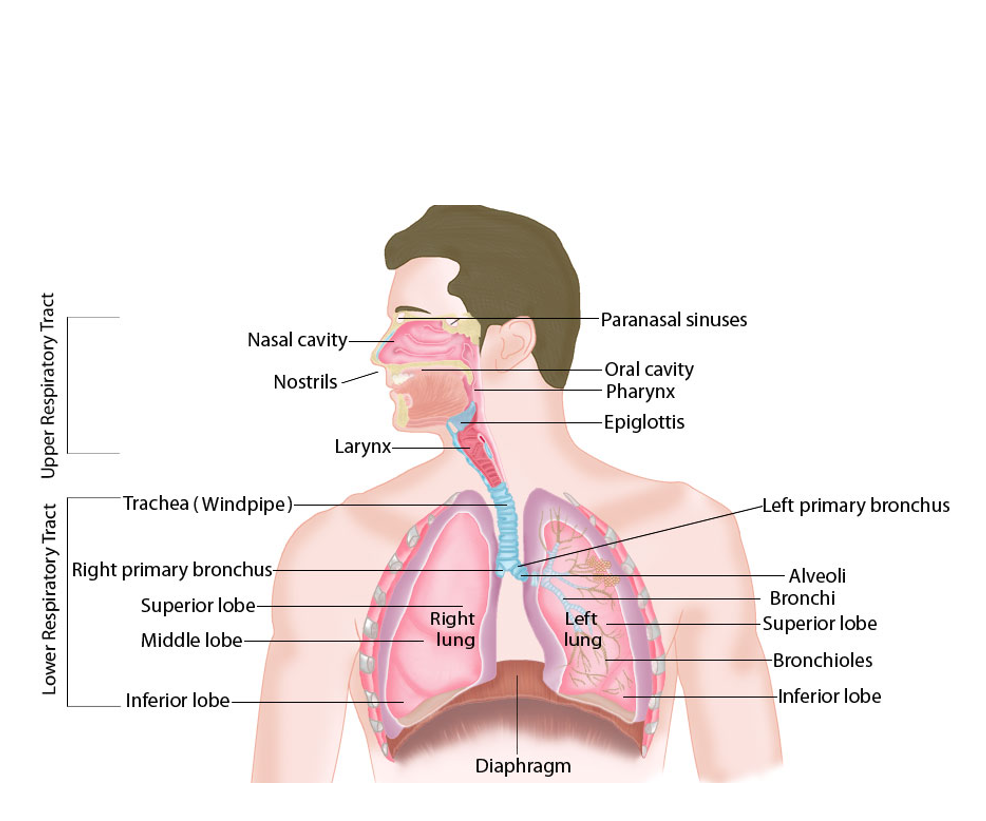
- Nose : Respiratory system ka pehla part hai jo ki externally visible hai but nose keval itni hi nahi hoti. Nose ka jo part aapke face par externally visible hai usko hum external nose kehte hai. Nose ki external tip me hyaline cartilage hota hai. Ek cartilaginous septum nose ko 2 parts me divide karta hai and nose ki external openings ko external nares/nostrils bola jata hai. Nose ke ander hair and mucous membranes hoti hai jinka main kaam hai : warming, moistening and filtering of incoming air, smell, modifying speech patterns. Posterior side me nasal cavity, internal nares ke through pharynx me open hoti hai.
- Pharynx : funnel shaped tube like structure hai jo internal nares se shuru hota hai and larynx ke cricoid cartilage (most inferior cartilage of the larynx) par end hota hai. Pharynx air and food ka common passageway hai. Pharynx ke further 3 parts hai but we do not need to go into that much detail. But ek cheez yaad rakhna ki pharynx ke last portion se 2 tubes originate hoti hai : samne yani ki chest ki taraf larynx me and iske peeche esophagus/food pipe so we say that its inferior end larynx opens into the esophagus (food tube) posteriorly and the larynx (voice box) anteriorly.
- Larynx : ek chota sa passageway hai jo pharynx ko trachea se connect karta hai. Isme 9 cartilages ke pieces hote hai. Yahi pe vocal cords hote hai jo voice produce karte hai.
→ Thyroid cartilage jiske adam’s apple bola jata hai, wo basically hyaline cartilage ki 2 fused plates se banta hai and larynx ki anterior side hota hai. Its triangular in shape. Important thing is, yeah hota dono males and females me hai but usually, males me androgens ke karan jyada prominent hota hai.
Epiglottis ke leaf like elastic cartilaginous structure hai jo thyroid cartilage ki anterior rim se juda hua hai. Ek end free hota hai jo ki upwards and downward movements show karta hai. Khana khate hue, pharynx and larynx elevate hote hai and epiglottis neeche ki taraf move karta hai and glottis ke uper lid ki taraf situated hojata hai toh glottis ko close kar deta hai. Glottis is opening of airways. Ab epiglottis iss glottis ko cover kar leta hai toh food and drinks ab keval oesophagus me hi ja sakte hai and yeah jaruri isiliye hai because agar food and water agar respiratory pathway me gya toh person generally choke kar jata hai and wo saans nahi le pata and yeah codition lethal bhi ho sakti hai if left untreated. - Trachea bhi ek pipe like structure hai jo larynx se shuru hoker T5 vertebrae ki position pe 2 tubes me divide hojata hai jinko hum right and left bronchi bolte hai. Trachea ki puri length and further structures tak, hyaline cartilage ki horizontal C shaped rings hoti hai. The open part of each C-shaped cartilage ring faces posteriorly toward the esophagus. Inn rings ka main kaam hai low pressure ki condition me trachea ki collapsing ko prevent karna.
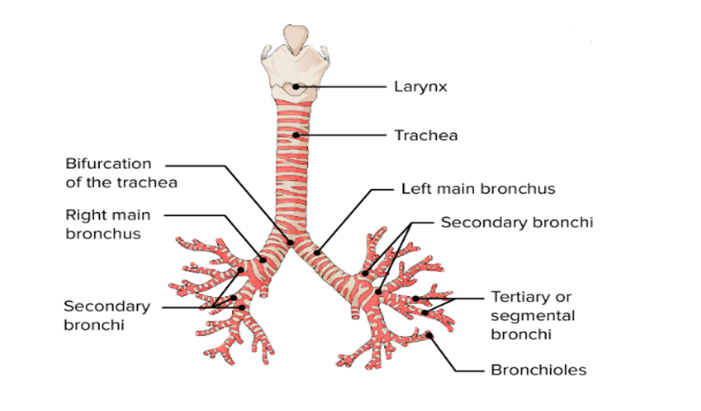
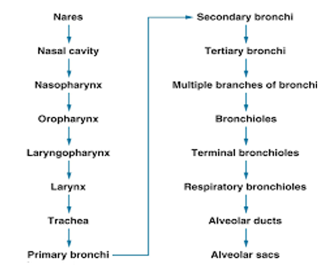
- Left and right bronchi apne apne respective lungs me enter karta hai. The right main bronchus is more vertical, shorter, and wider than the left toh usually jo cheez atakti hai toh wo right bronchus me atakne ke chance jyada hote hai. Lungs me enter karne ke baad bronchi, secondary then tertiary bronchi, then bronchioles to terminal bronchioles me aise further divide hote rehte hai and they end into alveoli (discussed shortly).
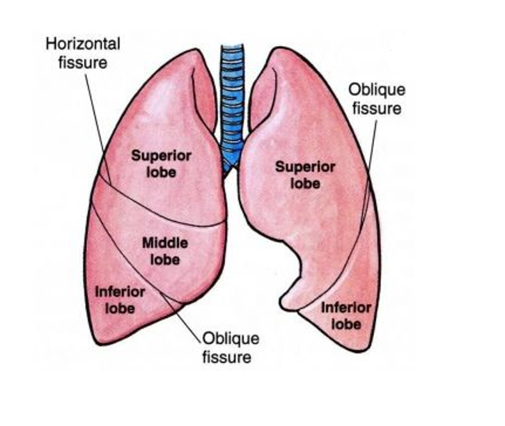
- Lungs : The right lung has three lobes; the left lung has two. Har ek lungs ke charo taraf ek double layered pleural membrane hoti hai. Isski outer layer protective hoti hai and inner layer lungs ke direct contact me hoti hai. In dono membrane ske beech e cavity hoti hai jisme fluid hoti hai jo lubrication ka function karti hai thus helps in reducing friction. Lungs ke neeche diaphragm naam ki muscle hoti hai jo thoracic cavity ko abdominal cavity se separate karti hai. Lungs ka bas concave hota hai. Left lung me cardiac notch hoti hai jisme heart ki apex situated hai, although separate hai dono.
- Alveoli : jo abhi uper bronchi and bronchioles padh rhe thy toh discuss kiya tha ki lungs me divide hote rehte hai finally ballon like blind sacs me terminate hote hai. Inhi ballon like structures ko hum alveolar sac kehte hai, jinme bahut sari alveoli hoti hai. Ek alveolus ki wall layer, single cell thick epithelium layer hai jisme 2 types ke cells hote hai jinko hum type I and type II alveolar cells kehte hai. Type I alveolar cell toh simple squamous epithelium cells hai ;- the main site of gaseous exchange; and type II alveolar cells ya fir septal cell, number me kam hote hai and yeah type I cells ke beech me situated htoe hai. Morphologically yeah rounded ya cuboidal epithelial cells hai and inpe microvilli present hoti hai. Yeah type II cells hi alveolar fluid secrete karte hai jiskne main component surfactants hote hai.
→ Surfactant surface tension ko reduce karne wale substances hai and in doing so they prevent collapsing of alveoli. Jhan tak baat lungs ki hai toh lungs ki alveoli me milne wale surfactant basically phospholipids and lipoproteins ka ek complex mixture hai. Alveoli me alveolar macrophages cells bhi hote hai jo phagocytic cells hai. - Respiratory Membrane : alveoli me gaseous exchange hota hai right? But kinke beech? Toh alveoli and alveoli ke pass wali capillary ke blood me gaseous exchange hota hai. Toh jo jo structures iss exchange me involved hai wo respiratory membrane bnate hai, jiske following components hai :
- Alveolar wall : single layer epithelium cells ki jisme type I and type II cells hote hai.
- Basement membrane : alveolar membrane and capillary membrane ke beech me hoti hai.
- Capillary endothelium layer, jo again single cell thick hoti hai.
- Lungs and bahar ke environment me pressure changes ke karan hai inhalation and exhalation ho pata hai.
- Inhalation : air breathe in karne ke liye lungs ke ander air pressure bahar ke atmospheric pressure se kam hona chaiye tabhi bahar se air lungs ke ander flow karegi. Toh inhalation ke waqt lungs ka size expand karke, lungs ki volume increase ho jati hai it leads to reduced pressure in the lungs. Yeah sab aapne chemistry me ideal gas me Boyle’s law vgarah padha hoga. Normal quiet inhalation ke waqt diaphragm ki muscle contract karti hai jiske karan yeah dome shaped muscle flat hojati hai jiske karan lungs ka inferior part jo ki concave tha wo bhi flat hojata hai toh lungs ka vertical diameter increase hojata hai which simply means ki lungs ki volume increase hogyi and pressure decrease hua. Yeah toh normal inspiration/inhalation.
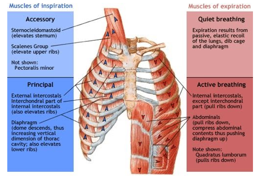
→ Diaphragm ke alawa jo important inalatory muscle hai wo hai, external intercostals. Jab yeah muscles contract karti hai toh ribs elevate hoti hai jiske karan lunga ka lateral diameter increase hota hai.
→ Deep inhalation ya forceful inhalation me baki accessory respiratory muscles bhi function karti hai jo ki normal quiet breathing ke time par function na ke brabar karti hai. Yeah accessory muscles basically sternum and first 5 ribs ko further elevate karke chest cavity ko further expand karti hai. Inke contraction ke karan jhaan ek hum normal breathing me 500mL air inhale karta hai vahi athletes vgrah forceful breathing ya exercise ke time par 2-3L air tak inhale kar sakte hai.
→ Inhalation ek active process hai kyunki muscle contraction ke liye ATP use hota hai. - Exhalation : ke waqt muscle relax karna shuru kardeti hai and iske karan chest cavity ka size decrease hojata hai jiske karan further chest cavity me present air ka pressure bahar ke atmospheric pressure ke mukable jyada hota hai toh air simple respiratory pathway ke through bahar ki taraf move kar jati hai.
→ Simple ya normal exhalation ek passive process hai.
→ Forceful exhalation ke waqt abdominal and internal intercostals contract karti hai jo thorax and abdominal region me pressure increase karti hai. Abdominal muscles ke contract karne se lower ribs neeche aati hai and diaphragm ko or uper tak jane ke liye force karti hai. Internal intercostals ke contraction se ribs neeche aati hai and helps in forceful exhalation/expiration.
Respiration ki process me 3 basic steps hoti hai :
Components of Human Respiratory System :
→ Respiratory system ke components hai : nose, pharynx/throat, larynx/voice box, trachea/wind pipe, bronchi & lungs.
Mechanism of Breathing :
HUMAN RESPIRATORY SYSTEM
- Respiratory tract
- Nostrils - A pair of external nostrils open out above the upper lips. External nostrils lead to the nasal chamber of the nasal cavity.
- Nasal Cavity - It opens to the outside through external nares(openings) and in the nasopharynx through internal nares. Nasal septum divides the nasal cavity in two chambers.
Nasal chamber is vascular, containing mucus gland cells and hair. - Pharynx - It is short, vertical, tube divisible into nasopharynx, oropharynx and laryngopharynx. Pharynx is a common passage for air and food. Nasopharynx opens through glottis of the larynx region into the trachea.
- Larynx - It is a cartilaginous box which helps in sound production and hence called sound box. It lies between 3rd to 6th cervical vertebrae in males and slightly higher in children and females.
Its size increases in males after puberty. It is made up of glottis, epiglottis and various cartilages. Epiglottis is a thin elastic cartilaginous flap which projects obliquely upward into the pharynx. During swallowing it covers glottis to prevent the entry of food into the larynx. The cavity of larynx contains 2 paired mucosal folds i.e. Upper folds/vestibular folds and lower folds/true vocal cords.Vocal cords are large and thicker in men. Pitch of sound depends upon thickness and tension of vocal cords. - Trachea(Windpipe) - It is a straight tube, extending up to the mid thoracic cavity. It divides at the level of 5th thoracic vertebrae into the right and left primary bronchi. The wall of trachea is fibrous and is supported by incomplete C- shaped cartilaginous rings. It is lined by ciliated epithelium which beat to push the mucus out.
- Bronchi - Carina is the point of branching of trachea. Bronchus divide into smaller bronchi which further divides into thin bronchioles. The trachea, bronchi and initial bronchioles are supported by incomplete cartilaginous rings.
- The branching network of bronchi, bronchioles and alveoli comprise the lungs. The system of branching and rebrancing of respiratory passageways is called respiratory tree.
- Alveoli-1: Each alveolus is thin walled, irregular, vascularised bag like structure for gaseous exchange. It contains the following types of cells - simple squamous epithelial cells,, type-II cells/pneumocyte cells and macrophages/dust cells. Type II alveolar cells are secretory and produce surfactant to prevent collapsing of lungs. Macrophages or dust cells eliminate particles and microbes that reach the alveoli. About 300 million alveoli are present in both lungs. These are primary site of exchange of gases in the human body.
- Functionally, the respiratory system consists of two parts:
- Conducting zone- It is the part starting from external nostrils up to the terminal bronchioles. Its function is to filter, warm and moisten air and conduct air to the lungs.
- Respiratory or exchange zone - It is the part starting from respiratory bronchiole to alveoli. These are sites of gas exchange between air and blood.
- Respiratory organs - Lungs
- A pair of lungs lie in the thoracic cavity which is an automatically airtight chamber on both the sides of the heart.Each lung is surrounded by double layer pleural membranes, outer parietal pleura thoracic wall k sath attached rehta hai aur inner layer visceral pleuron yeh lungs sath attached hota hai. The space between two layers is called pleural cavity. Pleural cavity contains pleural fluid which reduces friction and makes movement of lungs easy.
- Space between the two lungs is called mediastinum. It includes the pleural membrane of both the lungs. Iske andr heart, oesophagus, thoracic duct, thymus etc. hote hai. Mediastinal surface of each lung contain a region called hilus, jha se bronchi, blood vessel, lymph vessels, nerve enter aur exit krti h.
- Lungs are soft, spongy, elastic and pinkish at birth. Right lung is shorter as the diaphragm is raised on one side taki liver fit ho paye.
- Thoracic cage is formed by
a .Vertebral column on dorsal side
b. Sternum on ventral side
c .Ribs on lateral side
d. Dome shaped diaphragm on lower side
This anatomical setup of lungs in the thorax is such that any change in the volume of the thoracic cavity will be reflected in the lung(pulmonary) cavity. Such an arrangement is essential for breathing, as we cannot directly change the pulmonary volume.
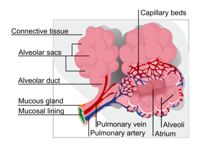
- The alveoli (singular: alveolus) are hollow air sacs which make the basic unit of respiration(Mtlab in chote chote sacs se hi milke poora lungs ka structure banta hai for respiration ).
- The alveolar walls are made up of collagen and elastic fibres which facilitate expansion during inspiration and return to the original shape during expiration. There are numerous capillaries within the alveolar walls where gas exchange occurs.
- Alveoli contain two major types of epithelial cells. The most abundant, type 1 pneumocytes (95%) are squamous cells in which gas exchange occurs. The remaining 5%, type 2 pneumocytes, are granular cells which secrete surfactant.
NOTE
Surfactant is a lipoprotein with a high phospholipid content which reduces surface tension. This increases pulmonary compliance, prevents atelectasis and aids recruitment of collapsed airways. - Alveolar macrophages are also located in the alveoli. They protect the alveoli from foreign material by engulfing it, including bacteria, dust and carbon particles.
- Gas exchange occurs in the lungs between alveolar air and blood of the pulmonary capillaries. For effective gas exchange to occur, alveoli must be ventilated and perfused. Ventilation (V) refers to the flow of air into and out of the alveoli, while perfusion (Q) refers to the flow of blood to alveolar capillaries.
- Alveoli are found at the terminal ends of the respiratory tree, clustered around alveolar sacs and alveolar ducts. Each alveolus is approximately 0.2 mm in diameter. There are around 300 million to 1 billion alveoli in the human lungs, covering an area of 70 square metres.
HUMAN RESPIRATORY SYSTEM
- Respiratory Tract :
- Nostrils - A pair of external nostrils open out above the upper lips. External nostrils lead to the nasal chamber of the nasal cavity.
- Nasal Cavity - It opens to the outside through external nares(openings) and in the nasopharynx through internal nares. Nasal septum divides the nasal cavity in two chambers.
Nasal chamber is vascular, containing mucus gland cells and hair. - Pharynx - It is short, vertical, tube divisible into nasopharynx, oropharynx and laryngopharynx. Pharynx is a common passage for air and food. Nasopharynx opens through glottis of the larynx region into the trachea.
- Larynx - It is a cartilaginous box which helps in sound production and hence called sound box. It lies between 3rd to 6th cervical vertebrae in males and slightly higher in children and females.
Its size increases in males after puberty. It is made up of glottis, epiglottis and various cartilages. Epiglottis is a thin elastic cartilaginous flap which projects obliquely upward into the pharynx. During swallowing it covers glottis to prevent the entry of food into the larynx. The cavity of larynx contains 2 paired mucosal folds i.e. Upper folds/vestibular folds and lower folds/true vocal cords.Vocal cords are large and thicker in men. Pitch of sound depends upon thickness and tension of vocal cords. - Trachea(Windpipe) - It is a straight tube, extending up to the mid thoracic cavity. It divides at the level of 5th thoracic vertebrae into the right and left primary bronchi. The wall of trachea is fibrous and is supported by incomplete C- shaped cartilaginous rings. It is lined by ciliated epithelium which beat to push the mucus out.
- Bronchi - Carina is the point of branching of trachea. Bronchus divide into smaller bronchi which further divides into thin bronchioles. The trachea, bronchi and initial bronchioles are supported by incomplete cartilaginous rings.
- The branching network of bronchi, bronchioles and alveoli comprise the lungs. The system of branching and rebrancing of respiratory passageways is called respiratory tree.
- Alveoli-1: Each alveolus is thin walled, irregular, vascularised bag like structure for gaseous exchange. It contains the following types of cells - simple squamous epithelial cells,, type-II cells/pneumocyte cells and macrophages/dust cells. Type II alveolar cells are secretory and produce surfactant to prevent collapsing of lungs. Macrophages or dust cells eliminate particles and microbes that reach the alveoli. About 300 million alveoli are present in both lungs. These are primary site of exchange of gases in the human body.
- Functionally, the respiratory system consists of two parts:
- Conducting zone- It is the part starting from external nostrils up to the terminal bronchioles. Its function is to filter, warm and moisten air and conduct air to the lungs.
- Respiratory or exchange zone - It is the part starting from respiratory bronchiole to alveoli. These are sites of gas exchange between air and blood.
- Respiratory organs - Lungs
- A pair of lungs lie in the thoracic cavity which is an automatically airtight chamber on both the sides of the heart.Each lung is surrounded by double layer pleural membranes, outer parietal pleura thoracic wall k sath attached rehta hai aur inner layer visceral pleuron yeh lungs sath attached hota hai. The space between two layers is called pleural cavity. Pleural cavity contains pleural fluid which reduces friction and makes movement of lungs easy.
- Space between the two lungs is called mediastinum. It includes the pleural membrane of both the lungs. Iske andr heart, oesophagus, thoracic duct, thymus etc. hote hai. Mediastinal surface of each lung contain a region called hilus, jha se bronchi, blood vessel, lymph vessels, nerve enter aur exit krti h.
- Lungs are soft, spongy, elastic and pinkish at birth. Right lung is shorter as the diaphragm is raised on one side taki liver fit ho paye.
- Thoracic cage is formed by
a .Vertebral column on dorsal side
b. Sternum on ventral side
c .Ribs on lateral side
d. Dome shaped diaphragm on lower sideThis anatomical setup of lungs in the thorax is such that any change in the volume of the thoracic cavity will be reflected in the lung(pulmonary) cavity. Such an arrangement is essential for breathing, as we cannot directly change the pulmonary volume.

- The alveoli (singular: alveolus) are hollow air sacs which make the basic unit of respiration(Mtlab in chote chote sacs se hi milke poora lungs ka structure banta hai for respiration ).
- Alveoli are found at the terminal ends of the respiratory tree, clustered around alveolar sacs and alveolar ducts. Each alveolus is approximately 0.2 mm in diameter. There are around 300 million to 1 billion alveoli in the human lungs, covering an area of 70 square metres.
- The alveolar walls are made up of collagen and elastic fibres which facilitate expansion during inspiration and return to the original shape during expiration. There are numerous capillaries within the alveolar walls where gas exchange occurs.
- Alveoli contain two major types of epithelial cells. The most abundant, type 1 pneumocytes (95%) are squamous cells in which gas exchange occurs. The remaining 5%, type 2 pneumocytes, are granular cells which secrete surfactant.
NOTE
Surfactant is a lipoprotein with a high phospholipid content which reduces surface tension. This increases pulmonary compliance, prevents atelectasis and aids recruitment of collapsed airways. - Alveolar macrophages are also located in the alveoli. They protect the alveoli from foreign material by engulfing it, including bacteria, dust and carbon particles.
- Gas exchange occurs in the lungs between alveolar air and blood of the pulmonary capillaries. For effective gas exchange to occur, alveoli must be ventilated and perfused. Ventilation (V) refers to the flow of air into and out of the alveoli, while perfusion (Q) refers to the flow of blood to alveolar capillaries.
HOW RESPIRATORY SYSTEM WORKS ?
- MECHANISM OF BREATHING
- Breathing involves two stages : inspiration during which atmospheric air is drawn in and expiration by which the alveolar air is released out.
- Inspiration can occur if the pressure within the lungs (intra pulmonary pressure) is less then the atmospheric pressure so there is a negative pressure in the lungs than the atmospheric pressure.
- Similarly, expiration takes place when the intra-pulmonary pressure is higher than the atmospheric pressure.
- INSPIRATION
- Inspiration is an active process.
- Normally it takes around 2 seconds.
- At the time of inspiration, contraction in diaphragm and external intercostal muscles takes place.
- Diaphragm becomes flat and is pushed towards the abdominal cavity.
- Sternum moves towards ventral and anterior direction.
- Ribs move towards outside and ventral side. As a result of all these reactions, the volume of thoracic cavity is increased. So the pressure of pleural fluid over lungs is decreased.
- Due to the spongy nature, lungs expand. As a result of this, air pressure in the lungs is decreased as compared to atmospheric pressure. Now air rushes from outside, through respiratory tract and fills the lungs.
- EXPIRATION
- Normal expiration is a passive activity. It takes around 3 seconds.
- During expiration, contraction (in any muscle) does not take place. During expiration, relaxation in diaphragm and external intercostal muscles takes place.
- As a result of this relaxation, diaphragm, sternum and ribs attain their actual (normal) position.
- Due to which the volume of thoracic cavity is decreased, and pressure of thoracic cage on lungs is increased.
- Thus air which was filled in lungs goes outside through respiratory tract. Normal breathing is also called Abdominal breathing.
- We have the ability to increase the strength of inspiration and expiration with the help of additional muscles. This is called forceful breathing.
- The cells in our bodies need oxygen to stay alive. Carbon dioxide is a by-product of respiration. The lungs and respiratory system allow oxygen in the air to be taken into the body, while also letting the body get rid of carbon dioxide in the air breathed out.
- When you breathe in, the diaphragm moves downward toward the abdomen, and the rib muscles pull the ribs upward and outward (making the chest cavity bigger and pulling air through the nose or mouth into the lungs).
- In exhalation, the diaphragm moves upward and the chest wall muscles relax, which causes the chest cavity to get smaller and push air out of the respiratory system through the nose or mouth.
- With each inhalation, air fills a large portion of the millions of alveoli. Oxygen diffuses from the alveoli to the blood through the capillaries lining the alveolar walls. Once in the bloodstream, oxygen gets picked up by the hemoglobin in red blood cells. This oxygen-rich blood then flows back to the heart, which pumps it through the arteries to oxygen needy tissues throughout the body.
- In the capillaries of the body tissues, oxygen is freed from the hemoglobin and moves into the cells.
- Carbon dioxide produced moves out of the cells into the capillaries, where most of it dissolves in the plasma of the blood.
- Blood rich in carbon dioxide then returns to the heart via the veins.
- From the heart, this blood is pumped to the lungs, where carbon dioxide passes into the alveoli to be exhaled.
HOW RESPIRATORY SYSTEM WORKS ?
- MECHANISM OF BREATHING :
- Breathing involves two stages : inspiration during which atmospheric air is drawn in and expiration by which the alveolar air is released out.
- Inspiration can occur if the pressure within the lungs (intra pulmonary pressure) is less then the atmospheric pressure so there is a negative pressure in the lungs than the atmospheric pressure.
- Similarly, expiration takes place when the intra-pulmonary pressure is higher than the atmospheric pressure.
- INSPIRATION
- Inspiration is an active process.
- Normally it takes around 2 seconds.
- At the time of inspiration, contraction in diaphragm and external intercostal muscles takes place.
- Diaphragm becomes flat and is pushed towards the abdominal cavity.
- Sternum moves towards ventral and anterior direction.
- Ribs move towards outside and ventral side. As a result of all these reactions, the volume of thoracic cavity is increased. So the pressure of pleural fluid over lungs is decreased.
- Due to the spongy nature, lungs expand. As a result of this, air pressure in the lungs is decreased as compared to atmospheric pressure. Now air rushes from outside, through respiratory tract and fills the lungs.
- EXPIRATION
- Normal expiration is a passive activity. It takes around 3 seconds.
- During expiration, contraction (in any muscle) does not take place. During expiration, relaxation in diaphragm and external intercostal muscles takes place.
- As a result of this relaxation, diaphragm, sternum and ribs attain their actual (normal) position.
- Due to which the volume of thoracic cavity is decreased, and pressure of thoracic cage on lungs is increased.
- Thus air which was filled in lungs goes outside through respiratory tract. Normal breathing is also called Abdominal breathing.
- We have the ability to increase the strength of inspiration and expiration with the help of additional muscles. This is called forceful breathing.
- The cells in our bodies need oxygen to stay alive. Carbon dioxide is a by-product of respiration. The lungs and respiratory system allow oxygen in the air to be taken into the body, while also letting the body get rid of carbon dioxide in the air breathed out.
- When you breathe in, the diaphragm moves downward toward the abdomen, and the rib muscles pull the ribs upward and outward (making the chest cavity bigger and pulling air through the nose or mouth into the lungs).
- In exhalation, the diaphragm moves upward and the chest wall muscles relax, which causes the chest cavity to get smaller and push air out of the respiratory system through the nose or mouth.
- With each inhalation, air fills a large portion of the millions of alveoli. Oxygen diffuses from the alveoli to the blood through the capillaries lining the alveolar walls. Once in the bloodstream, oxygen gets picked up by the hemoglobin in red blood cells. This oxygen-rich blood then flows back to the heart, which pumps it through the arteries to oxygen needy tissues throughout the body.
- In the capillaries of the body tissues, oxygen is freed from the hemoglobin and moves into the cells.
- Carbon dioxide produced moves out of the cells into the capillaries, where most of it dissolves in the plasma of the blood.
- Blood rich in carbon dioxide then returns to the heart via the veins.
- From the heart, this blood is pumped to the lungs, where carbon dioxide passes into the alveoli to be exhaled.
CONTROL OF RESPIRATORY RATE
- Breathing is an automatic and rhythmic act produced by networks of neurons in the hindbrain (thepons and medulla).
- The neural networks direct muscles that form the walls of the thorax and abdomen and produce pressure gradients that move air into and out of the lungs.
- The respiratory rhythm and the length of each phase of respiration are set by reciprocal stimulatory and inhibitory interconnection of these brain-stem neurons.
- An important characteristic of the human respiratory system is its ability to adjust breathing patterns to changes in both the internal and the external environment.
- Ventilatory rate (minute volume) is tightly controlled and determined primarily by blood levels of carbon dioxide as determined by metabolic rate.
- Chemoreceptors can detect changes in blood pH that require changes in involuntary respiration to correct.The apneustic (stimulating) and pnuemotaxic (limiting) centers of the pons work together to control rate of breathing.
- The medulla sends signals to the muscles that initiate inspiration and expiration and controls nonrespiratory air movement reflexes, like coughing and sneezing.
- The motor cortex within the cerebral cortex of the brain controls voluntary respiration (the ascending respiratory pathway).
- Voluntary respiration may be overridden by aspects of involuntary respiration, such as chemoreceptor stimulus, and hypothalamus stress response.
- The phrenic nerves, vagus nerves, and posterior thoracic nerves are the major nerves involved in respiration.
- Voluntary respiration is needed to perform higher functions, such as voice control.
Regulation Of Respiration
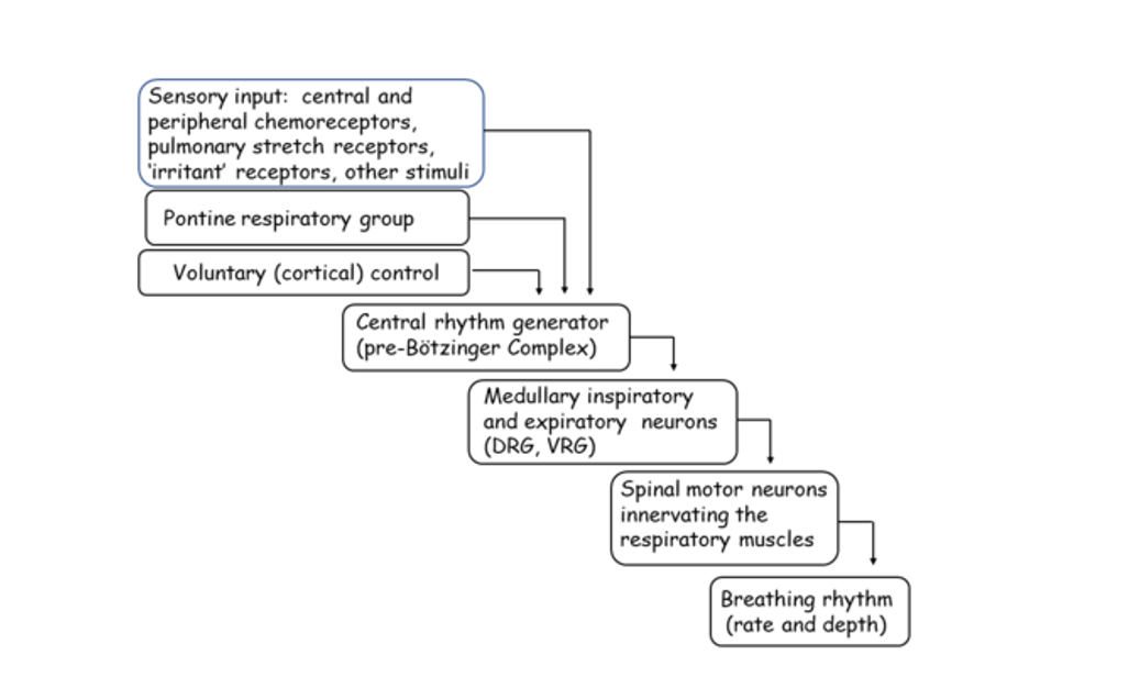
- Brain stem me respiratory center located hota hai jo nerve impulses bhej kar breathing pattern ko modulate karta hai. Yeah respiratory center ke 2 main parts hai : medullary respiratory center and pontine respiratory center.
- Isme further 2 areas hote hai : Dorsal respiratory Group inspiration ko control karta hai jabki ventral respiratory group expiration ke control karta hai.
- Normal breathing ke waqt dorsal group phrenic nerves ke through diaphragm muscles tak impulses bhejta hai and intercostal nerves ke through external intercostal muscles tak. Jab dorsal group se impulses nahi aati toh muscle relax karti hai and that initiates expiration.
- Normal quiet inhalation nearly 2 seconds tak last karti hai and normal quiet exhalation 3 seconds tak.
- Ventral group neurons agar accessory inhalation muscles ko stimulate karte hai toh forceful inhalation hota hai and agar yeah neuron accessory muscles of exhalation ko stimulate karte hai toh forceful exhalation hota hai.
- Iska pneumotaxic center basically upper pons me situated hai. yeah sxpiratory center ko stimulate karta hai and inspiration center ko inhibit karta hai.
- Lower pons me apneustic center hota hai jo inspiration ko stimulate karta hai.
- Voluntary control of breathing me cerebral cortex ka role hai. Jaise aap swimming and dive karte hue respiration ko control karte hai toh cerebral cortex ka role hai lekin iska matlab yeah ki unlimited amount of time tak aap control kar sakte hai ya aap inhale hi karte jayenge ya forceful expiration karte hi rahenge. Yeh limitation basically chemoreceptors generates karte hai jo ki next point me padhenge.
- Chemoreceptor Regulation of Breathing : chemoreceptors CO2, O2 and H+ ko sense karte hai and then respiratory control center tak appropriate signals transmit karte hai. 2 types ke chemoreceptors hote hai : central chemoreceptors medulla oblongata ke pass located hai and they respond to changes in H+ concentration or PCO2, or both, in cerebrospinal fluid. Carotid arteries and aorta me bhi chemoreceptors hote hai jinko hum peripheral chemoreceptors kehte hai and these are sensitive to changes in PO2, H+, and PCO2 in the blood.
→ Jab PCO2 increase hota hai toh central chemoreceptors are stimulated and respond vigorously to the resulting increase in H+ level. The peripheral chemoreceptors also are stimulated by both the high PCO2 and the rise in H+. In addition, the peripheral chemoreceptors (but not the central chemoreceptors) respond to a deficiency of O2. - Stimuli that increase breathing rate and depth are :
- Voluntary hyperventilation controlled by cerebral cortex and anticipation of activity by stimulation of limbic system.
- Increase in arterial blood PCO2 above 40 mmHg (causes an increase in H+) detected by peripheral and central chemoreceptors.
- Decrease in arterial blood PO2 from 105 mmHg to 50 mmHg.
- Increased activity of proprioceptors.
- Increase in body temperature.
- Prolonged pain.
- Decrease in blood pressure.
- Stretching of anal sphincter.
- Stimuli that decrease breathing rate & depth :
- Voluntary hypoventilation controlled by cerebral cortex.
- Decrease in arterial blood PCO2 below 40 mmHg (causes a decrease in H+) detected by peripheral and central chemoreceptors.
- Decrease in arterial blood PO2 below 50 mmHg.
- Decreased activity of proprioceptors.
- Decrease in body temperature (decreases respiration rate), sudden cold stimulus (causes apnea).
- Severe pain (causes apnea). Increase in blood pressure.
- Irritation of pharynx or larynx by touch or chemicals (causes brief apnea followed by coughing or sneezing).
Medullary Respiratory Center :
Pontine Center :
Regulation of Respiratory Center :
RESPIRATORY VOLUME AND CAPACITIES
- RESPIRATORY VOLUME
- The volume of air involved in breathing movements can be estimated by using a spirometer which helps in clinical assessment of pulmonary functions.
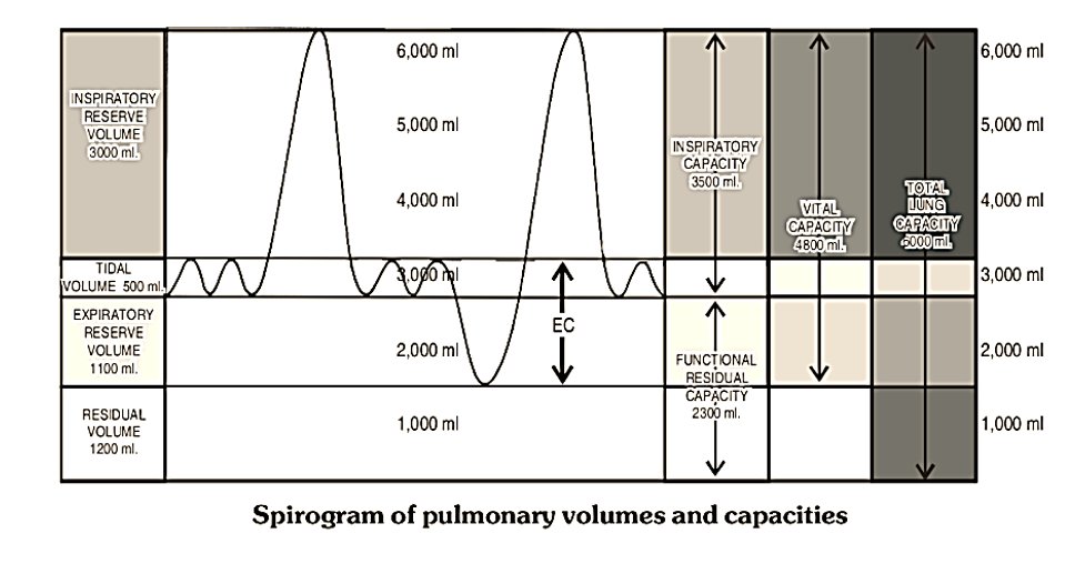
- Tidal volume [T.V.] - It is amount of air inspired or expired during normal breathing. Its value for man is 500 ml. Whole inspired air does not reach up to lungs. The portion of air which remains in the respiratory tract is called Anatomical dead-space volume. Its value for man is 150 ml .A healthy man can inspire or expire approximately 6000 to 8000 ml of air per minute.
- Inspiratory Reserve Volume [I.R.V.] - Volume of air a person can inspire by forcible inspiration over tidal volume. It's value is 2500 to 3000 ml
- Expiratory reserve volume [E.R.V.]- It is the amount of air expired over tidal volume by most forceful expiration.Its value is 1000-1100 ml.
- Residual volume - [R.V.]- It is the amount of air that remains inside lungs after forceful expiration. Residual volume can not be given out of lungs. Its value is 1100-1200 ml.
- Inspiratory capacity - [I.C.] - Total volume of air a person can inspire after a normal expiration. I.C. = I.R.V. + T.V. = 3000 ml + 500 ml I.C. = 3500 ml
- Expiratory capacity (EC) : Total volume of air a person can expire after a normal inspiration. E.C. = TV + ERV
- Functional Residual capacity [FRC] - It is the amount of air that normally remains inside lungs after normal expiration. In it expiratory reserve volume and residual volume are included FRC = ERV + RV = 1000 ml + 1200 ml FRC = 2200 to 2500 ml
- Vital capacity - [V.C.] - It is the amount of air that can be expired by most forceful expiration after a deepest inspiration. Inspiratory reserve volume, expiratory reserve volume and tidal volume are included in it. V.C. = IRV + ERV + TV = 3000 ml + 1000 ml + 500 ml = 4500 ml (4300 to 4800 ml)
- Total lung capacity - Total volume of air that can accomodated in the lungs at the end of forced inspiration. TLC = IRV + TV + ERV + RV = 3000 + 500 + 1000 + 1200 TLC = 6000 ml (approx) (5700 to 6000 ml)
PULMONARY CAPACITIES
CONTROL OF RESPIRATORY RATE
- Breathing is an automatic and rhythmic act produced by networks of neurons in the hindbrain (thepons and medulla).
- The neural networks direct muscles that form the walls of the thorax and abdomen and produce pressure gradients that move air into and out of the lungs.
- The respiratory rhythm and the length of each phase of respiration are set by reciprocal stimulatory and inhibitory interconnection of these brain-stem neurons.
- An important characteristic of the human respiratory system is its ability to adjust breathing patterns to changes in both the internal and the external environment.
- Ventilatory rate (minute volume) is tightly controlled and determined primarily by blood levels of carbon dioxide as determined by metabolic rate.
- Chemoreceptors can detect changes in blood pH that require changes in involuntary respiration to correct.The apneustic (stimulating) and pnuemotaxic (limiting) centers of the pons work together to control rate of breathing.
- The medulla sends signals to the muscles that initiate inspiration and expiration and controls nonrespiratory air movement reflexes, like coughing and sneezing.
- The motor cortex within the cerebral cortex of the brain controls voluntary respiration (the ascending respiratory pathway).
- Voluntary respiration may be overridden by aspects of involuntary respiration, such as chemoreceptor stimulus, and hypothalamus stress response.
- The phrenic nerves, vagus nerves, and posterior thoracic nerves are the major nerves involved in respiration.
- Voluntary respiration is needed to perform higher functions, such as voice control.
EXCHANGE OF GASES
- Gases, primarily O2 and CO2 Alveoli me exchange hoti hai. Alveoli’s are richly supplied with blood so the blood coming to the lungs has high amount of CO2 and low amount of O2. This blood at the alveoli is oxygenated which simply means that it dumps its CO2 into alveoli and takes O2 so that it becomes oxygen rich or oxygenated. This blood is then sent to the heart and heart pumps it to all the other organs and tissues and this cycle continues. Exchange of gases occurs by simple diffusion mainly based on pressure/concentration gradient. Solubility of the gases as well as the thickness of the membranes involved in diffusion are also some important factors that can affect the rate of diffusion. Pressure contributed by an individual gas in a mixture of gases is called partial pressure and is represented as pO2 for oxygen and pCO2 for carbon dioxide. Partial pressures of these two gases in the atmospheric air and the two sites of diffusion are given in Table below and in the following figures. The data given in the table clearly indicates a concentration gradient for oxygen from alveoli to blood and blood to tissues. Similarly, a gradient is present for CO2 in the opposite direction, i.e., from tissues to blood and
blood to alveoli. As the solubility of CO2 is 20-25 times higher than that of O2, the amount of CO2 that can diffuse through the diffusion membrane per unit difference in
partial pressure is much higher compared to that of O2. The diffusion membrane is
made up of three major layers namely, the thin squamous epithelium of alveoli, the endothelium of alveolar capillaries and the basement substance in between them. However, its total thickness is much less than a millimetre. Therefore, all the factors in our body are favourable for diffusion of O2 from alveoli to tissues and that of CO2 from tissues to alveoli.
- Pehli layer hai alveoli ki alveolar wall.
- Uske baad dusri layer hai basement membrane jo ki is case me again dekho bahiut hi patli hai which helps in diffusion. Agar yeah layer moti hoti toh diffusion ki process ko hinder karti.
- Next, third and final layer hai Single cells ki capillary layer.

FIG : Partial Pressures (in mm Hg) of Oxygen and Carbon dioxide at Different Parts Involved in Diffusion in Comparison to those in Atmosphere
NCERT me iss section me 2 figures had and ek yeah uper wali table. Yeah 3 cheez samjhli ache se toh matlab aadha chapter samjhlo ho gaya toh ab ek baar apun uper table me dekhte hai hai kya usme…
Explanation of Partial pressure table : Apun ne padh lia partial pressure wo pressure hai jo koi bhi gas ek mixture me gases me apna pressure lgati hai. Ab jab human breath in and breath out karenge toh hum air se keval oxygen ya CO2 ka exchange ni karte. Air is a mixture of gases and hum wo mixture inhale karte hain. Gases ke uss mixture se lungs ki aleoli me humara blood keval oxygen ko lega and CO2 ko dedega isilie hum keval yhaan par O2 and CO2 se concerned hai. Atmosphere ki air me oxygen jyada hai as compared to CO2 toh oxygen ka partial pressure yani ki pO2 > pCO2. Ab aleoli me aao, alveoli me bhi pO2 > pCO2 or lungs me blood aaya hai that blood is deoxygenated yani ki usme pCO2>pO2. Toh gases ka diffusion high partial pressure se low partial pressure ki taraf hoga yani ki alveoli se O2 blood capillaries ke blood me jayegi and blood se CO2 lung alveoli me jayega. Yahi funda pure lungs ki sabhi alveoli me ho raha hai jiske karan ab lungs me CO2 rich air hai so we have to exhale yani humne saans chor di and do remember yeah itna sab keval itni der me ho raha hai jitni der me tumne ek saans li or chor di ani inhale and exhale kiya single breath. Har breath me repeat hota hai. Neeche jo figure given hai usme bhi yahi sab ho raha hai toh have a look at this picture and isko dimag me ache se utar lena ki kabhi nahi bhulna uper wali table and yeah neeche wale 2 diagram :
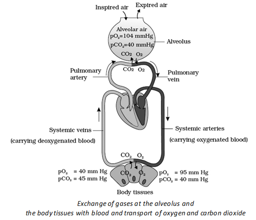
FIG : Diagrammatic representation of exchange of gases at the alveolus and the body tissues with blood and transport of oxygen and carbon dioxide
Now we only have to take a look at one last diagram jo alveoli and blood supply ko thoda clear kar dega so have a look :
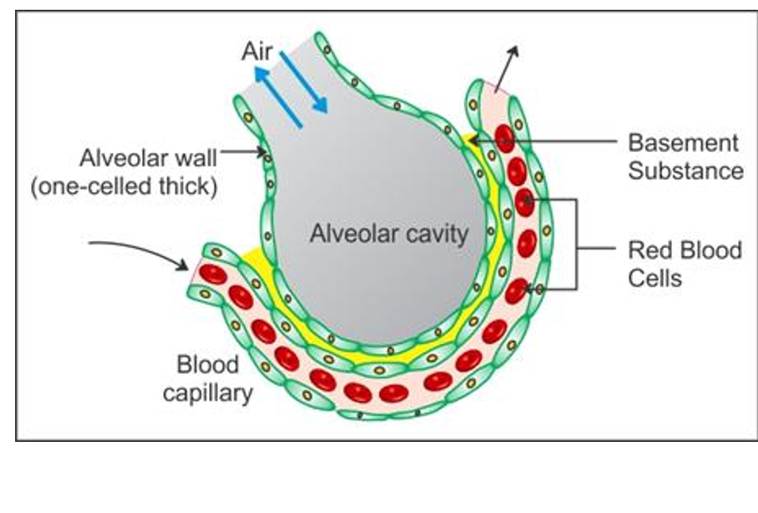
FIG : A Diagram of a section of an alveolus with a pulmonary capillary.
Explanation : generally humne padha tha ki diffusion slow process hai but diffusion also depends on the thickness of the surface or membrane across which it is happening. In this lung ki alveoli keval ek cell jitni moti hai and and same goes for thickness of blood capillary. Ab yhaan jo exhange hai gases ka wo 3 layers se ho kar jayega which are :
Jitni, patli yeah 3 layers hongi diffusion utna hi efficient hoga toh that’s it about the internal structure of alveoli with a pulmonary capillary.
Lung Volumes & Lung Capacities
- Rest ke dauran ek adult healthy individual ek minute me 12 bar saans leta hai. These values are for normal inhalation and normal exhalatioon.
- Tidal volume : ek breath ki volume. Yani ki ek normal saans ander lete hue ya normal exhalation ke waqt kitni air flow kar rhi hai wo. Iski value hoti hai 500mL.
- Anatomical dead space : yeah basicially respiratiory pathway ka wo space hai jhan par gaseous exchange toh nahi hota lekin kuch amount air ki isme hamesha rehti hi hai, jaise ki bronchi, bronchioles, traches etc. nearly 150mL air inme rehti hai and 500 me baki bachi hui 350mL hi gaseous exchange ke liye alveoli tak pahuchti hai.
- Inspirtaory Reserve Volume (IRV) : forceful inhalation ke time par jitni air inhale ki wo hai IRV. Males ke iski value hai 3100mL jabki females me iski value hai 1900mL. NCERT says that this averages 2500 mL to 3000 mL. (Go with NCERT)
- Expiratory reserve volume (ERV) : air ki wo amount jo aap forcefully exhale kar sakte ho. Males me iski value 1200mL hoti hai and females me around 700mL.
- Residual Volume : forceful exhalation ke baad bhi kuch air lungs and baki pathways me reh jati hai jiske aap exhale nahi kar sakte. Issi ko hum residual volume/RV kehte hai. Males me iski value 1200mL hai and femals me 1100mL.
- Inspiratory capacity (IC) is the sum of tidal volume and inspiratory reserve volume (500 mL + 3100 mL = 3600 mL in males and 500 mL + 1900 mL = 2400 mL in females).
- Functional residual capacity (FRC) is the sum of residual volume and expiratory reserve volume (1200 mL + 1200 mL = 2400 mL in males and 1100 mL + 700 mL = 1800 mL in females).
- Vital capacity (VC) is the sum of inspiratory reserve volume, tidal volume, and expiratory reserve volume (4800 mL in males and 3100 mL in females).
- Finally, total lung capa city (TLC) is the sum of vital capacity and residual volume (4800 mL + 1200 mL = 6000 mL in males and 3100 mL + 1100 mL = 4200 mL in females).
Lung Volumes :
Lung Capacities :
TRANSPORT OF GASES
- In the previous section we learned how gases are exchanged between alveoli and capillaries so in this section we will how and in which form these gases are transported. We have 2 subsections in this section : Transport of O2 and Transport of CO2.
- Mostly jitni bhi O2 hai wo haemoglobin ke bind hokr oxyhaemoglobin (HbO2) ki form me hi transport ki jati hai.
- Each haemoglobin molecule can carry a maximum of four molecules of O2. Binding of oxygen with haemoglobin is primarily related to partial pressure of O2. Partial pressure of CO2, hydrogen ion concentration and temperature are the other factors which can interfere with this binding.
- 100mL of blood carries 19.4mL or 20mL of O2. out of this nearly 20 mL, tissue take 5ml/4.6mL O2 and now this venous blood still has 14.4mL O2 which means that our tissue can only take 25% of O2 that blood carries to the cells/tissues.
- 1gm of Hb transports 1.34mL of O2
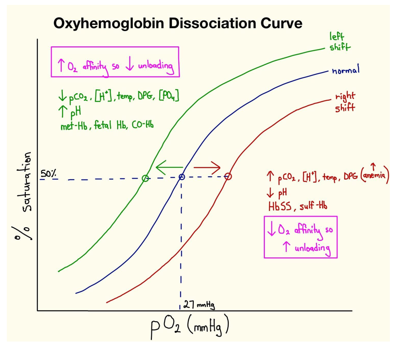
- O2-Hb Curve is a S-shaped curve or sigmoid curve as shown below :
- Bahut sare factors hai jo Hb ke sath O2 ki binding ko affect karte hain.
- Jaise ki agar CO2 ki concentration ya pCO2 badh jayega toh HbO2 dissociated into Hb + O2 yani ki graph shift to right. Issi phenomenon ko hum Bohr’s effect kehte hai or we can define Bohr’s effect as Loading of CO2 causes unloading of O2
- APan jab yeah kahenge abse ki curve shifts to right uska matlab hai ki HbO2 dissociate ho raha hai Hb + O2 me. ok
- Increase in pH shifts curve to the right.
- Increase in temperature, shifts curve to right.
- P50 wo partial pressure hai O2 ka jis waqt Hb 50% saturated hai O2 se which from the graph comes out to be nearly 27mm Hg. Affinity of Hb is inversely proportional to P50level.
- Fetal haemoglobin ka structure adult se thoda alag hota hai and it has 2 alpha and 2 gamma chains while haemoglobin of adult human has 2 alpha and 2 beta polypeptide chains. Fetal Hb 20mmHg pe 50% saturated hota hai jabki adult Hb 20 mm Hg of pO2 pe keval 30-35% saturated hai which indicates ki fetal Hb has more affinity for O2.
- Myoglobin does not show Bohr’s effect as a result, even at 40 mm Hg O2 it is 95% saturated and only when pO2 falls below 5mmHg, it becomes less than 60% saturated.
- 7% CO2 ka transport plasma me dissolved form me hota hai, 70% or CO2 is transported in the form of HCO3- ki form me hota hai and baki bachi hui 20-25% CO2 Hb ke sath transport ki jati hai.
- CO2 is 20 to 25 more soluble than O2 isiliye or plasma vgarah me dissolve hokr transport kari jati hai jabki O2 ke transport ke liye we specifically need Haemoglobin
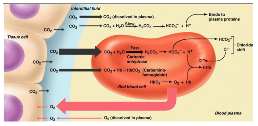
- RBC ke ander CO2 ko H2O ke sath milakr, carbonic anhydrase naam ka enzyme H2CO3 (carbonic acid) ki formation me role play karta hai and iss enzyme ke karan hi yeah reaction bahut fast hoti hai. Enzyme ki absence me this reaction very much slow on its own.
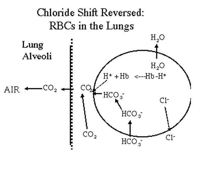
- Carbonic acid abdi jaldi bicarbonate ion and ek proton me tut jata hai. Bicarbonate ions move out of the RBC lekin ussi waqt ions ke balance ko maintain karne ke liye RBC ke ander utne hi Chloride ions move karte hain jitne ki bicarbonate ions bahar gye thy. Issi ki hum Chloride Shift ya Hamburger Effect kehte hain.
- Toh RBC me ab HBO2 tutega into Hb and O2. O2 tissue ne utha li and further tissue ne CO2 ko RBC me dump kar dia. Ab RBC me CO2 ki amount kafi jyada hai toh some CO2 will combine to HB to form carbaminohemoglobin (HbCO2) or yeah CO2 issi form me lungs ki alveoli tak jati hai. Alveole me HbCO2 se CO2 alag hojati hai and alveoli me diffuse kar jayegi or alveoli se O2 aakr Hb ke sath judkar oxyhameoglobin (HBO2) banati hai ji firse tissue ke pass iss oxygen ka transport HbO2 ki form me hoga and uper likhi hui story bar bar repeat hoti rehti hai.
- Yani ki tissues ke paas blood me jo uper wale diagram me reactions dikhae hai wo lung ki alveoli me jakr reverse hogyi. So the reversed reactions are shown below with the help of a diagram :
Transport of O2
Transport of Carbon dioxide
This ends our section on transport of gases.
RESPIRATORY VOLUME AND CAPACITIES
- RESPIRATORY VOLUMES :
- The volume of air involved in breathing movements can be estimated by using a spirometer which helps in clinical assessment of pulmonary functions.
- Tidal volume [T.V.] - It is amount of air inspired or expired during normal breathing. Its value for man is 500 ml. Whole inspired air does not reach up to lungs. The portion of air which remains in the respiratory tract is called Anatomical dead-space volume. Its value for man is 150 ml .A healthy man can inspire or expire approximately 6000 to 8000 ml of air per minute.
- Inspiratory Reserve Volume [I.R.V.] - Volume of air a person can inspire by forcible inspiration over tidal volume. It's value is 2500 to 3000 ml
- Expiratory reserve volume [E.R.V.]- It is the amount of air expired over tidal volume by most forceful expiration.Its value is 1000-1100 ml.
- Residual volume - [R.V.]- It is the amount of air that remains inside lungs after forceful expiration. Residual volume can not be given out of lungs. Its value is 1100-1200 ml.
- Inspiratory capacity - [I.C.] - Total volume of air a person can inspire after a normal expiration. I.C. = I.R.V. + T.V. = 3000 ml + 500 ml
I.C. = 3500 ml - Expiratory capacity (EC) : Total volume of air a person can expire after a normal inspiration.
E.C. = TV + ERV - Functional Residual capacity [FRC] - It is the amount of air that normally remains inside lungs after normal expiration. In it expiratory reserve volume and residual volume are included
FRC = ERV + RV = 1000 ml + 1200 ml
FRC = 2200 to 2500 ml - Vital capacity - [V.C.] - It is the amount of air that can be expired by most forceful expiration after a deepest inspiration. Inspiratory reserve volume, expiratory reserve volume and tidal volume are included in it.
V.C. = IRV + ERV + TV
= 3000 ml + 1000 ml + 500 ml
= 4500 ml (4300 to 4800 ml) - Total lung capacity - Total volume of air that can accomodated in the lungs at the end of forced inspiration.
TLC = IRV + TV + ERV + RV
= 3000 + 500 + 1000 + 1200
TLC = 6000 ml (approx) (5700 to 6000 ml)
PULMONARY CAPACITIES :

REGULATION OF RESPIRATION
- This deals with the regulation centers and other regulatory mechanisms to regulate the rate of respiration. We have 2 types of regulation : Nervous regulation of Respiration by Brain and Chemical Regulation.
- Brain ke 2 parts : Medulla Oblongata and Pons Varoli respiration ke rate ko regulate karte hai.
- Medulla Oblongata has inspiratory center which control inspiration, on its dorsal surface and expiration center on its ventral side.
- Inspiratory centre has motos inspiratory neurons. They send impulses to diaphragm- the main inspiratory muscle via Phrenic nerve
- Expiratory center has both inspiratory and expiratory neurons which are only activated during forceful expiration.
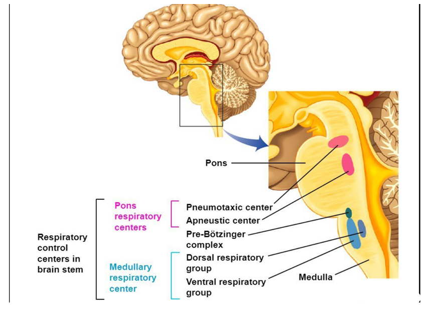
- Pneumotaxic center is located dorsally in the upper pons part and it stimulates expiration while inhibiting inspiration.
- Apneustic center is located dorsally in the lower pons region and it stimulates inspiration. It promote inhalation by a constant stimulation of the neurons in the medulla oblongata.
- The chemical regulatory mechanism keeps the alveolar pCO2 at constant value of 40 mm Hg. if arterial pO2 falls or arterial pCO2 rises, this is sensed by special chemoreceptors and which stimulates respiratory center to increase the rate and depth of respiration. These chemoreceptors are known as respiratory chemoreceptors which are of two types : peripheral chemoreceptors (which are found in carotid and aortic artery ) and central chemoreceptors (found in Medulla)
- Medullary receptors or the central receptors that we talked about in the previous point are located on the ventral surface of Medulla. They get stimulated by increase in H+ concentration in CSF and interstitial fluid.
Nervous Regulation of Respiration
Pons Varolii has 2 centers : Pneumotaxic center and apneustic centres
Chemotaxis Center
Exchange of Gases
External Respiration :
- External respiration ka matlab hai alveoli se O2 ka pulmonary blood me diffuse hona and uss blood se CO2 ka alveoli me diffuse hona.
- Firse thodi si cheistry yaad karo, states of matter wala chapter jhan par partial pressure ka concept padha tha. Air bhi different gases ka mixture hai and har gas apna pressure exert karti hai jo ki partial pressure kehlata hai. Toh blood and alveoli me O2 and CO2 ka yeah partial pressure hi different hota hai jiske karan yeah external respiration yani ki pulmonary exchange possible hai.
- Alveoli me oxygen ka partial pressure yani ki PO2 = 105 mmHg hai and pulmonary capillaries me PO2 = 40 mm Hg. toh naturally O2 alveoli se pulmonary capillaries ki taraf diffuse karegi.
- Same situation hai CO2 sath. PCO2 of deoxygenated blood = 45 mm Hg and PCO2 in alveoli = 40 mm Hg. Toh CO2 deoxygenated blood se alveoli ki taraf diffuse karti hai. Oxygenated blood returning to the le% side of the heart in the pulmonary veins thus has a PCO2 of 40 mmHg.
- Tissues and systemic capillaries ke beech gaseous exchange hi internal respiration kehlata hai.
- Cells and tissue constantly oxygen use kar rhe hai toh unme oxygen ka partial pressure low hoga. PO2 in tissues = 40 mm Hg and PO2 systemic capillaries me hai = 100 mm Hg toh oxygen gase blood se tisues me diffuse kar jati hai.
- Tissues me PCO2 = 45mm Hg but systemic capillaries me PCO2 = 40 mm Hg toh CO2 tissues se blood me diffuse kar jati hai.
- In a person at rest, tissue cells, on average, need only 25% of the available O2 in oxygenated blood; despite its name, deoxygenated blood retains 75% of its O2 content. During exercise, more O2 diffuses from the blood into metabolically active cells, such as contracting skeletal muscle fibers. Active cells use more O2 for ATP production, causing the O2 content of deoxygenated blood to drop below 75%.
DISORDERS OF RESPIRATORY SYSTEM
- Asthma is a difficulty in breathing causing wheezing due to inflammation of bronchi and bronchioles.
- Bronchitis is a disorder of bronchi in which there is regular swelling and itching of bronchi and is characterised by regular coughing.
- Emphysema is a chronic disorder in which alveolar walls are damaged due to which respiratory surface is decreased. One of the major causes of this is cigarette smoking.
- Occupational Respiratory Disorders: In certain industries, especially those involving grinding or stone- breaking, so much dust is produced that the defense mechanism of the body cannot fully cope with the situation.Long exposure can give rise to inflammation leading to fibrosis (proliferation of fibrous tissues) and thus causing serious lung damage. Workers in such industries should wear protective masks.
EXCHANGE OF GASES
- Gases, primarily O2 and CO2 Alveoli me exchange hoti hai. Alveoli’s are richly supplied with blood so the blood coming to the lungs has high amount of CO2 and low amount of O2. This blood at the alveoli is oxygenated which simply means that it dumps its CO2 into alveoli and takes O2 so that it becomes oxygen rich or oxygenated. This blood is then sent to the heart and heart pumps it to all the other organs and tissues and this cycle continues. Exchange of gases occurs by simple diffusion mainly based on pressure/concentration gradient. Solubility of the gases as well as the thickness of the membranes involved in diffusion are also some important factors that can affect the rate of diffusion. Pressure contributed by an individual gas in a mixture of gases is called partial pressure and is represented as pO2 for oxygen and pCO2 for carbon dioxide. Partial pressures of these two gases in the atmospheric air and the two sites of diffusion are given in Table below and in the following figures. The data given in the table clearly indicates a concentration gradient for oxygen from alveoli to blood and blood to tissues. Similarly, a gradient is present for CO2 in the opposite direction, i.e., from tissues to blood and blood to alveoli. As the solubility of CO2 is 20-25 times higher than that of O2, the amount of CO2 that can diffuse through the diffusion membrane per unit difference in partial pressure is much higher compared to that of O2. The diffusion membrane is made up of three major layers namely, the thin squamous epithelium of alveoli, the endothelium of alveolar capillaries and the basement substance in between them. However, its total thickness is much less than a millimetre. Therefore, all the factors in our body are favourable for diffusion of O2 from alveoli to tissues and that of CO2 from tissues to alveoli.
- Pehli layer hai alveoli ki alveolar wall.
- Uske baad dusri layer hai basement membrane jo ki is case me again dekho bahiut hi patli hai which helps in diffusion. Agar yeah layer moti hoti toh diffusion ki process ko hinder karti.
- Next, third and final layer hai Single cells ki capillary layer.

FIG : Partial Pressures (in mm Hg) of Oxygen and Carbon dioxide at Different Parts Involved in Diffusion in Comparison to those in Atmosphere
NCERT me iss section me 2 figures had and ek yeah uper wali table. Yeah 3 cheez samjhli ache se toh matlab aadha chapter samjhlo ho gaya toh ab ek baar apun uper table me dekhte hai hai kya usme…
Explanation of Partial pressure table : Apun ne padh lia partial pressure wo pressure hai jo koi bhi gas ek mixture me gases me apna pressure lgati hai. Ab jab human breath in and breath out karenge toh hum air se keval oxygen ya CO2 ka exchange ni karte. Air is a mixture of gases and hum wo mixture inhale karte hain. Gases ke uss mixture se lungs ki aleoli me humara blood keval oxygen ko lega and CO2 ko dedega isilie hum keval yhaan par O2 and CO2 se concerned hai. Atmosphere ki air me oxygen jyada hai as compared to CO2 toh oxygen ka partial pressure yani ki pO2 > pCO2. Ab aleoli me aao, alveoli me bhi pO2 > pCO2 or lungs me blood aaya hai that blood is deoxygenated yani ki usme pCO2>pO2. Toh gases ka diffusion high partial pressure se low partial pressure ki taraf hoga yani ki alveoli se O2 blood capillaries ke blood me jayegi and blood se CO2 lung alveoli me jayega. Yahi funda pure lungs ki sabhi alveoli me ho raha hai jiske karan ab lungs me CO2 rich air hai so we have to exhale yani humne saans chor di and do remember yeah itna sab keval itni der me ho raha hai jitni der me tumne ek saans li or chor di ani inhale and exhale kiya single breath. Har breath me repeat hota hai. Neeche jo figure given hai usme bhi yahi sab ho raha hai toh have a look at this picture and isko dimag me ache se utar lena ki kabhi nahi bhulna uper wali table and yeah neeche wale 2 diagram :

FIG : Diagrammatic representation of exchange of gases at the alveolus and the body tissues with blood and transport of oxygen and carbon dioxide
Now we only have to take a look at one last diagram jo alveoli and blood supply ko thoda clear kar dega so have a look :

FIG : A Diagram of a section of an alveolus with a pulmonary capillary.
Explanation : generally humne padha tha ki diffusion slow process hai but diffusion also depends on the thickness of the surface or membrane across which it is happening. In this lung ki alveoli keval ek cell jitni moti hai and and same goes for thickness of blood capillary. Ab yhaan jo exhange hai gases ka wo 3 layers se ho kar jayega which are :
Jitni, patli yeah 3 layers hongi diffusion utna hi efficient hoga toh that’s it about the internal structure of alveoli with a pulmonary capillary.
SUMMARY
- Cells utilise oxygen for metabolism and produce energy along with substances like carbon dioxide which is harmful. Animals have evolved different mechanisms for the transport of oxygen to the cells and for the removal of carbon dioxide from there.
- Humans a well developed respiratory system comprising two lungs and associated air passages to perform this function.
- The first step in respiration is breathing by which atmospheric air is taken in (inspiration) and the alveolar air is released out (expiration).
- Exchange of O2 and CO2 between deoxygenated blood and alveoli, transport of these gases throughout the body by blood, exchange of O2 and CO2 between the oxygenated blood and tissues and utilisation of O2 by the cells (cellular respiration) are the other steps involved.
- Inspiration and expiration are carried out by creating pressure gradients between the atmosphere and the alveoli with the help of specialised muscles – intercostals and diaphragm. Volumes of air involved in these activities can be estimated with the help of spirometer and are of clinical significance.
- Exchange of O2 and CO2 at the alveoli and tissues occur by diffusion. Rate of diffusion is dependent on the partial pressure gradients of O2 (pO2 ) and CO2 (pCO2 ), their solubility as well as the thickness of the diffusion surface. These factors in our body facilitate diffusion of O2 from the alveoli to the deoxygenated blood as well as from the oxygenated blood to the tissues. The factors are favourable for the diffusion of CO2 in the opposite direction, i.e., from tissues to alveoli. Oxygen is transported mainly as oxyhaemoglobin. In the alveoli where pO2 is higher, O2 gets bound to haemoglobin which is easily dissociated at the tissues where pO2 is low and pCO2 and H+ concentration are high. Nearly 70 per cent of carbon dioxide is transported as bicarbonate (HCO3 – ) with the help of the enzyme carbonic anhydrase. 20-25 per cent of carbon dioxide is carried by haemoglobin as carbamino-haemoglobin. In the tissues where pCO2 is high, it gets bound to blood whereas in the alveoli where pCO2 is low and pO2 is high, it gets removed from the blood. Respiratory rhythm is maintained by the respiratory centre in the medulla region of brain.
- A pneumotaxic centre in the pons region of the brain and a chemosensitive area in the medulla can alter respiratory mechanism.
Transport of Gases
- Transport of Oxygen
- 98.5% O2 Hb ke sath bind karke transport ki jati hai. Keval bachi hui 1.5% hi blood plasma me dissolved form me hoti hai.
- Har 100mL oxygenated blood me 20mL gaseous oxygen hoti hai.
- Hemoglobin ke heme portion me 4 iron atoms hote hai and har ek iron atom 1 O2 ke molecule se bind karta hai toh every Hb molecule can carry 4 molecules of O2
- Hb + O2 → Hb-O2 (oxyhemoglobin)
- Higher the PO2, the more O2 combines with Hb.
- Percent saturation of Hb and PO2 ke beech ki relationship oxygen-dissociation curve se pta lagti. Keval yahi nahi, baki cheezein bhi bta sakte hai.
- Hb is 75% saturated with O2 at PO2 =40mmHg, which is the average PO2 of tissues in a person at rest.
- When the PO2 is between 60 and 100 mmHg, hemoglobin is 90% or more saturated with O2.
- 40-20mmHg PO2 ki values ke paas, Hb se oxygen kafi quickly dissociated karta hai and dissociation ke baad hi tissues me oxygen diffuse kar pati hai. Toh basically Hb ka kaam yeah hua ki lungs se oxygen ko har tissues tak transport karna. Tissue me low oxygen partial pressure ke response me oxygen, Hb se dissociate hokar tissues me diffuse kar jati hai.
- Factors affecting affinity of Hb for O2 :
- Acidity/pH : as pH decreases, dissociation of oxygen from Hb increases. Jo metabolically active cells hai unme lactic acid and carbonic acid banta rehta hai toh unka pH low hai and this helps in unloading of O2. Bohr’s Effect : An increase in H+ in blood causes O2 to unload from hemoglobin, and the binding of O2 to hemoglobin causes unloading of H+ from hemoglobin.
→ Graph shift : normal se low pH par Hb-O2 curve RHS ki taraf shift hoga and high pH/alkalinity ke time par curve LHS ki taraf shift hoga. - PCO2 : Jab CO2 ka partial pressure normal se high hota hai toh graph RHS ki taraf shift hoga else to the right in case of low PCO2.
- Temperature : high temperature dissociation ko promote karta hai yani ki graph RHS ki taraf shift hoga.
- 2,3-bisphosphoglycerate (BPG)/diphosphoglycerate (DPG): yeah Hb ki oxygen ke liye affinity kam karta hai. And plant physiology ke padha tha ki BPG basically glycolysis ke dauran banta hai. Toh jab BPG ki amount jyada hone lagti hai toh dissociation aasani se hoga yani ki graph RHS ki taraf shift hoga.
- Acidity/pH : as pH decreases, dissociation of oxygen from Hb increases. Jo metabolically active cells hai unme lactic acid and carbonic acid banta rehta hai toh unka pH low hai and this helps in unloading of O2. Bohr’s Effect : An increase in H+ in blood causes O2 to unload from hemoglobin, and the binding of O2 to hemoglobin causes unloading of H+ from hemoglobin.
- CO2 ka transport 3 main forms me hota hai :
- Dissolved CO2 : in plasma : 7%.
- Carbamino compounds : 23%, it combines with the amino groups of amino acids and proteins in blood to form carbamino compounds.
→ Hb + CO2 → Hb-CO2 (carbaminohemoglobin) - Bicarbonate ions (HCO3-) : 70%.
→ CO2 + H2O → H2CO3 (carbonic acid); yeah reaction carbonic anhydrase catalyse karta hai. Carbonic acid, hydrogen ion + bicarbonate ion ke sath equilibrium me exist karta hai.
- Chloride shift : Jaise hi CO2 blood me jati hai, tissues me bicarbonate accumulate hona shuru kardeti hai toh thodi bahut bicarbonate ions, down the gradient, RBC ke bahar diffuse kar jati hai and chloride ions (Cl−) RBCs ke ander move karti hai electrical balance ko maintain karne ke liye. Issi phenomenon ko chloride shift kehte hai. The net e! ect of these reactions is that CO2 is removed from tissue cells and transported in blood plasma as HCO3-. As blood passes through pulmonary capillaries in the lungs, all of these reactions reverse and CO2 is exhaled.
- Haldane Effect : The lower the amount of oxyhemoglobin (Hb–O2), the higher the CO2 -carrying capacity of the blood,
Carbon Dioxide Transport
TRANSPORT OF GASES
- In the previous section we learned how gases are exchanged between alveoli and capillaries so in this section we will how and in which form these gases are transported. We have 2 subsections in this section : Transport of O2 and Transport of CO2.
- Mostly jitni bhi O2 hai wo haemoglobin ke bind hokr oxyhaemoglobin (HbO2) ki form me hi transport ki jati hai.
- Each haemoglobin molecule can carry a maximum of four molecules of O2. Binding of oxygen with haemoglobin is primarily related to partial pressure of O2. Partial pressure of CO2, hydrogen ion concentration and temperature are the other factors which can interfere with this binding.
- 100mL of blood carries 19.4mL or 20mL of O2. out of this nearly 20 mL, tissue take 5ml/4.6mL O2 and now this venous blood still has 14.4mL O2 which means that our tissue can only take 25% of O2 that blood carries to the cells/tissues.
- 1gm of Hb transports 1.34mL of O2
- O2-Hb Curve is a S-shaped curve or sigmoid curve as shown below :
- Bahut sare factors hai jo Hb ke sath O2 ki binding ko affect karte hain.
- Jaise ki agar CO2 ki concentration ya pCO2 badh jayega toh HbO2 dissociated into Hb + O2 yani ki graph shift to right. Issi phenomenon ko hum Bohr’s effect kehte hai or we can define Bohr’s effect as Loading of CO2 causes unloading of O2
- APan jab yeah kahenge abse ki curve shifts to right uska matlab hai ki HbO2 dissociate ho raha hai Hb + O2 me. ok
- Increase in pH shifts curve to the right.
- Increase in temperature, shifts curve to right.
- P50 wo partial pressure hai O2 ka jis waqt Hb 50% saturated hai O2 se which from the graph comes out to be nearly 27mm Hg. Affinity of Hb is inversely proportional to P50level.
- Fetal haemoglobin ka structure adult se thoda alag hota hai and it has 2 alpha and 2 gamma chains while haemoglobin of adult human has 2 alpha and 2 beta polypeptide chains. Fetal Hb 20mmHg pe 50% saturated hota hai jabki adult Hb 20 mm Hg of pO2 pe keval 30-35% saturated hai which indicates ki fetal Hb has more affinity for O2.
- Myoglobin does not show Bohr’s effect as a result, even at 40 mm Hg O2 it is 95% saturated and only when pO2 falls below 5mmHg, it becomes less than 60% saturated.
- 7% CO2 ka transport plasma me dissolved form me hota hai, 70% or CO2 is transported in the form of HCO3- ki form me hota hai and baki bachi hui 20-25% CO2 Hb ke sath transport ki jati hai.
- CO2 is 20 to 25 more soluble than O2 isiliye or plasma vgarah me dissolve hokr transport kari jati hai jabki O2 ke transport ke liye we specifically need Haemoglobin
- RBC ke ander CO2 ko H2O ke sath milakr, carbonic anhydrase naam ka enzyme H2CO3 (carbonic acid) ki formation me role play karta hai and iss enzyme ke karan hi yeah reaction bahut fast hoti hai. Enzyme ki absence me this reaction very much slow on its own.
- Carbonic acid abdi jaldi bicarbonate ion and ek proton me tut jata hai. Bicarbonate ions move out of the RBC lekin ussi waqt ions ke balance ko maintain karne ke liye RBC ke ander utne hi Chloride ions move karte hain jitne ki bicarbonate ions bahar gye thy. Issi ki hum Chloride Shift ya Hamburger Effect kehte hain.
- Toh RBC me ab HBO2 tutega into Hb and O2. O2 tissue ne utha li and further tissue ne CO2 ko RBC me dump kar dia. Ab RBC me CO2 ki amount kafi jyada hai toh some CO2 will combine to HB to form carbaminohemoglobin (HbCO2) or yeah CO2 issi form me lungs ki alveoli tak jati hai. Alveole me HbCO2 se CO2 alag hojati hai and alveoli me diffuse kar jayegi or alveoli se O2 aakr Hb ke sath judkar oxyhameoglobin (HBO2) banati hai ji firse tissue ke pass iss oxygen ka transport HbO2 ki form me hoga and uper likhi hui story bar bar repeat hoti rehti hai.
- Yani ki tissues ke paas blood me jo uper wale diagram me reactions dikhae hai wo lung ki alveoli me jakr reverse hogyi. So the reversed reactions are shown below with the help of a diagram :
Transport of O2
Transport of Carbon dioxide

This ends our section on transport of gases.
REGULATION OF RESPIRATION
- This deals with the regulation centers and other regulatory mechanisms to regulate the rate of respiration. We have 2 types of regulation : Nervous regulation of Respiration by Brain and Chemical Regulation.
- Brain ke 2 parts : Medulla Oblongata and Pons Varoli respiration ke rate ko regulate karte hai.
- Medulla Oblongata has inspiratory center which control inspiration, on its dorsal surface and expiration center on its ventral side.
- Inspiratory centre has motos inspiratory neurons. They send impulses to diaphragm- the main inspiratory muscle via Phrenic nerve
- Expiratory center has both inspiratory and expiratory neurons which are only activated during forceful expiration.
- Pneumotaxic center is located dorsally in the upper pons part and it stimulates expiration while inhibiting inspiration.
- Apneustic center is located dorsally in the lower pons region and it stimulates inspiration. It promote inhalation by a constant stimulation of the neurons in the medulla oblongata.
- The chemical regulatory mechanism keeps the alveolar pCO2 at constant value of 40 mm Hg. if arterial pO2 falls or arterial pCO2 rises, this is sensed by special chemoreceptors and which stimulates respiratory center to increase the rate and depth of respiration. These chemoreceptors are known as respiratory chemoreceptors which are of two types : peripheral chemoreceptors (which are found in carotid and aortic artery ) and central chemoreceptors (found in Medulla)
- Medullary receptors or the central receptors that we talked about in the previous point are located on the ventral surface of Medulla. They get stimulated by increase in H+ concentration in CSF and interstitial fluid.
Nervous Regulation of Respiration
Pons Varolii has 2 centers : Pneumotaxic center and apneustic centres

Chemotaxis Center
DISORDERS OF RESPIRATORY SYSTEM
- Asthma is a difficulty in breathing causing wheezing due to inflammation of bronchi and bronchioles.
- Bronchitis is a disorder of bronchi in which there is regular swelling and itching of bronchi and is characterised by regular coughing.
- Emphysema is a chronic disorder in which alveolar walls are damaged due to which respiratory surface is decreased. One of the major causes of this is cigarette smoking.
- Occupational Respiratory Disorders: In certain industries, especially those involving grinding or stone- breaking, so much dust is produced that the defense mechanism of the body cannot fully cope with the situation.Long exposure can give rise to inflammation leading to fibrosis (proliferation of fibrous tissues) and thus causing serious lung damage. Workers in such industries should wear protective masks.
SUMMARY
- Cells utilise oxygen for metabolism and produce energy along with substances like carbon dioxide which is harmful. Animals have evolved different mechanisms for the transport of oxygen to the cells and for the removal of carbon dioxide from there.
- Humans a well developed respiratory system comprising two lungs and associated air passages to perform this function.
- The first step in respiration is breathing by which atmospheric air is taken in (inspiration) and the alveolar air is released out (expiration).
- Exchange of O2 and CO2 between deoxygenated blood and alveoli, transport of these gases throughout the body by blood, exchange of O2 and CO2 between the oxygenated blood and tissues and utilisation of O2 by the cells (cellular respiration) are the other steps involved.
- Inspiration and expiration are carried out by creating pressure gradients between the atmosphere and the alveoli with the help of specialised muscles – intercostals and diaphragm. Volumes of air involved in these activities can be estimated with the help of spirometer and are of clinical significance.
- Exchange of O2 and CO2 at the alveoli and tissues occur by diffusion. Rate of diffusion is dependent on the partial pressure gradients of O2 (pO2 ) and CO2 (pCO2 ), their solubility as well as the thickness of the diffusion surface. These factors in our body facilitate diffusion of O2 from the alveoli to the deoxygenated blood as well as from the oxygenated blood to the tissues. The factors are favourable for the diffusion of CO2 in the opposite direction, i.e., from tissues to alveoli. Oxygen is transported mainly as oxyhaemoglobin. In the alveoli where pO2 is higher, O2 gets bound to haemoglobin which is easily dissociated at the tissues where pO2 is low and pCO2 and H+ concentration are high. Nearly 70 per cent of carbon dioxide is transported as bicarbonate (HCO3– ) with the help of the enzyme carbonic anhydrase. 20-25 per cent of carbon dioxide is carried by haemoglobin as carbamino-haemoglobin. In the tissues where pCO2 is high, it gets bound to blood whereas in the alveoli where pCO2 is low and pO2 is high, it gets removed from the blood. Respiratory rhythm is maintained by the respiratory centre in the medulla region of brain.
- A pneumotaxic centre in the pons region of the brain and a chemosensitive area in the medulla can alter respiratory mechanism.
