CELL CYCLE AND CELL DIVISION for Class 11 BIOLOGY
INTRODUCTION
- Continuity to life is an important characteristic of living organisms. It is achieved through the process of reproduction. The reproduction may be asexual or sexual both of these are involved in the division and replication of cells. Even the growth and development of every living organism depends upon the growth and division of its cells, for example in the unicellular organisms, cell division means the reproduction by which mother cells produce two or more new cells. In multicellular organisms also, a new individual developed from a single cell - the zygote, by cell division. So by this process zygote is transferred into an adult which has millions of cells. It also forms the basis of replacement of old and worn out cells in the process of repair and regeneration, so cell division is the central tool life of the cell.
- Two basic processes in cell reproduction:
-- Cell growth period of synthesis and duplication of various components of cell
--Cell division adult cell divides into two daughter cells.
INTRODUCTION
- Continuity to life is an important characteristic of living organisms. It is achieved through the process of reproduction. The reproduction may be asexual or sexual both of these are involved in the division and replication of cells .Even the growth and development of every living organism depends upon the growth and division of its cells, for example in the unicellular organisms ,cell division means the reproduction by which mother cells produce two or more new cells . In multicellular organisms also, a new individual developed from a single cell - the zygote , by cell division.So by this process zygote is transferred into an adult which has millions of cells . It also forms the basis of replacement of old and worn out cells in the process of repair and regeneration, so cell division is the central tool life of the cell.
- Two basic processes in cell reproduction:
-- Cell growth period of synthesis and duplication of various components of cell
--Cell division adult cell divides into two daughter cells.
CHAPTER WEIGHTAGE
The chapter is quite important w.r .t the entire syllabus as it forms the base of the biology as we learn here division and cell cycle . This chapter along with biomolecules and cells, is a very important unit whole together . This chapter will not only fetch you brownie marks in school but in NEET also . Number of questions expected from the entire unit is about 10-12.
Chapter Weightage
- The chapter is quite important w.r .t the entire syllabus as it forms the base of the biology as we learn here division and cell cycle. This chapter along with biomolecules and cells, is a very important unit whole together. This chapter will not only fetch you brownie marks in school but in NEET also. Number of questions expected from the entire unit is about 10-12 so kafi important chapter hai boards and competitive exams ke point of view se bhi. MCQs se lekar short answer type and long answer type sabhi type se questions aate hai iss chapter se including label the diagram type questions.
CELL CYCLE
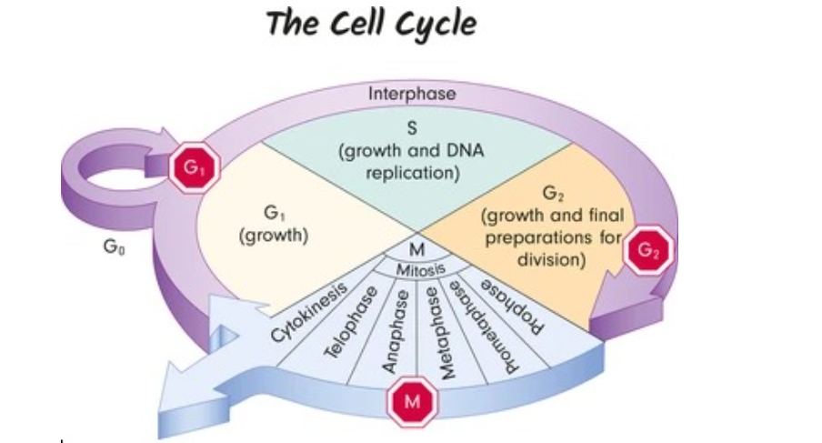
- All those changes which occur during cell growth and cell division are collectively called cell cycle .It is the sum of those events by which a cell duplicate its genome and synthesizes cell contents and eventually divides into two daughter cells.
- These events are genetically controlled and divided into two parts:
- Interphase
- M phase
- On the basis of synthetic activities this is divided into three sub phases:
- G1 or gap 1 phase or post mitotic or pre synthetic phase for first growth phase:It is the period between mitosis and initiation of DNA replication .It is characterized by the increase of cell size and nucleus due to higher rate of biosynthesis .
-It is involved in pooling of amino acids for protein synthesis.
-Pooling of nucleotides for the synthesis of RNA and DNA.
-Synthesis of enzymes and energy molecules to unzip the synthesis of DNA molecules.
-Synthesis of macromolecules like RNAs, carbohydrates, lipids and proteins as the cell is metabolically active .
-Chromosome are fully extended but no DNA duplication occurs - S or synthetic phaseDNA replicates so chromosomes are formed of two sister chromatids joined at centromere. synthesis of histones which is basic protein, each cell with double the amount 4C DNA but chromosome number remains the same.
- G2 phase (2nd Gap phase) or Post DNA synthesis phase (Pre mitosis phase):Actual preparation (Final preparation) of M-phase occurs during this phase. Special materials
required for M-phase are synthesized in G2 phase. eg. Tubulin protein. –(Required for formation of spindle fibres). Cell growth continues.
-Synthesis of spindle proteins
-Synthesis of RNA molecules
-Synthesis and storage of ATP molecule
Duplication of Mitochondria plastid and centrioles
-Damaged DNA is prepared in G2. - G0phase : cells in the adult animals do not appear to exhibit division (e.g., heart cells) and many other cells divide only occasionally, as needed to replace cells that have been lost because of injury or cell death. These cells that do not divide further exit the G1 phase to enter an inactive stage called the quiescent stage (G0) of the cell cycle.Cells in this stage remain metabolically active but no longer proliferate (divide) unless called on to do so depending on the requirement of the organism.
- M phase or mitotic phase: It follows the interphase and involves the separation of sister chromatids and their Re-distribution to daughter cells. It also involves the orderly distribution of cell or galleries and macro biomolecules to the daughter cells. So after M phase is completed, the hereditary information is distributed in the daughter cells. Now these consist of two phases: karyokinesis (division of nucleus)and cytokinesis (division of cytoplasm).
NOTE:
Duration of cell cycle it varies from organism to organism and also with the cell type it depends upon the type of cell external factors such as temperature, food, oxygen.The time period of the cell cycle is specific for different organisms and in under environmental conditions also ,for example 20 minutes for a bacterial cell, 90 minutes for yeast ,8 to 10 hours for intestinal epithelial cell, 12 hours for onion root tip cell and 24 hours for a human cell in culture . - G1 or gap 1 phase or post mitotic or pre synthetic phase for first growth phase:It is the period between mitosis and initiation of DNA replication .It is characterized by the increase of cell size and nucleus due to higher rate of biosynthesis .
INTERPHASE
: It is a period between the end of one cell division to the beginning of next cell division, so it is called inter-Mitosis.It is normally called resting phase but is actually highly metabolic active or energy phase in which cell prepare itself for next cell division by a number of biosynthetic activities so is called preparatory phase. Interphase last for about 95% of the cell cycle.CELL CYCLE
- All those changes which occur during cell growth and cell division are collectively called cell cycle .It is the sum of those events by which a cell duplicate its genome and synthesizes cell contents and eventually divides into two daughter cells.
- These events are genetically controlled and divided into two parts:
- Interphase
- M phase
- On the basis of synthetic activities this is divided into three sub phases:
- G1 or gap 1 phase or post mitotic or pre synthetic phase for first growth phase:It is the period between mitosis and initiation of DNA replication .It is characterized by the increase of cell size and nucleus due to higher rate of biosynthesis .
-It is involved in pooling of amino acids for protein synthesis.
-Pooling of nucleotides for the synthesis of RNA and DNA.
-Synthesis of enzymes and energy molecules to unzip the synthesis of DNA molecules.
-Synthesis of macromolecules like RNAs, carbohydrates, lipids and proteins as the cell is metabolically active .
-Chromosome are fully extended but no DNA duplication occurs - S or synthetic phaseDNA replicates so chromosomes are formed of two sister chromatids joined at centromere. synthesis of histones which is basic protein, each cell with double the amount 4C DNA but chromosome number remains the same.
- G2 phase (2nd Gap phase) or Post DNA synthesis phase (Pre mitosis phase):Actual preparation (Final preparation) of M-phase occurs during this phase. Special materials
required for M-phase are synthesized in G2 phase. eg. Tubulin protein. –(Required for formation of spindle fibres). Cell growth continues.
-Synthesis of spindle proteins
-Synthesis of RNA molecules
-Synthesis and storage of ATP molecule
Duplication of Mitochondria plastid and centrioles
-Damaged DNA is prepared in G2. - G0phase : cells in the adult animals do not appear to exhibit division (e.g., heart cells) and many other cells divide only occasionally, as needed to replace cells that have been lost because of injury or cell death. These cells that do not divide further exit the G1 phase to enter an inactive stage called the quiescent stage (G0) of the cell cycle.Cells in this stage remain metabolically active but no longer proliferate (divide) unless called on to do so depending on the requirement of the organism.
- M phase or mitotic phase: It follows the interphase and involves the separation of sister chromatids and their Re-distribution to daughter cells. It also involves the orderly distribution of cell or galleries and macro biomolecules to the daughter cells. So after M phase is completed, the hereditary information is distributed in the daughter cells. Now these consist of two phases: karyokinesis (division of nucleus)and cytokinesis (division of cytoplasm).
NOTE:
Duration of cell cycle it varies from organism to organism and also with the cell type it depends upon the type of cell external factors such as temperature, food, oxygen.The time period of the cell cycle is specific for different organisms and in under environmental conditions also ,for example 20 minutes for a bacterial cell, 90 minutes for yeast ,8 to 10 hours for intestinal epithelial cell, 12 hours for onion root tip cell and 24 hours for a human cell in culture . - G1 or gap 1 phase or post mitotic or pre synthetic phase for first growth phase:It is the period between mitosis and initiation of DNA replication .It is characterized by the increase of cell size and nucleus due to higher rate of biosynthesis .
INTERPHASE
: It is a period between the end of one cell division to the beginning of next cell division, so it is called inter-Mitosis.It is normally called resting phase but is actually highly metabolic active or energy phase in which cell prepare itself for next cell division by a number of biosynthetic activities so is called preparatory phase. Interphase last for about 95% of the cell cycle.Cell Cycle
- Beta, cell division har cell ke survival ke liye zaruri hai qnki har cell ki ek limited life span hai toh har cell ko divide hona padega taki uske baad new cells uski jagah le sake. Issi division ke dauran cell me bahut sare serial biochemical reactions hote hai and wo har baar jab bhi cell divide hoga toh repeat honge toh inhi events ke cycle ko hum cell cycle kehte hain.
- Jo main events hai cell cycle ke wo hai : cell division, DNA ki replication and cell ki growth.
- 2 bahut hi prominent phases hai ek cell ki life me, wo hain : Interphase and M phase. Interphase wo phase hai jab cell grow karega, division ke liye jaruri nutrients or raw materials ka accumulation and storage karega. M phase wo actual phase hai jhan par ek cell divide karega and it’s the shortest phase. Matlab ek cell apni puri life ka 90% se jyada samay interphase me gujarega and baki bache hue time me wo divide karta hai.
- Interphase bhi further 3 subphases me divided hai : G1, S and G2 phases. G1 me cell metabolically active hoga and grow karta jata hai lekin DNA ka replication nhi hoga. S phase ka naam pda hai : ‘synthesis’ se. iss phase me DNA ki synthesis hoti hai. Yani ki cell me jitna bhi DNA hai wo amount me double hojayega. Ek diploid cell ke DNA content ko hum 2C se represent karte hain toh S phase me wo double hokr 4C hojayega lekin yaad rakhna beta ki number of chromosomes abhi bhi utne hi honge jitney pehle thy, yani ki agar G1 phase me koi cell diploid hai toh wo S phase ke baad bhi diploid hi rahega. Cytoplasm me centriole bhi duplicate karti hai.
- G2 phase me un proteins ki synthesis hogi jo cell ko M phase me chahiye honge.
- Quiescent stage : kuch cells aise hote hai jo further divide nhi karte hai kisi ek function ko assume kar lete hai jaise ki neurons hogye, muscle cells generally divide nhi karte, RBCs divide nhi karte toh yeah cells G1 ke baad cell cycle se bahar nikal kar ek inactive stage me jate hai jhaan par yeah divide nhi karenge. Ussi stage ko quiescent stage ya G0 stage bola jata hai.
- M stage stands for mitotic stage jhaan par ek cell, daughter cells me split karta hai. Iss phase me bhi further 4 stages hai : prophase, metaphase, anaphase and telophase. Ab kis stage me kya hota hai wo aage padhenge.
REGULATION OF CELL CYCLE / CHECKPOINTS
- Cell cycle is run by a group of special proteins "Cyclins and Cdks”.
- The activity of enzymes, known as cyclin dependent kinases. (Cdk's) regulates the cell cycle. Kinase is an enzyme that removes a phosphate group from ATP & add to another protein.
- The kinases involved in the cell cycle are called Cdks because they are activated when they are combined with a key protein called cyclin.
- The stage of regulation of cell cycle Is G1 phase during which a cell may follow three paths :
- Start a cell cycle and enter in S phase and finally divide.
- It makes me get arrested in the G1 phase at a specific point which is also known as the G1 checkpoint.
- It may stop division and enter G0 stage or quiescent stage
- Once the cell passes the G1 checkpoint there is no looking back and cell enters as face once the DNA replication has begin the cell has to complete its new cycle to the initiation of cell division is triggered by the doubling of DNA in S phase even before the start of the M phase
- NOTE : There are two checkpoints in the cell cycle :
- First checkpoint is G1 cyclin which acts as the S phase inhibitor so operates between G1 and S phase interphase.
- Second checkpoint of the cell cycle is mitotic cyclin which operates between the G2 phase and the M phase of the interphase.
Cell Cycle
- All those changes which occur during cell growth and cell division are collectively called cell cycle .It is the sum of those events by which a cell duplicate its genome and synthesizes cell contents and eventually divides into two daughter cells.
- These events are genetically controlled and divided into two parts:
- Interphase
- M phase
- On the basis of synthetic activities this is divided into three sub phases:
- G1 or gap 1 phase or post mitotic or pre synthetic phase for first growth phase:It is the period between mitosis and initiation of DNA replication .It is characterized by the increase of cell size and nucleus due to higher rate of biosynthesis .
-It is involved in pooling of amino acids for protein synthesis.
-Pooling of nucleotides for the synthesis of RNA and DNA.
-Synthesis of enzymes and energy molecules to unzip the synthesis of DNA molecules.
-Synthesis of macromolecules like RNAs, carbohydrates, lipids and proteins as the cell is metabolically active .
-Chromosome are fully extended but no DNA duplication occurs - S or synthetic phaseDNA replicates so chromosomes are formed of two sister chromatids joined at centromere. Synthesis of histones which is basic protein, each cell with double the amount 4C DNA but chromosome number remains the same.
- G2 phase (2nd Gap phase) or Post DNA synthesis phase (Pre mitosis phase):Actual preparation (Final preparation) of M-phase occurs during this phase. Special materials required for M-phase are synthesized in G2 phase. eg. Tubulin protein. –(Required for formation of spindle fibres). Cell growth continues.
-Synthesis of spindle proteins
-Synthesis of RNA molecules
-Synthesis and storage of ATP molecule
Duplication of Mitochondria plastid and centrioles
-Damaged DNA is prepared in G2. - G0 phase : cells in the adult animals do not appear to exhibit division (e.g., heart cells) and many other cells divide only occasionally, as needed to replace cells that have been lost because of injury or cell death. These cells that do not divide further exit the G1 phase to enter an inactive stage called the quiescent stage (G0) of the cell cycle.Cells in this stage remain metabolically active but no longer proliferate (divide) unless called on to do so depending on the requirement of the organism.
- M phase or mitotic phase: It follows the interphase and involves the separation of sister chromatids and their Re-distribution to daughter cells. It also involves the orderly distribution of cell or galleries and macro biomolecules to the daughter cells. So after M phase is completed, the hereditary information is distributed in the daughter cells. Now these consist of two phases: karyokinesis (division of nucleus)and cytokinesis (division of cytoplasm).
NOTE:
Duration of cell cycle it varies from organism to organism and also with the cell type it depends upon the type of cell external factors such as temperature, food, oxygen.The time period of the cell cycle is specific for different organisms and in under environmental conditions also ,for example 20 minutes for a bacterial cell, 90 minutes for yeast ,8 to 10 hours for intestinal epithelial cell, 12 hours for onion root tip cell and 24 hours for a human cell in culture . - G1 or gap 1 phase or post mitotic or pre synthetic phase for first growth phase:It is the period between mitosis and initiation of DNA replication .It is characterized by the increase of cell size and nucleus due to higher rate of biosynthesis .
INTERPHASE :
It is a period between the end of one cell division to the beginning of next cell division, so it is called inter-Mitosis.It is normally called resting phase but is actually highly metabolic active or energy phase in which cell prepare itself for next cell division by a number of biosynthetic activities so is called preparatory phase. Interphase last for about 95% of the cell cycle.AMITOSIS
- It is also called direct or incipient cell division . It is the simplest type of the cell-division in which there is no synthesis of spindle ,no condensation of chromatin fibers and no disappearance of nuclear membrane.
- It is a simple method of cell division which is also called direct cell division. In this division there is no differentiation of chromosomes and spindle. The nuclear envelope does not degenerate.
- The nucleus elongates and constricts in the middle to form two daughter nuclei. This is followed by a centripetal constriction of the cytoplasm to form two daughter cells. Eg. Prokaryotes and some unicellular eukaryotes.
AMITOSIS
- It is also called direct or incipient cell division . It is the simplest type of the cell-division in which there is no synthesis of spindle ,no condensation of chromatin fibers and no disappearance of nuclear membrane.
- It is a simple method of cell division which is also called direct cell division. In this division there is no differentiation of chromosomes and spindle. The nuclear envelope does not degenerate.
- The nucleus elongates and constricts in the middle to form two daughter nuclei. This is followed by a centripetal constriction of the cytoplasm to form two daughter cells. Eg. Prokaryotes and some unicellular eukaryotes.
Regulation Of Cell Cycle
- Cell cycle is run by a group of special proteins "Cyclins and Cdks”.
- The activity of enzymes, known as cyclin dependent kinases. (Cdk's) regulates the cell cycle. Kinase is an enzyme that removes a phosphate group from ATP & add to another protein.
- The kinases involved in the cell cycle are called Cdks because they are activated when they are combined with a key protein called cyclin.
- The stage of regulation of cell cycle Is G1 phase during which a cell may follow three paths :
- Start a cell cycle and enter in S phase and finally divide.
- It makes me get arrested in the G1 phase at a specific point which is also known as the G1 checkpoint.
- It may stop division and enter G0 stage or quiescent stage
- Once the cell passes the G1 checkpoint there is no looking back and cell enters as face once the DNA replication has begin the cell has to complete its new cycle to the initiation of cell division is triggered by the doubling of DNA in S phase even before the start of the M phase
- NOTE : There are two checkpoints in the cell cycle :
- First checkpoint is G1 cyclin which acts as the S phase inhibitor so operates between G1 and S phase interphase.
- Second checkpoint of the cell cycle is mitotic cyclin which operates between the G2 phase and the M phase of the interphase.
MITOSIS
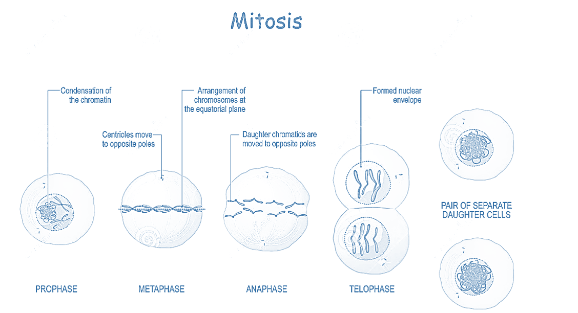
- This is the most dramatic period of the cell cycle, involving a major reorganisation of virtually all components of the cell. Since the number of chromosomes in the parent and progeny cells is the same, it is also called as equational division.
- Though for convenience mitosis has been divided into four stages of nuclear division, it is very essential to understand that cell division is a progressive process and very clear-cut lines cannot be drawn between various stages.
- The M-phase start with nuclear division, corresponding to the separation of the daughter chromosome (Karyokinesis) and usually ends with division of cytoplasm (cytokinesis).
- Mitosis is divided into the following four stages :-
- Prophase
- Metaphase
- Anaphase
- Telophase
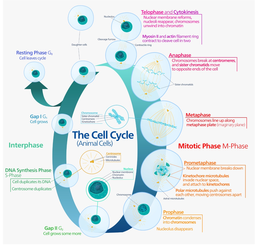
- Prophase which is the first stage of karyokinesis of mitosis follows the S and G2 phases of interphase.
- In the S and G2 phases the new DNA molecules formed are not distinct but intertwined.
- Prophase is marked by the initiation of condensation of chromosomal material.
- The chromosomal material becomes untangled during the process of chromatin condensation.
- The centriole, which had undergone duplication during the S phase of interphase, now begins to move towards opposite poles of the cell.
- Formation of astral ray occurs due to gelation of proteins around centrioles in animal cells.
- Anastral and Amphiastral Mitosis : In higher plants, centrioles are absent and no asters are formed. Mitosis without asters is known as anastral mitosis. In animals, the asters are present and the mitosis is described as amphiastral or astral mitosis.
- The completion of prophase can thus be marked by the following characteristic events:
- Chromosomal material condenses to form compact mitotic chromosomes. Chromosomes are seen to be composed of two chromatids attached together at the centromere.
- Centrosome which had undergone duplication during interphase, begins to move towards opposite poles of the cell. Each centrosome radiates out microtubules called asters. The two asters of centrosome together with spindle fibres form a mitotic apparatus.
- Cell at the end of prophase when viewed under the microscope, do not show golgi complexes, endoplasmic reticulum, nucleolus and nuclear envelope.
- The complete disintegration of the nuclear envelope marks the start of the second phase of mitosis, hence the chromosomes are spread through the cytoplasm of the cell.
- By this stage, condensation of chromosomes is completed and they can be observed clearly under the microscope.This then, is the stage at which morphology of chromosomes is most easily studied.
- At this stage, metaphase chromosome is made up of two sister chromatids, which are held together by the centromere. Small disc-shaped structures at the surface of the centromeres are called kinetochores. These structures serve as the sites of attachment of spindle fibres (formed by the microtubules) to the chromosomes that are moved into position at the centre of the cell.
- Hence, the metaphase is characterised by all the chromosomes coming to lie at the equator with one chromatid of each chromosome connected by its kinetochore to spindle fibres from one pole and its sister chromatid connected by its kinetochore to spindle fibres from the opposite pole. The plane of alignment of the chromosomes at metaphase is referred to as the metaphase plate.
- Chromosomal fibres (discontinuous/kinetochore which run from pole to centromere) and supporting fibres (continuous/non-kinetochore, which run from pole to pole) arrange in the cell.
- Centromere lies at the equator and the arms of chromosomes remain directed towards the poles.
- The key features of metaphase are:
- Spindle fibres attach to kinetochores of chromosomes.
- Chromosomes are moved to spindle equator and get aligned along metaphase plate through spindle fibres to both poles.
- Centromere of each chromosome splits simultaneously lengthwise (division of centromere). Sister chromatids separate from each other and separated each chromatid is now referred to as an individual chromosome.
- Number of chromosomes becomes double in the cell.
- As each chromosome moves away from the equatorial plate, the centromere of each chromosome is towards the pole and hence at the leading edge, with the arms of the chromosome trailing behind.
- The two new daughter chromosomes begin moving toward opposite ends of the cell as their kinetochore microtubule shorten due to depolymerisation of tubulin protein towards kinetochore end. Because these microtubules are attached at the centromere region, the centromeres are pulled ahead of the arms. (Pulling)
- The cell elongates as the nonkinetochore microtubules lengthen.
- Anaphase stage is characterised by the following key events:
- Centromeres split and chromatids separate.
- Chromatids (now referred as chromosomes) move to opposite poles.
- At the beginning of the final stage of karyokinesis, i.e., telophase, the chromosomes that have reached their respective poles decondense and lose their individuality. The individual chromosomes can no longer be seen and chromatin material tends to collect at each of the two poles. This is the stage which shows the following key events:
- Chromosomes cluster at opposite spindle poles and their identity is lost as discrete elements.
- Nuclear envelope develops around the chromosome clusters at each pole forming two daughter nuclei.
- Nucleolus, golgi complex and ER reform.
Prophase :
Metaphase :
Anaphase :
Telophase (Reverse prophase) :
MITOSIS
- This is the most dramatic period of the cell cycle, involving a major reorganisation of virtually all components of the cell. Since the number of chromosomes in the parent and progeny cells is the same, it is also called as equational division.
- Though for convenience mitosis has been divided into four stages of nuclear division, it is very essential to understand that cell division is a progressive process and very clear-cut lines cannot be drawn between various stages.
- The M-phase start with nuclear division, corresponding to the separation of the daughter chromosome (Karyokinesis) and usually ends with division of cytoplasm (cytokinesis).
- Mitosis is divided into the following four stages :-
- Prophase
- Metaphase
- Anaphase
- Telophase
- Prophase which is the first stage of karyokinesis of mitosis follows the S and G2 phases of interphase.
- In the S and G2 phases the new DNA molecules formed are not distinct but intertwined.
- Prophase is marked by the initiation of condensation of chromosomal material.
- The chromosomal material becomes untangled during the process of chromatin condensation.
- The centriole, which had undergone duplication during the S phase of interphase, now begins to move towards opposite poles of the cell.
- Formation of astral ray occurs due to gelation of proteins around centrioles in animal cells.
- Anastral and Amphiastral Mitosis : In higher plants, centrioles are absent and no asters are formed. Mitosis without asters is known as anastral mitosis. In animals, the asters are present and the mitosis is described as amphiastral or astral mitosis.
- The completion of prophase can thus be marked by the following characteristic events:
- Chromosomal material condenses to form compact mitotic chromosomes. Chromosomes are seen to be composed of two chromatids attached together at the centromere.
- Centrosome which had undergone duplication during interphase, begins to move towards opposite poles of the cell. Each centrosome radiates out microtubules called asters. The two asters of centrosome together with spindle fibres form a mitotic apparatus.
- Cell at the end of prophase when viewed under the microscope, do not show golgi complexes, endoplasmic reticulum, nucleolus and nuclear envelope.
- The complete disintegration of the nuclear envelope marks the start of the second phase of mitosis, hence the chromosomes are spread through the cytoplasm of the cell.
- By this stage, condensation of chromosomes is completed and they can be observed clearly under the microscope.This then, is the stage at which morphology of chromosomes is most easily studied.
- At this stage, metaphase chromosome is made up of two sister chromatids, which are held together by the centromere. Small disc-shaped structures at the surface of the centromeres are called kinetochores. These structures serve as the sites of attachment of spindle fibres (formed by the microtubules) to the chromosomes that are moved into position at the centre of the cell.
- Hence, the metaphase is characterised by all the chromosomes coming to lie at the equator with one chromatid of each chromosome connected by its kinetochore to spindle fibres from one pole and its sister chromatid connected by its kinetochore to spindle fibres from the opposite pole. The plane of alignment of the chromosomes at metaphase is referred to as the metaphase plate.
- Chromosomal fibres (discontinuous/kinetochore which run from pole to centromere) and supporting fibres (continuous/non-kinetochore, which run from pole to pole) arrange in the cell.
- Centromere lies at the equator and the arms of chromosomes remain directed towards the poles.
- The key features of metaphase are:
- Spindle fibres attach to kinetochores of chromosomes.
- Chromosomes are moved to spindle equator and get aligned along metaphase plate through spindle fibres to both poles.
- Centromere of each chromosome splits simultaneously lengthwise (division of centromere). Sister chromatids separate from each other and separated each chromatid is now referred to as an individual chromosome.
- Number of chromosomes becomes double in the cell.
- As each chromosome moves away from the equatorial plate, the centromere of each chromosome is towards the pole and hence at the leading edge, with the arms of the chromosome trailing behind.
- The two new daughter chromosomes begin moving toward opposite ends of the cell as their kinetochore microtubule shorten due to depolymerisation of tubulin protein towards kinetochore end. Because these microtubules are attached at the centromere region, the centromeres are pulled ahead of the arms. (Pulling)
- The cell elongates as the nonkinetochore microtubules lengthen.
- Anaphase stage is characterised by the following key events:
- Centromeres split and chromatids separate.
- Chromatids (now referred as chromosomes) move to opposite poles.
- At the beginning of the final stage of karyokinesis, i.e., telophase, the chromosomes that have reached their respective poles decondense and lose their individuality. The individual chromosomes can no longer be seen and chromatin material tends to collect at each of the two poles. This is the stage which shows the following key events:
- Chromosomes cluster at opposite spindle poles and their identity is lost as discrete elements.
- Nuclear envelope develops around the chromosome clusters at each pole forming two daughter nuclei.
- Nucleolus, golgi complex and ER reform.

Prophase :
Metaphase :
Anaphase :
Telophase (Reverse prophase) :
Amitosis & Mitosis
- Cell division generally 2 type ka hota hai : equational and reductional jinko hum mitosis and meiosis bolte hai, respectively. Ek or term hai jo kabhi kabhi ek primitive cell division ki form ke liye use karte hai, wo term hai AMITOSIS. Amitosis sabse simple cell division ki process hai jisme typical cell division wale events jasie ki spindle fibres ki formation ya chromatin fibres ki condensation nhi hogi, nuclear envelope bhi dissolve nhi hoga balki throughout the cell division wo intact rehta hai. Amitosis prokaryotes and kuch unicellular eukaryotes me dekhne ko milti hai.
- Mitosis equational division hai yani ki ek cell 2 daughter cells me divide hoga and dono daughter cells me genetic content double hone ke baad equal -equal dia jayega. In short, mitosis me ploidy level change nhi hogi. Ek haploid cell se 2 haploid daughter cells banege jabki ek diploid parental cell se 2 diploid daughter cells hi banege. Interphase me cell prepare karega khud ko and jab M phase aayega toh division hoga. Right? Ab M phase ke upper further 4 subphases btaye thy. Ab unko eke k karke sequence me discuss karenge.
Prophase - M phase ki pehli stage hi prophase hai jhaan par cell ke genetic material jo ab tak chromatin fibre ki form me tha wo condense hona shuru hota hai, nuclear envelope degenerate krne shuru kar deta hai. Interphase ke S phase ke daruan jo centriole ek jagah par duplicate hue thy wo ab cell ke opposite poles ki taraf chalna shuru kar dete hai. Toh prophase khatam hote hote nuclear envelope sara hi dissolve ho chuka hoga and chromatin fibers condense hokr chromosomes banne shuru hojate hai.
- Jis waqt nuclear envelope puri tarah disintegrate ho chuka hota hai tab prophase complete hokr next phase yani ki metaphase shuru hojata hai. Issi phase ke daruan chromosomes sabse jyada condensed hote hai toh unki morphology padhne ka sabse sahi time yhi hai. Chromosomes par kinetochores bhi visible hojate hai. Kinetochore basically primary constriction hai jo ek protein complex hai. Yahi wo location jhaan spindle fibres chromosome se attach honge. Centrioles se spindle fibres nikalna shuru hote hai chromosome ke kinetochore par attach hokr, chromosomes ko equator par align kar dete hai jiske karan equator par condensed chromosomes ek plat like appearance denge jisko hum metaphasic plate kehte hai.
- Jab metaphasic plate ban chuki hai toh uske kuch samay baad hi dono opposite poles se spindle fibres chromosomes ko apni apni taraf khichna shuru kar dete hai. balako, yaad rakho ki ek chromosome me 2 sister chromatids kinetochores ki location par attached hote hai. Toh jab ek chromosome ko spindle opposite direction me pull karte hai toh wo ek chromosome 2 sister chromatids me split hokr opposite poles ki taraf move karna shuru kar dete hai. Toh jab spindle in chromatids ko apni taraf kheechte hai toh centrosome sabse aage hota hai and chromosomes ki arms peeche peeche chalti hui dikhti hai. Toh yeah splitting event or sister chromatids ka opposite pole ki taraf move karna hi anaphase ki pehchaan hai.
- Ab isse pehle wale phase tak genetic material split hokr ek hi cells 2 opposite pole ki taraf ja chuka lekin ab iss phase me wo firse decondense hona shuru hoga. Genetic material ke charo taraf firse nuclear envelope aa jayega and cell organelles jaise ki nucleolus, golgi complex and endoplasmic reticulum bhi cells me ban jate hai.
- Animal and plant cell me mitosis ke case me karyokinesis tak similarities hai lekin cytokinesis thodi si alag ho jati hai dono ke case me.
- Animal cell ke case me wo middle se constrict hona shuru hojate hai or cell ke center tak constrict hota hi jata hai jaise hum kisi chote balloon ko beech se pinch karde or jab humari ek finger dusri finger se mile, pinching karte hue toh humare pass (hypothetically) socho ki 2 naye chotu balloons aa jaye. Theek ussi tarah animal cell me plasma membrane me furrow formation hoti hai jo cell ke middle tak continued rehti hai and iske end me humare pas totally 2 nye daughter cells hote hai.
- Ab tak yeah padha ki meiosis hoti kaise hai. Ab yeah dekh lete hai ki hoti kyu hai? Yani ki zarurat kya hai? Toh beta mitosis me ek cell apne jaise hi genetically similar cells paida karta hai. Toh jo bhi jakham bharne ke liye new cells ki jarurat ho ya kuch regenerate karwana ho jaise ki skin to huss case me mitosis help karti hai. Grow karne me, chahe animal cells ho ya plant cells ki unlimited growth wo sab mitosis ke karan hi hota hai.
MITOSIS
Metaphase
Anaphase
Telophase
Ab tak jo hua usme ek hi cell me nuclear material ka division hua alongwith of course the cell organelles. Toh issi nucleus ke division ko hum karyokinesis kahte hai. Ab iske baad keval cytoplasm bhi agar 2 freshly formed daughter nuclei ke sath divide ho jaye toh hume 2 complete new, individual daughter cells milenge. Toh next step hai Cytokinesis, yani ki cytoplasm ka division.
Cytokinesis
Significance of Mitosis
CYTOKINESIS
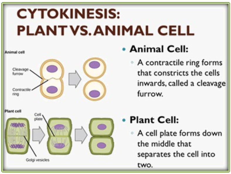
- Mitosis accomplishes not only the segregation of duplicated chromosome into daughter nuclei (Karyokinesis) but the cell itself is divided into two daughter cells by the separation of cytoplasm called cytokinesis at the end of which cell division gets completed.
- In animals cytokinesis occurs by constriction & furrow formation. Microtubules and microfilaments arrange on the equator to form midbody and at the periphery of the equator a contractile ring is formed that is made up of actin and myosin protein. Due to interaction between actin and myosin ring contract, thus a furrow forms from outside to inside in the cell. Furrow deepens continuously and ultimately a cell divides into two daughter cells. In animals cytokinesis occurs in centripetal order.
- Cytokinesis in plants takes place by cell plate formation because constriction is not possible due to presence of the rigid cell wall. Many golgi vesicles and spindle microtubules arrange themselves on equator to form phragmoplast. Fragments of ER may also deposit in phragmoplast. Membranes of golgi vesicles fuse to form a plate-like structure called a cell plate. Golgi vesicles secrete calcium and magnesium pectate. Further cell plates are modified into middle lamella. In plants, cytokinesis occurs in centrifugal order (cell plate formation is from center to periphery).
- In some organisms karyokinesis is not followed by cytokinesis as a result of which a multinucleate condition arises leading to the formation of syncytium (e.g., liquid endosperm in coconut).

Amitosis & Mitosis
- It is also called direct or incipient cell division . It is the simplest type of the cell-division in which there is no synthesis of spindle, no condensation of chromatin fibers and no disappearance of nuclear membrane.
- It is a simple method of cell division which is also called direct cell division. In this division there is no differentiation of chromosomes and spindle. The nuclear envelope does not degenerate.
- The nucleus elongates and constricts in the middle to form two daughter nuclei. This is followed by a centripetal constriction of the cytoplasm to form two daughter cells. Eg. Prokaryotes and some unicellular eukaryotes.
Amitosis
MITOSIS
- This is the most dramatic period of the cell cycle, involving a major reorganisation of virtually all components of the cell. Since the number of chromosomes in the parent and progeny cells is the same, it is also called as equational division.
- Though for convenience mitosis has been divided into four stages of nuclear division, it is very essential to understand that cell division is a progressive process and very clear-cut lines cannot be drawn between various stages.
- The M-phase start with nuclear division, corresponding to the separation of the daughter chromosome (Karyokinesis) and usually ends with division of cytoplasm (cytokinesis).
- Mitosis is divided into the following four stages :
- Prophase
- Metaphase
- Anaphase
- Telophase
- Prophase which is the first stage of karyokinesis of mitosis follows the S and G2 phases of interphase.
- In the S and G2 phases the new DNA molecules formed are not distinct but intertwined.
- Prophase is marked by the initiation of condensation of chromosomal material.
- The chromosomal material becomes untangled during the process of chromatin condensation.
- The centriole, which had undergone duplication during the S phase of interphase, now begins to move towards opposite poles of the cell.
- Formation of astral ray occurs due to gelation of proteins around centrioles in animal cells.
- Anastral and Amphiastral Mitosis : In higher plants, centrioles are absent and no asters are formed. Mitosis without asters is known as anastral mitosis. In animals, the asters are present and the mitosis is described as amphiastral or astral mitosis.
- The completion of prophase can thus be marked by the following characteristic events:
- Chromosomal material condenses to form compact mitotic chromosomes. Chromosomes are seen to be composed of two chromatids attached together at the centromere.
- Centrosome which had undergone duplication during interphase, begins to move towards opposite poles of the cell. Each centrosome radiates out microtubules called asters. The two asters of centrosome together with spindle fibres form a mitotic apparatus.
- Cell at the end of prophase when viewed under the microscope, do not show golgi complexes, endoplasmic reticulum, nucleolus and nuclear envelope.
- The complete disintegration of the nuclear envelope marks the start of the second phase of mitosis, hence the chromosomes are spread through the cytoplasm of the cell.
- By this stage, condensation of chromosomes is completed and they can be observed clearly under the microscope.This then, is the stage at which morphology of chromosomes is most easily studied.
- At this stage, metaphase chromosome is made up of two sister chromatids, which are held together by the centromere. Small disc-shaped structures at the surface of the centromeres are called kinetochores. These structures serve as the sites of attachment of spindle fibres (formed by the microtubules) to the chromosomes that are moved into position at the centre of the cell.
- Hence, the metaphase is characterised by all the chromosomes coming to lie at the equator with one chromatid of each chromosome connected by its kinetochore to spindle fibres from one pole and its sister chromatid connected by its kinetochore to spindle fibres from the opposite pole. The plane of alignment of the chromosomes at metaphase is referred to as the metaphase plate.
- Chromosomal fibres (discontinuous/kinetochore which run from pole to centromere) and supporting fibres (continuous/non-kinetochore, which run from pole to pole) arrange in the cell.
- Centromere lies at the equator and the arms of chromosomes remain directed towards the poles.
- The key features of metaphase are:
- Spindle fibres attach to kinetochores of chromosomes.
- Chromosomes are moved to spindle equator and get aligned along metaphase plate through spindle fibres to both poles.
- Centromere of each chromosome splits simultaneously lengthwise (division of centromere). Sister chromatids separate from each other and separated each chromatid is now referred to as an individual chromosome.
- Number of chromosomes becomes double in the cell.
- As each chromosome moves away from the equatorial plate, the centromere of each chromosome is towards the pole and hence at the leading edge, with the arms of the chromosome trailing behind.
- The two new daughter chromosomes begin moving toward opposite ends of the cell as their kinetochore microtubule shorten due to depolymerisation of tubulin protein towards kinetochore end. Because these microtubules are attached at the centromere region, the centromeres are pulled ahead of the arms. (Pulling)
- The cell elongates as the nonkinetochore microtubules lengthen.
- Anaphase stage is characterised by the following key events:
- Centromeres split and chromatids separate.
- Chromatids (now referred as chromosomes) move to opposite poles.
- At the beginning of the final stage of karyokinesis, i.e., telophase, the chromosomes that have reached their respective poles decondense and lose their individuality. The individual chromosomes can no longer be seen and chromatin material tends to collect at each of the two poles. This is the stage which shows the following key events:
- Chromosomes cluster at opposite spindle poles and their identity is lost as discrete elements.
- Nuclear envelope develops around the chromosome clusters at each pole forming two daughter nuclei.
- Nucleolus, golgi complex and ER reform.

Prophase :
Metaphase :
Anaphase :
Telophase (Reverse prophase) :
CYTOKINESIS
- Mitosis accomplishes not only the segregation of duplicated chromosome into daughter nuclei (Karyokinesis) but the cell itself is divided into two daughter cells by the separation of cytoplasm called cytokinesis at the end of which cell division gets completed.
- In animals cytokinesis occurs by constriction & furrow formation. Microtubules and microfilaments arrange on the equator to form midbody and at the periphery of the equator a contractile ring is formed that is made up of actin and myosin protein. Due to interaction between actin and myosin ring contract, thus a furrow forms from outside to inside in the cell. Furrow deepens continuously and ultimately a cell divides into two daughter cells. In animals cytokinesis occurs in centripetal order.
- Cytokinesis in plants takes place by cell plate formation because constriction is not possible due to presence of the rigid cell wall. Many golgi vesicles and spindle microtubules arrange themselves on equator to form phragmoplast. Fragments of ER may also deposit in phragmoplast. Membranes of golgi vesicles fuse to form a plate-like structure called a cell plate. Golgi vesicles secrete calcium and magnesium pectate. Further cell plates are modified into middle lamella. In plants, cytokinesis occurs in centrifugal order (cell plate formation is from center to periphery).
- In some organisms karyokinesis is not followed by cytokinesis as a result of which a multinucleate condition arises leading to the formation of syncytium (e.g., liquid endosperm in coconut).

SIGNIFICANCE OF MITOSIS :
- Development of an organism occurs by mitosis. Every organism starts its life from a single cell i.e. zygote. Repeated mitosis in zygote leads to the formation of the whole body.
- The growth of multicellular organisms is due to mitosis.
- Cell growth results in disturbing the ratio between the nucleus and the cytoplasm. It therefore becomes essential for the cell to divide to restore the nucleo-cytoplasmic ratio.
- A very significant contribution of mitosis is cell repair. The cells of the upper layer of the epidermis, cells of the lining of the gut, and blood cells are being constantly replaced.
- Mitotic divisions in the meristematic tissues – the apical and the lateral cambium, result in a continuous growth of plants throughout their life.
SIGNIFICANCE OF MITOSIS
- Development of an organism occurs by mitosis. Every organism starts its life from a single cell i.e. zygote. Repeated mitosis in zygote leads to the formation of the whole body.
- The growth of multicellular organisms is due to mitosis.
- Cell growth results in disturbing the ratio between the nucleus and the cytoplasm. It therefore becomes essential for the cell to divide to restore the nucleo-cytoplasmic ratio.
- A very significant contribution of mitosis is cell repair. The cells of the upper layer of the epidermis, cells of the lining of the gut, and blood cells are being constantly replaced.
- Mitotic divisions in the meristematic tissues – the apical and the lateral cambium, result in a continuous growth of plants throughout their life.
Meiosis
- Beta meiosis meioum naam ke Greek shabd se liya gya hai jiska matlab hi hai ‘to lessen’ yani ki kam karna, yahan par hamara matlab hai number of chromosome ko kam karna ya reduce karna jiske karan iss cell division ko hum reductional division bhi keh dete hai kyunki isme daughter cell me number of chromosomes parent cell ke comparison me half hote hai.
- Meiosis ka interphase bhi lagbhag mitotic interphase ke similar hi hai. Ek aadha difference hai wo yeah ki ek toh meiosis ke interphase me G2 phase bahut hi chota hota and even absent bhi ho sakta hai. Lekin S-phase comparatively prolonged hota hai jisme DNA synthesis hogi jiske baad har ek chromosome pe 2 chromatids jinko sister chromatids bola jata hai, wo synthesize honge.
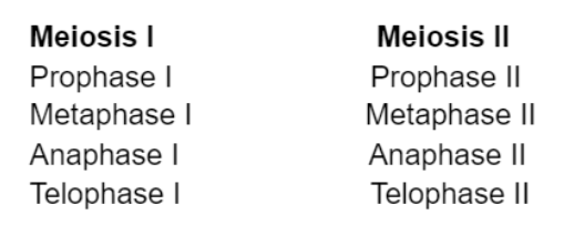
- Meiosis me 2 M phases hote hai : Meiosis – I and Meiosis – II. Dono M phases ke beech ka chota phase jisme koi DNA replication nhi hoga wo interkinesis kehlata hai. Dono M phases ka apna khud ka karyokinesis and cytokinesis hoga. Toh ek cell pehle Meiosis I me enter karta hai ek parent cell se 2 daughter cell bnate hai jinme number of chromosomes half ho chuke hote hai. Next, dono daughter cells fir Meiosis II me enter karte hai and unse 4 daughter cell milte hai. Meiosis II reductional division nahi hota balki mitosis ki tarah yeah equational division hota hai.
- Meiosis I ko reductional division kha jata hai and isme bhi karyokinesis ke vhi basic 4 phases : prophase-I, metaphase-I, anaphase-I and telophase-I hote hai jinke baad cytokinesis I hoga aur fir iske baad cell interkinesis me enter karega. Meiosis I and actual mitosis me kafi differences hai jo hum abhi dekhenge and ek toh yeah ki iska Prophase -I bhi further subphases me divided hota hai.
- Mitosis ke comparison me meiosis ka prophase bahut hi lamba and complicated sa hai. Iske bhi further 5 sub phases hai :
- Leptotene : Same vhi ki chromosomes start to condense lekin kafi animal cells me chromosomes ke end nuclear membrane ki internal side se connected hote hai jisko karan iss stage ko ‘bouquet stage’ bhi bol dia jata hai. Centrioles opposite poles pe move karna shuru kar dete hai and unse astral rays nikalna shuru hojati hai. Aster = centriole pair + astral rays. Iss stages me replicated chromatids ek dusre se distinguishable nahi hote.
- Zygotene : diploid cells me chromosome ka ek set papa se aata hai jinko hum paternal chromosomes kehte hai and ek set mother se aata hai jinko hum maternal chromosomes kehte hai. Toh ek hi chromosome ki 2 copies hoti hai, for ex : humare paas 2 chromosome number 1 honge – ek jo papa se aaya hai and ek jo mom se aaya hai toh ais eek type ke 2 chromosomes ko hum homologous chromosomes kehte hai. Zygotene stage me aise homologous chromosomes ki pairing shuru hojati hai jisko hum synapsis kehte hai. Synapsis ke dauran homologus chromosomes me ribonucleoprotein (tripartite structure : 2 vertically aligned lateral elements + 1 horizontal central structure) ke jariye se homologous chromosomes ke beech me physical connection hota hai and 2 homologous chromosome jab synapsis ke baad apas me jude hue hote hai to uss structure ko hum bivalent ya synaptonemal complex kehte hai. So agar kisi cell me 46 chromosomes hai toh beta usme 23 bivalents banege zygotene stage me.
- Pachytene : yeah prophase-I ki sabse badi stage hai kyunki ek cell iss stage me kuch ghanto se lekr kuch dino takh bhi reh sakta hai. Iss phase me Synaptonemal comples ke beech me recombination hoti hai with the help of recombinase enzyme, yani ki ek bivalent me jo 2 non-sister chromatids thy unki segment ka exchange hota hai, toh iss stage ki characteristic step hui : recombination (between non sister chromatids arms of a bivalent chromosome).
- Diplotene : iss stage me bivalents ke beech me jo ribonucleoprotein ka physical connection establish hua tha recombination ko facilitate karwane ke liye ab wo dissolve hona shuru ho jata hai and it dissolves partially in this stage yani ki jhaan par recombination hua tha us jagah ko chor kar baki jagaho se ribonucleoprotein dissolve ho jata hai jsike karan iss stage me ek characteristic X shape ban jati hai non sister chromatids me and isko hum Chiasmata kehte hai. Chiasmata beta ya toh chromosomes ke terminal part par ho sakta hai ya beech me toh chromosomes cross like ya ring like ya chain like dikhte hai.
→ kuch animal cells jaise hi human female ke oocytes me diplotene stage me oocyte arrest hojata hai and issi stage me wo 11-13 saal tak reh sakta hai. Lekin diplotene stage metabolically bhi active hoti hai toh chromosome decondense hokr RNA synthesis me part lete hai toh iss decondensation ke karan chromosomes ki shape ek brush ke bristles jaisi dikhti hai toh inko hum lampbrush chromosomes bhi keh dete hai. - Diakinesis : iss stage me chiasmata terminals ki taraf move karna shuru karta hai toh iss process ko hum terminalisation kehte hai. Chiasmata disappears and nucleolus starts to degenerate alongwith nuclear membrane.
- Isme spindle fibers chromosomes ke kinetochores par attach ho jate hai and chromosomes moves towards equator and metaphasic plate ki formation ho jati hai. Yaad rakhna ki yha par diploid organism hai toh chromosomes ke 2 set hai toh double metaphasic plate ki formation hogi.
- Same vahi isme spindle fibre short hona shuru honge jiski vajah se homologous chromosomes me se ek ek chromosome opposite pole ki taraf move karna shuru kar denge. Homologous chromosomes ke beech ka connection/junction iss stage me tut jata hai and separated chromosomes yani ki univalent / dyads opposite poles ki tarah move karte hai and number of chromosomes iss stage me half ho gya. Issi separation ki process ko disjunction bhi kha gya hai.
- Opposite poles par chromosomes decondense kar jate hai, nucelolus, nuclear membrane and cell organelle bnti hai and usually next step hogi : cytokinesis jisme 2 daughter cells ban jayenge.
- Meiosis me beta homologous chromosomes ka separation hota hai yani ki number of chromosomes daughter cell me half ho jayenge. Yeah sexual reproduction ke liye jaruri hai.
- crossing over ke karan new variations introduce hoti hai.
- paternal and maternal chromosomes ka independent assortment hoga daughter cell me jo firse variations introduce karta hai.
- Disjunction ke daruan disturbance hone pe genetic mutations ho sakti hai.
- Gamete formation
- Interphase II ya interkinesis me DNA ki replication nhi hoti lekin RNA and protein production hota hai. Centrosomes bhi replicate ho sakte hai.
- Meiosis II ek equational division hai ya fir homotypic, jiska matlab hai ki isme chromosome numbers me reduction nhi hota lekin ab iss phase me ek hi chromosome ke sister chromatid separate honge.
→ Chromatids differ in number of linkage groups due to crossing over/recombination - Prophase II : kafi chota sa phase hai yeah. Centrioles astar (in animal cells) develop karke opposite poles ki taraf move karna shuru kar dete hai. Nucelolus and nuclear envelope degenerate karna shuru kar dete hai.
Kabhi kabhi kisi kisi cell me telophase I puri taraf se gayab hota hai to uss cell me prophase II bhi absent hi hoga. - Metaphase II : spindle fibres (amphiastral animals me and anastral plant cells me) bante hai, kinetochore pe attach honge and metaphasic plate ki formation hogi.
- Anaphase II : splitting of chromosome jiske karan sister chromatids opposite poles ki taraf move karna shuru kar dete hai.
- Telophase II : chromosomes decondense to form chromatin fibers. Nucleolus and nuclear membrane ke sath sath baki cell organelles bhi reappear hojati hai. Spindle fibers degenerate hojate hai.
- Cytokinesis II : cytoplasm divide hota hai and ek cell se 2 daughter cell bante hai lekin yaad rakhna ki ek single diploid cell jab meiosis me enter hua that oh meiosis ke baad 2 daughter cells meiosis II me enter hote hai jinse total 4 daughter cells milenge toh meiosis me ek diploid cell se 4 haploid cells milte hai.
- Gametes ki formation ke liye, chromosome number maintain karne ke liye, chromosomes ki assortment ke liye, variations introduce karwane ke liye and mutations.
Meiosis I
Prophase – I
Metaphase – I
Anaphase – I
Telophase – I
Significance of Meiosis – I
Meiosis – II
Significance
MEIOSIS
- The production of offspring by sexual reproduction includes the fusion of two gametes, each with a complete haploid set of chromosomes. Gametes are formed from specialised diploid cells.
- This specialised kind of cell division that reduces the chromosome number by half results in the production of haploid daughter cells. This kind of division is called meiosis.
- Meiosis ensures the production of haploid phase in the life cycle of sexually reproducing organisms whereas fertilisation restores the diploid phase.
- We come across meiosis during gametogenesis in plants and animals. This leads to the formation of haploid gametes.
- The key features of meiosis are as follows:
- Meiosis involves two sequential cycles of nuclear and cell division called meiosis I and meiosis II but only a single cycle of DNA replication.
- Meiosis I is initiated after the parental chromosomes have replicated to produce identical sister chromatids at the S phase.
- Meiosis involves pairing of homologous chromosomes and recombination between non-sister chromatids of homologous chromosomes.
- Four haploid cells are formed at the end of meiosis II. Meiotic events can be grouped under the following phases:
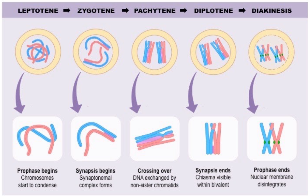
- Prophase I : Prophase of the first meiotic division is typically longer and more complex when compared to prophase of mitosis. It has been further subdivided into the following five phases based on chromosomal behaviour, i.e., Leptotene, Zygotene, Pachytene, Diplotene and Diakinesis.
-
Leptotene (Leptonema thread stage)
- During the leptotene stage the chromosomes become gradually visible under the light microscope. Chromosomes appear thin, uncoiled, elongated and beaded.
- These beads are also called chromomeres which represent genes.
- The compaction of chromosomes continues throughout leptotene.
- Zygotene ( Zygonema thread stage)
- In this stage homologous chromosomes come closer and form pairs. This pairing process is called Synapsis or Syndesis or Zipping. Now each pair is Bivalent.
- Synapsis is accompanied by the formation of a complex structure called synaptonemal complex.
- The first two stages of prophase I are relatively short-lived compared to the next stage that is pachytene.
-
Leptotene (Leptonema thread stage)
MEIOSIS I
- During this stage, the four chromatids of each bivalent chromosome become distinct and clearly appear as tetrads. This stage is characterised by the appearance of recombination nodules, the sites at which crossing over occurs between non-sister chromatids of the homologous chromosomes.
- Crossing over is the exchange of genetic material between two homologous chromosomes. Crossing over is also an enzyme-mediated process and the enzyme involved is called recombinase. Crossing over leads to recombination of genetic material on the two chromosomes.
- Recombination between homologous chromosomes is completed by the end of pachytene, leaving the chromosomes linked at the sites of crossing over.
- The beginning of diplotene is recognised by the dissolution of the synaptonemal complex and the tendency of the recombined homologous chromosomes of the bivalents to separate from each other except at the sites of crossovers. These X-shaped structures are called chiasmata.
- In oocytes of some vertebrates, diplotene can last for months or years.
- Nucleolus and nuclear membrane start degenerating.
- The final stage of meiotic prophase I is diakinesis. This is marked by terminalisation of chiasmata.
- During this phase the chromosomes are fully condensed and the meiotic spindle is assembled to prepare the homologous chromosomes for separation.
- By the end of diakinesis, the nucleolus disappears and the nuclear envelope also breaks down.
- Synaptonemal complex- Zygotene
- Recombination nodule - Pachytene
- Beginning of dissolution of synaptonemal complex- Diplotene
- Terminalisation of chiasmata - Diakinesis
- Separation of homologous chromosome - Anaphase I
- Formation of dyad of cells - Telophase I
MEIOSIS II
It is also called homotypic division because the number of chromosomes remains the same.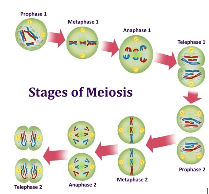
Meiosis
- The production of offspring by sexual reproduction includes the fusion of two gametes, each with a complete haploid set of chromosomes. Gametes are formed from specialised diploid cells.
- This specialised kind of cell division that reduces the chromosome number by half results in the production of haploid daughter cells. This kind of division is called meiosis.
- Meiosis ensures the production of haploid phase in the life cycle of sexually reproducing organisms whereas fertilisation restores the diploid phase.
- We come across meiosis during gametogenesis in plants and animals. This leads to the formation of haploid gametes.
- The key features of meiosis are as follows:
- Meiosis involves two sequential cycles of nuclear and cell division called meiosis I and meiosis II but only a single cycle of DNA replication.
- Meiosis I is initiated after the parental chromosomes have replicated to produce identical sister chromatids at the S phase.
- Meiosis involves pairing of homologous chromosomes and recombination between non-sister chromatids of homologous chromosomes.
- Four haploid cells are formed at the end of meiosis II. Meiotic events can be grouped under the following phases:
- Prophase I : Prophase of the first meiotic division is typically longer and more complex when compared to prophase of mitosis. It has been further subdivided into the following five phases based on chromosomal behaviour, i.e., Leptotene, Zygotene, Pachytene, Diplotene and Diakinesis.
- Leptotene (Leptonema thread stage)
- During the leptotene stage the chromosomes become gradually visible under the light microscope. Chromosomes appear thin, uncoiled, elongated and beaded.
- These beads are also called chromomeres which represent genes.
- The compaction of chromosomes continues throughout leptotene.
- Zygotene ( Zygonema thread stage)
- In this stage homologous chromosomes come closer and form pairs. This pairing process is called Synapsis or Syndesis or Zipping. Now each pair is Bivalent.
- Synapsis is accompanied by the formation of a complex structure called synaptonemal complex.
- The first two stages of prophase I are relatively short-lived compared to the next stage that is pachytene.
- Leptotene (Leptonema thread stage)
MEIOSIS I
- During this stage, the four chromatids of each bivalent chromosome become distinct and clearly appear as tetrads. This stage is characterised by the appearance of recombination nodules, the sites at which crossing over occurs between non-sister chromatids of the homologous chromosomes.
- Crossing over is the exchange of genetic material between two homologous chromosomes. Crossing over is also an enzyme-mediated process and the enzyme involved is called recombinase. Crossing over leads to recombination of genetic material on the two chromosomes.
- Recombination between homologous chromosomes is completed by the end of pachytene, leaving the chromosomes linked at the sites of crossing over.
- The beginning of diplotene is recognised by the dissolution of the synaptonemal complex and the tendency of the recombined homologous chromosomes of the bivalents to separate from each other except at the sites of crossovers. These X-shaped structures are called chiasmata.
- In oocytes of some vertebrates, diplotene can last for months or years.
- Nucleolus and nuclear membrane start degenerating.
- The final stage of meiotic prophase I is diakinesis. This is marked by terminalisation of chiasmata.
- During this phase the chromosomes are fully condensed and the meiotic spindle is assembled to prepare the homologous chromosomes for separation.
- By the end of diakinesis, the nucleolus disappears and the nuclear envelope also breaks down.
- Synaptonemal complex- Zygotene
- Recombination nodule - Pachytene
- Beginning of dissolution of synaptonemal complex- Diplotene
- Terminalisation of chiasmata - Diakinesis
- Separation of homologous chromosome - Anaphase I
- Formation of dyad of cells - Telophase I
MEIOSIS II
It is also called homotypic division because the number of chromosomes remains the same.
SIGNIFICANCE OF MEIOSIS :
- Meiosis is the mechanism by which conservation of specific chromosome number of each species is achieved across generations in sexually reproducing organisms.
- Helps in formation of gametes thar are essential for sexual reproduction.
- It also increases the genetic variability in the population of organisms from one generation to the next. Variations are very important for the process of evolution.
SIGNIFICANCE OF MEIOSIS
- Meiosis is the mechanism by which conservation of specific chromosome number of each species is achieved across generations in sexually reproducing organisms.
- Helps in formation of gametes thar are essential for sexual reproduction.
- It also increases the genetic variability in the population of organisms from one generation to the next. Variations are very important for the process of evolution.
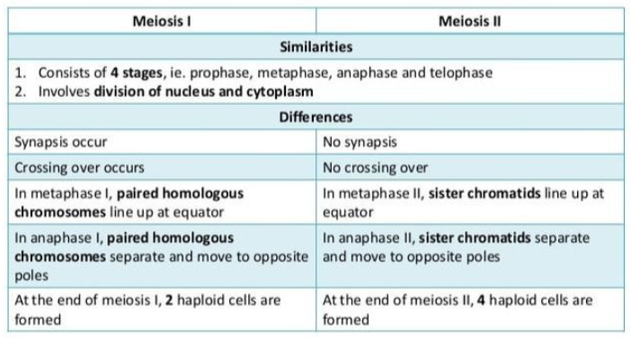
Difference Between Mitosis And Meiosis
DIFFERENCE BETWEEN MITOSIS AND MEIOSIS
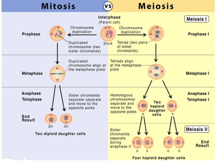
DIFFERENCE BETWEEN MITOSIS AND MEIOSIS


Summary
- According to the cell theory, cells arise from preexisting cells. The process by which this occurs is called cell division.
- Any sexually reproducing organism starts its life cycle from a single-celled zygote. Cell division does not stop with the formation of the mature organism but continues throughout its life cycle.
- The stages through which a cell passes from one division to the next is called the cell cycle.
- Cell cycle is divided into two phases called (i) Interphase – a period of preparation for cell division, and (ii) Mitosis (M phase) – the actual period of cell division.
- Interphase is further subdivided into G1 , S and G2 . G1 phase is the period when the cell grows and carries out normal metabolism. Most of the organelle duplication also occurs during this phase. S phase marks the phase of DNA replication and chromosome duplication. G2 phase is the period of cytoplasmic growth. Mitosis is also divided into four stages namely prophase, metaphase, anaphase and telophase.
- Chromosome condensation occurs during prophase. Simultaneously, the centrioles move to the opposite poles. The nuclear envelope and the nucleolus disappear and the spindle fibres start appearing.
- Metaphase is marked by the alignment of chromosomes at the equatorial plate. During anaphase the centromeres divide and the chromatids start moving towards the two opposite poles.
- Once the chromatids reach the two poles, the chromosomal elongation starts, the nucleolus and the nuclear membrane reappear. This stage is called the telophase.
- Nuclear division is then followed by the cytoplasmic division and is called cytokinesis. Mitosis thus, is the equational division in which the chromosome number of the parent is conserved in the daughter cell.
- In contrast to mitosis, meiosis occurs in the diploid cells, which are destined to form gametes. It is called the reduction division since it reduces the chromosome number by half while making the gametes.
- In sexual reproduction when the two gametes fuse the chromosome number is restored to the value in the parent. Meiosis is divided into two phases – meiosis I and meiosis II. In the first meiotic division the homologous chromosomes pair to form bivalents, and undergo crossing over.
- Meiosis I has a long prophase, which is divided further into five phases. These are leptotene, zygotene, pachytene, diplotene and diakinesis. During metaphase I the bivalents arrange on the equatorial plate. This is followed by anaphase I in which homologous chromosomes move to the opposite poles with both their chromatids. Each pole receives half the chromosome number of the parent cell.
- In telophase I, the nuclear membrane and nucleolus reappear. Meiosis II is similar to mitosis. During anaphase II the sister chromatids separate. Thus at the end of meiosis four haploid cells are formed.
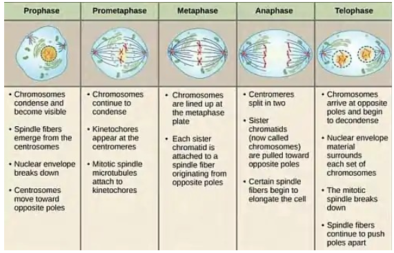
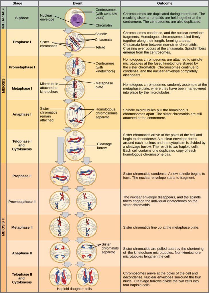
SUMMARY
- According to the cell theory, cells arise from preexisting cells. The process by which this occurs is called cell division.
- Any sexually reproducing organism starts its life cycle from a single-celled zygote. Cell division does not stop with the formation of the mature organism but continues throughout its life cycle.
- The stages through which a cell passes from one division to the next is called the cell cycle.
- Cell cycle is divided into two phases called (i) Interphase – a period of preparation for cell division, and (ii) Mitosis (M phase) – the actual period of cell division.
- Interphase is further subdivided into G1 , S and G2 . G1 phase is the period when the cell grows and carries out normal metabolism. Most of the organelle duplication also occurs during this phase. S phase marks the phase of DNA replication and chromosome duplication. G2 phase is the period of cytoplasmic growth. Mitosis is also divided into four stages namely prophase, metaphase, anaphase and telophase.
- Chromosome condensation occurs during prophase. Simultaneously, the centrioles move to the opposite poles. The nuclear envelope and the nucleolus disappear and the spindle fibres start appearing.
- Metaphase is marked by the alignment of chromosomes at the equatorial plate. During anaphase the centromeres divide and the chromatids start moving towards the two opposite poles.
- Once the chromatids reach the two poles, the chromosomal elongation starts, the nucleolus and the nuclear membrane reappear. This stage is called the telophase.
- Nuclear division is then followed by the cytoplasmic division and is called cytokinesis. Mitosis thus, is the equational division in which the chromosome number of the parent is conserved in the daughter cell.
- In contrast to mitosis, meiosis occurs in the diploid cells, which are destined to form gametes. It is called the reduction division since it reduces the chromosome number by half while making the gametes.
- In sexual reproduction when the two gametes fuse the chromosome number is restored to the value in the parent. Meiosis is divided into two phases – meiosis I and meiosis II. In the first meiotic division the homologous chromosomes pair to form bivalents, and undergo crossing over.
- Meiosis I has a long prophase, which is divided further into five phases. These are leptotene, zygotene, pachytene, diplotene and diakinesis. During metaphase I the bivalents arrange on the equatorial plate. This is followed by anaphase I in which homologous chromosomes move to the opposite poles with both their chromatids. Each pole receives half the chromosome number of the parent cell.
- In telophase I, the nuclear membrane and nucleolus reappear. Meiosis II is similar to mitosis. During anaphase II the sister chromatids separate. Thus at the end of meiosis four haploid cells are formed.


SUMMARY
- According to the cell theory, cells arise from preexisting cells. The process by which this occurs is called cell division.
- Any sexually reproducing organism starts its life cycle from a single-celled zygote. Cell division does not stop with the formation of the mature organism but continues throughout its life cycle.
- The stages through which a cell passes from one division to the next is called the cell cycle.
- Cell cycle is divided into two phases called (i) Interphase – a period of preparation for cell division, and (ii) Mitosis (M phase) – the actual period of cell division.
- Interphase is further subdivided into G1 , S and G2 . G1 phase is the period when the cell grows and carries out normal metabolism. Most of the organelle duplication also occurs during this phase. S phase marks the phase of DNA replication and chromosome duplication. G2 phase is the period of cytoplasmic growth. Mitosis is also divided into four stages namely prophase, metaphase, anaphase and telophase.
- Chromosome condensation occurs during prophase. Simultaneously, the centrioles move to the opposite poles. The nuclear envelope and the nucleolus disappear and the spindle fibres start appearing.
- Metaphase is marked by the alignment of chromosomes at the equatorial plate. During anaphase the centromeres divide and the chromatids start moving towards the two opposite poles.
- Once the chromatids reach the two poles, the chromosomal elongation starts, the nucleolus and the nuclear membrane reappear. This stage is called the telophase.
- Nuclear division is then followed by the cytoplasmic division and is called cytokinesis. Mitosis thus, is the equational division in which the chromosome number of the parent is conserved in the daughter cell.
- In contrast to mitosis, meiosis occurs in the diploid cells, which are destined to form gametes. It is called the reduction division since it reduces the chromosome number by half while making the gametes.
- In sexual reproduction when the two gametes fuse the chromosome number is restored to the value in the parent. Meiosis is divided into two phases – meiosis I and meiosis II. In the first meiotic division the homologous chromosomes pair to form bivalents, and undergo crossing over.
- Meiosis I has a long prophase, which is divided further into five phases. These are leptotene, zygotene, pachytene, diplotene and diakinesis. During metaphase I the bivalents arrange on the equatorial plate. This is followed by anaphase I in which homologous chromosomes move to the opposite poles with both their chromatids. Each pole receives half the chromosome number of the parent cell.
- In telophase I, the nuclear membrane and nucleolus reappear. Meiosis II is similar to mitosis. During anaphase II the sister chromatids separate. Thus at the end of meiosis four haploid cells are formed.


