CELL: THE UNIT OF LIFE for Class 11 BIOLOGY
INTRODUCTION
- The rise of cytology is connected to rise of microscopy because for visualization of cell microscope is required therefore cytology started with the discovery of microscope. A Dutch father-son team named Hans and Zacharias Janssen invented the first so-called compound microscope in the late 16th century when they discovered that, if they put a lens at the top and bottom of a tube and looked through it, objects on the other end became magnified.

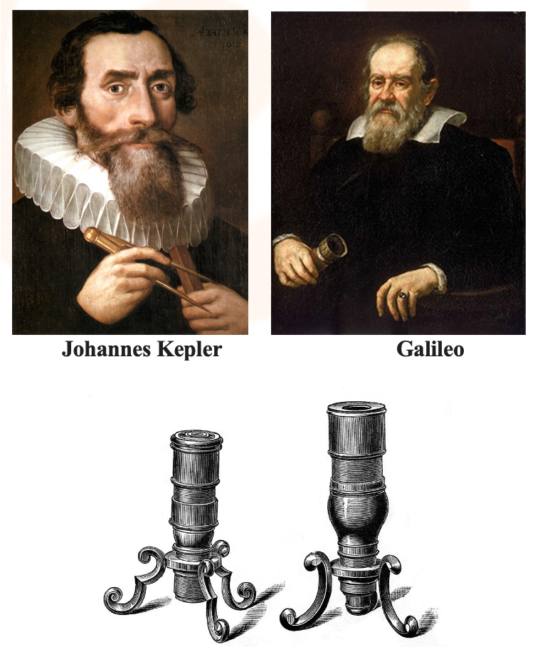
Johannes Kepler, an astronomer, suggested in 1611 that using convex lenses within the eyepiece and a convex lens in the objective would help improve the image. However, Kepler never attempted to make this microscope. Galileo constructed the first microscope on the suggestion of Keplar and his friend Faber is coined the name “Microscope" in 1624. Galileo Microscope had a power of 30X.
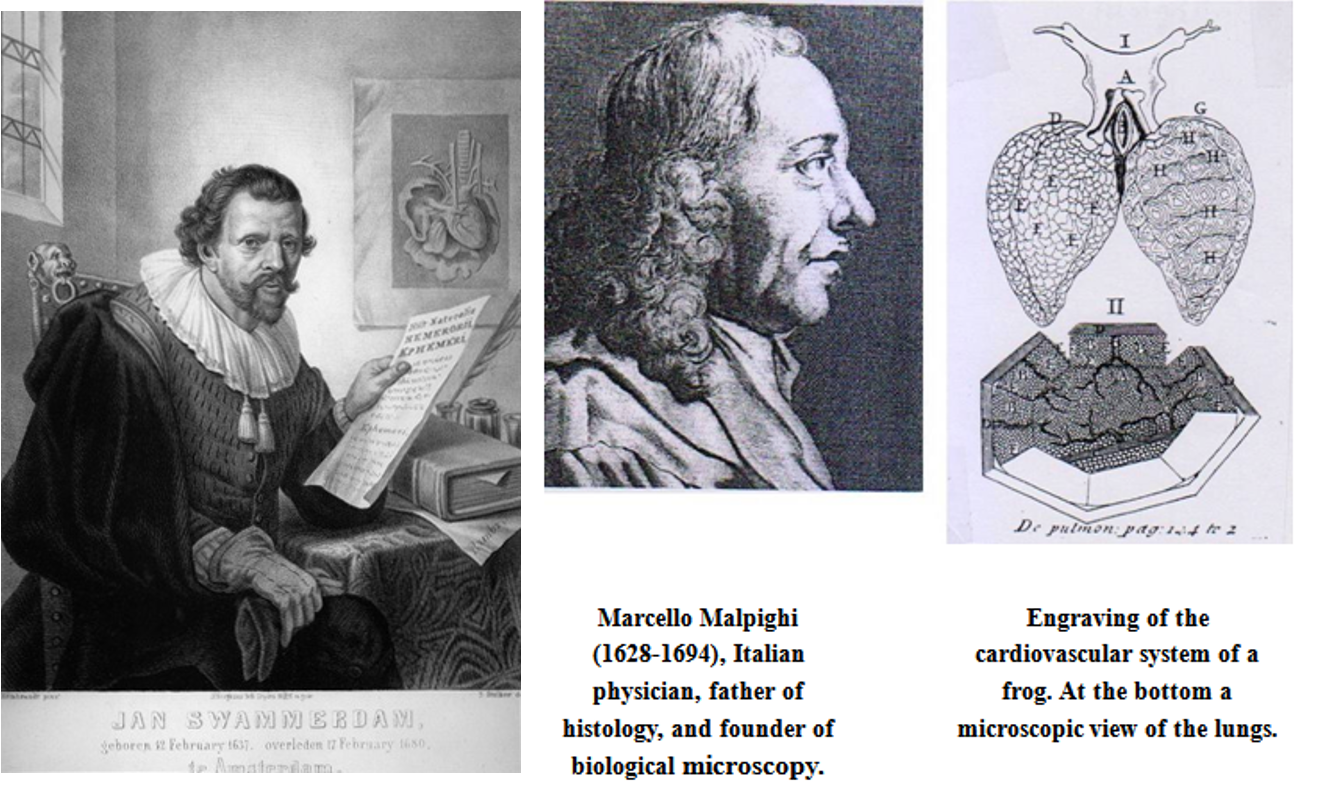
Jan Swammerdam (February 12, 1637 – February 17, 1680) was a Dutch biologist and microscopist. In 1658, he was the first to observe and describe red blood cells. He was one of the first people to use the microscope in dissections, and his techniques remained useful for hundreds of years. Marcello Malpighi, Italian physician and biologist who, in developing experimental methods to study living things, founded the science of microscopic anatomy. In 1660 he started studies on tissues by making slides.
The next major development came with the publication of ‘Micrographia’ in 1665 by the English natural philosopher Robert Hooke (1635-1703). This first illustrated microscopy book contained detailed engravings including the eye of a fly and a complete flea. In this book he also presented structure of Cork Cambium and proposed that it is made up of small chambers called Cell. Hooke described these structures as resembling “Honey-comb,” and as “small Boxes or Bladders of Air,” noting that each “Cavern, Bubble, or Cell” is distinct from the others (in Latin, “cell” literally means “small room”). They likely appeared to Hooke to be filled with air because the cork cells were dead, with only the rigid cell walls providing the structure.

Antonie van Leeuwenhoek, sometimes hailed as “the Father of Microbiology,” and Microscopy is typically credited as the first person to have created microscopes powerful enough to view microbes. Born in the city of Delft in the Dutch Republic, van Leeuwenhoek began his career selling fabrics. However, he later became interested in lens making (perhaps to look at threads) and his innovative techniques produced microscopes that allowed him to observe microorganisms as no one had before. In 1674, he described his observations of single-celled organisms such as Bacteria, Protozoa, Sperms and many more, whose existence was previously unknown, in a series of letters to the Royal Society of London.

- Alfonso Corti (1772) - Observed that all cells contain a living substance.
- The nucleolus was first discovered in eel cells in 1774 by Felice Fontana.
- In 1809 Lamarck in his book Philosphic Zoologiue laid the foundation of cell theory when he proposed that “No body has life without cell and their products”.
- In 1831 Robert Brown discovered Nucleus in orchid root cells.
- Felix Dujardin (1836) - Observed living substance in amoeba and called it ‘Sarcode’.
- In 1838 Theodore Schwann in animals and M.J. Scheilden in plants independently proposed that animals and plants are made up of cells and their products.
- In 1839 Theodore Schwann and M.J. Scheilden proposed cell theory jointly.
- Johannes Purkinje (1839) - Called the jelly-like living substance as protoplasm.
- In 1840 Huxley proposed that Protoplasm is the physical basis of life.
INTRODUCTION
- The rise of cytology is connected to rise of microscopy because for visualization of cell microscope is required therefore cytology started with the discovery of microscope. A Dutch father-son team named Hans and Zacharias Janssen invented the first so-called compound microscope in the late 16th century when they discovered that, if they put a lens at the top and bottom of a tube and looked through it, objects on the other end became magnified.

Johannes Kepler, an astronomer, suggested in 1611 that using convex lenses within the eyepiece and a convex lens in the objective would help improve the image. However, Kepler never attempted to make this microscope. Galileo constructed the first microscope on the suggestion of Keplar and his friend Faber is coined the name “Microscope" in 1624. Galileo Microscope had a power of 30X.

Jan Swammerdam (February 12, 1637 – February 17, 1680) was a Dutch biologist and microscopist. In 1658, he was the first to observe and describe red blood cells. He was one of the first people to use the microscope in dissections, and his techniques remained useful for hundreds of years. Marcello Malpighi, Italian physician and biologist who, in developing experimental methods to study living things, founded the science of microscopic anatomy. In 1660 he started studies on tissues by making slides.

The next major development came with the publication of ‘Micrographia’ in 1665 by the English natural philosopher Robert Hooke (1635-1703). This first illustrated microscopy book contained detailed engravings including the eye of a fly and a complete flea. In this book he also presented structure of Cork Cambium and proposed that it is made up of small chambers called Cell. Hooke described these structures as resembling “Honey-comb,” and as “small Boxes or Bladders of Air,” noting that each “Cavern, Bubble, or Cell” is distinct from the others (in Latin, “cell” literally means “small room”). They likely appeared to Hooke to be filled with air because the cork cells were dead, with only the rigid cell walls providing the structure.

Antonie van Leeuwenhoek, sometimes hailed as “the Father of Microbiology,” and Microscopy is typically credited as the first person to have created microscopes powerful enough to view microbes. Born in the city of Delft in the Dutch Republic, van Leeuwenhoek began his career selling fabrics. However, he later became interested in lens making (perhaps to look at threads) and his innovative techniques produced microscopes that allowed him to observe microorganisms as no one had before. In 1674, he described his observations of single-celled organisms such as Bacteria, Protozoa, Sperms and many more, whose existence was previously unknown, in a series of letters to the Royal Society of London.

- Alfonso Corti (1772) - Observed that all cells contain a living substance.
- The nucleolus was first discovered in eel cells in 1774 by Felice Fontana.
- In 1809 Lamarck in his book Philosphic Zoologiue laid the foundation of cell theory when he proposed that “No body has life without cell and their products”.
- In 1831 Robert Brown discovered Nucleus in orchid root cells.
- Felix Dujardin (1836) - Observed living substance in amoeba and called it ‘Sarcode’.
- In 1838 Theodore Schwann in animals and M.J. Scheilden in plants independently proposed that animals and plants are made up of cells and their products.
- In 1839 Theodore Schwann and M.J. Scheilden proposed cell theory jointly.
- Johannes Purkinje (1839) - Called the jelly-like living substance as protoplasm.
- In 1840 Huxley proposed that Protoplasm is the physical basis of life.
THE CELL THEORY
- The cell theory was jointly put forward by Schleiden and Schwann (1839) in their paper “Microscopic investigations on the similarity of structure and growth in animals and plants : As follows,
- All organisms are made of cell and their products.
- All cells are fundamentally similar and made up of similar kind of chemicals.
- All cells are structurally similar and similar kind of reactions occur in them.
- All cells arise from pre existing cells by division.
In 1855, a German medical doctor named Rudolph Virchow observed, under the microscope, cells dividing. He reasoned that all cells come from other pre-existing cells by cell division or Omnis cellule ‘e’ cellule.
Haeckel (1866) established that nucleus stores and transmits hereditary material. Cell theory was modified accordingly and was renamed as cell doctrine or cell principle.

SIZE OF CELL
- Generally cell size ranges between 0.2 µm-20 µm Smallest cell: PPLO (Pleuropneumonia Like Organism) : Mycoplasma gallisepticum: 0.1 µm
- Bacteria: Unicellular : About 10 times smaller than Animal cell
- Escherichia coli : Rod shaped Bacteria : 1-2 micron long
- Protozoa: Amoeba proteus : 220–760 µm
- Generally plant cells are larger than animal cell
- Generally Animal cell range from 10 µm to 30 µm & plant cell 10 µm
- Exceptions: Ostrich egg cell: 15.2 cm in diameter.
- Acetabularia a unicellular green alga is about 10 cms in length
- Nerve cells are the longest cell. The longest of nerve cells in any species are be found in the Colossal squid, with some nerve cells estimated to be over 10 meters in length.
Cell size

CELL SHAPE
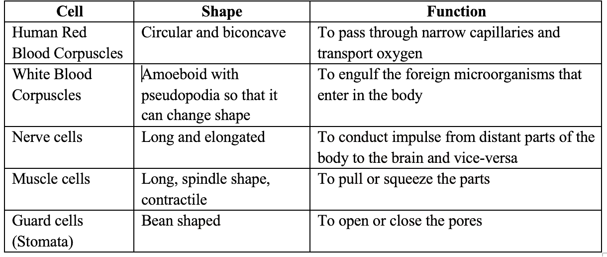
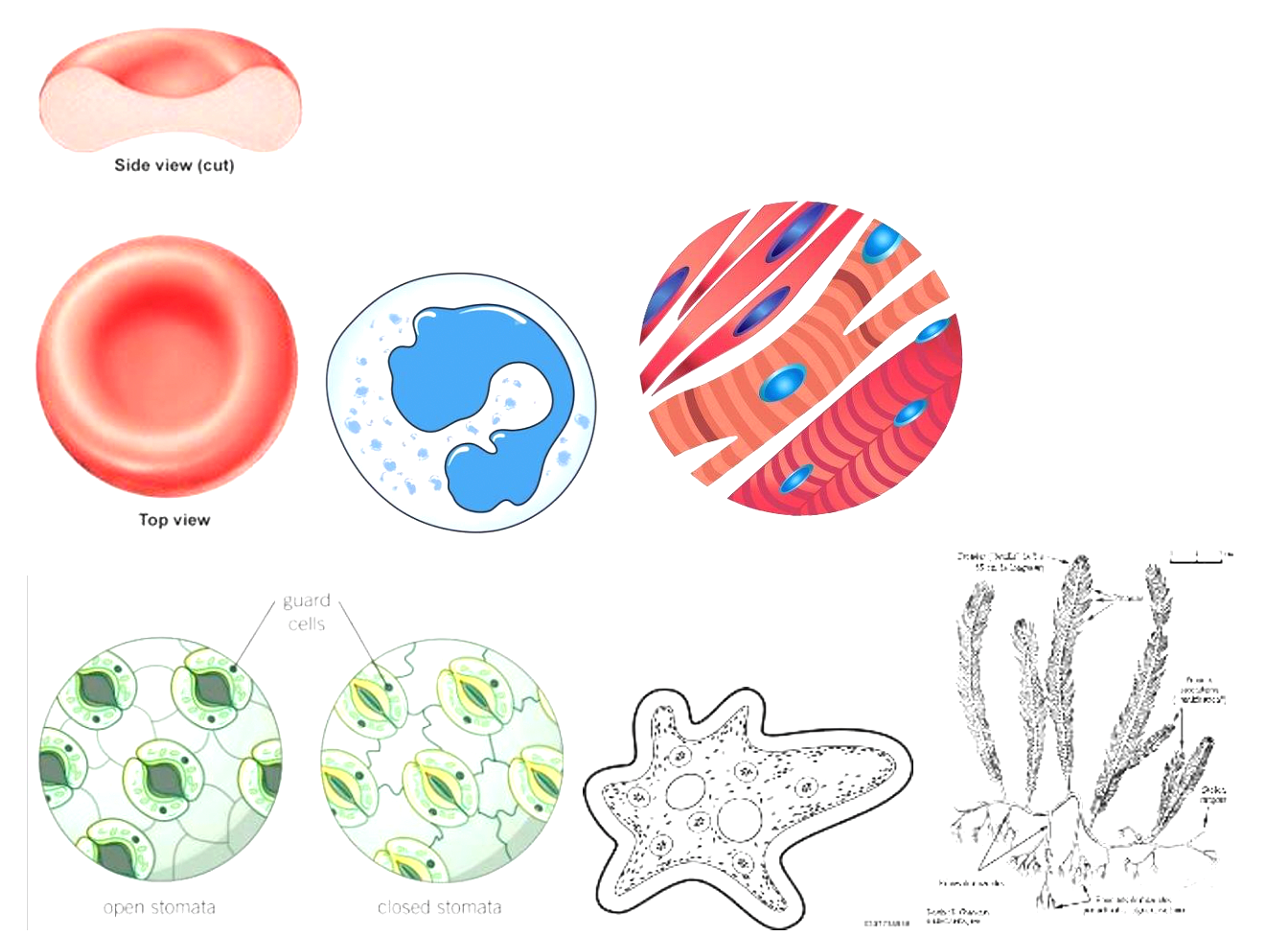
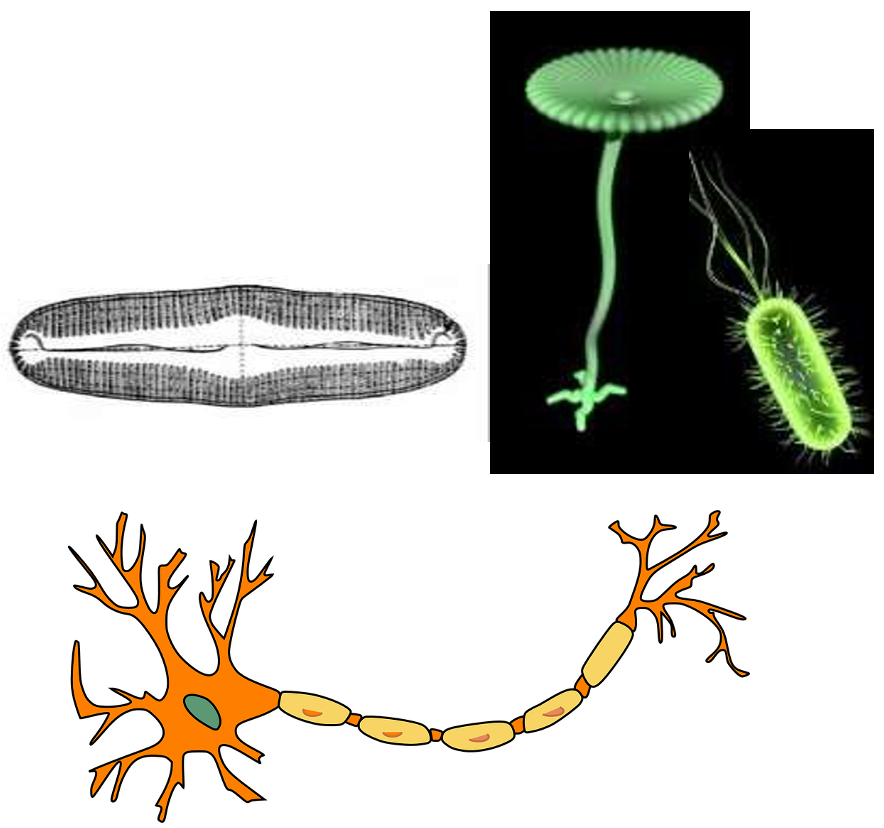
- There is great variability in cell shape i.e. spherical, polygonal, disc like, cuboidal, columnar, spindle like or fiber like. Generally cells are spherical but in multicellular forms due to pressure become polyhedral. In plants as cells have cell wall appear hexagonal or polygonal . Some cells variable shape or they keep on changing shape such as Amoeba and leucocytes. Generally shape of animals cells are various depending upon their function while plants cells are rectangular or cube shaped.
Nucleo-cytoplasmic Ratio or Kernplasm ratio:
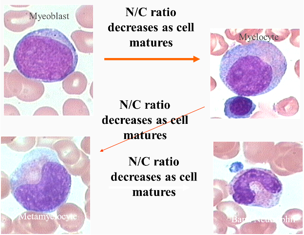
- The ratio in between Nucleus and Cytoplasm is called Nucleocytoplasmic ratio or Kernplasmic ratio or N/C ratio. It is very important property of a cell and tells about physiological nature of the cell, If N/C ratio is equal to 1 or N/C = 1 then cell is normal
- If N/C ratio is greater than 1 or N/C > 1 then cell is metabolically more active or young cell and will divide.
- If N/C ratio is lesser than 1 or N/C < 1 then cell is metabolically less active or dying.
Surface area to volume ratio or SA/V ratio:
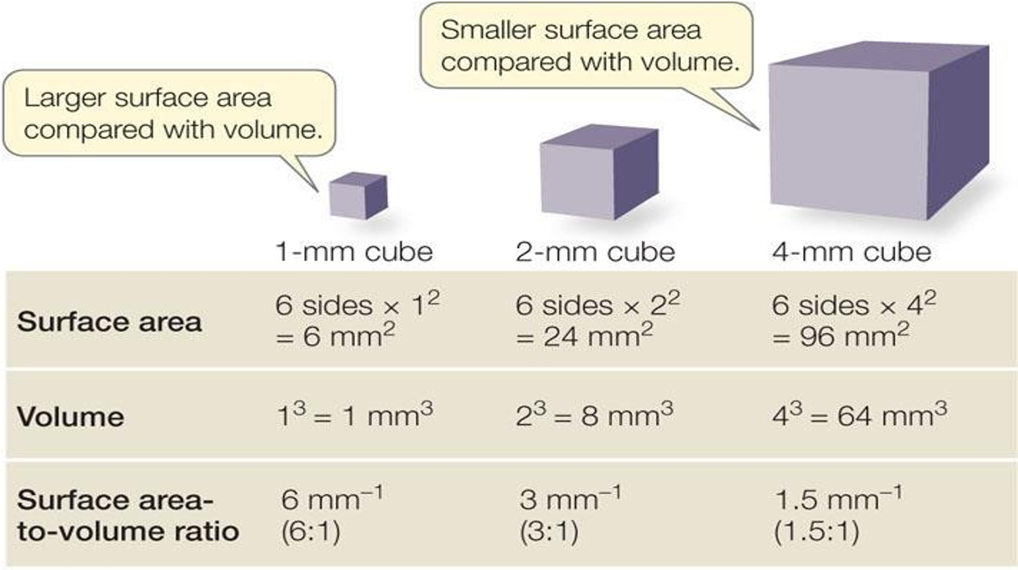
- Surface area to volume ratio is an important property of a cell and actually tell us how cell interacts with the environment. Surface area to volume ration could have three values,
- Sa/V ratio is greater than 1 or Sa/V > 1, It is usually present in smaller cells, which have larger surface area.
- Sa/V ratio is lesser than 1 or Sa/V < 1, It is usually present in larger cells which have smaller surface area.
Sa/V ratio is equal to 1 or Sa/V = 1, It is usually present in normal size cells.
Factors which determine the size of cell:
- The factors which set the limit of cell size or volume are,
⁕
- Nucleocytoplasmic ratio which determines the range of control of metabolic activities by nucleus.
- ⁕Rate of metabolic activity .
- ⁕Ratio of surface area to the volume of the cell.
Metabolically active cells are usually smaller due to higher N/C ratio and higher surface volume ratio. The bigger sized nucleus will allow the nucleus to have better control of metabolic activities. The high surface area volume ratio will allow quicker exchange of materials between the cell and its outside environment. Surface volume ratio decreases with the increase in cell size or volume. Therefore larger cells tend to become less efficient. In order to overcome this deficiency, larger cells are either cylindrical in shape or possess several extensions of the cell membrane such as microvilli, axons, dendrons, pseudopodia, etc. are such extensions. They are found in all those cells which are active in absorption. Membrane infoldings also occur in transfer cells found in plants in the region of absorption or secretion of nutrients.
Cellular totipotency
- Cellular totipotency refers that vegetative cells of a plant body have got all the potentialities to grow into a new plant. Cultured plant cells, are used to obtain new information on the mechanism of gene function during cell reproduction, cell growth and differentiation and when the cells start to synthesize secondary substances. Isolated protoplasts and cultured cells are regarded as the basis of genetic engineering in plants.
History
- Gottlieb Haberlandt (1902), a German botanist, was first to grow on culture, cells isolated from plant. Although he was unsuccessful in his attempts to culture cells of leaves, he gave the concept of totipotency.
- Robbins and Kotte in (1922) were partially successful in growing isolated cells.
- White (1932) raised the ovule culture for a long period by changing the medium regularly with vitamin B. Gautheret, Nobecourt and White (1939) got success in raising the callus cultures of differentiated cells excised from mesophyll, pith, endosperm and secondary phloem.
Concept of totipotency
- Skoog & Miller - differentiation of callus into tissues.
- Steward's experiment : Steward et al. showed the phenomenon of cellular totipotency in carrot cultures. Small fragments (phloem) of mature carrot roots were placed in liquid medium, in special containers and growth factors like 'coconut milk' was added. F. C. Steward developed complete carrot plant.
- Steward (1963), Halperin and Wetharell (1964) put callus on high auxin medium and found out they develop into embryo like structures which undergoes somatic embryogenesis. These embryos were called as Embryoids or Artificial seeds.
- Vasiland Hildebrandt developed a special technique in 1965 to raise whole plants of tobacco from a single cell. Guha and Maheshwari (1965) cultured the anthers of Datura and obtained androgenic haploids called as Androids. Cellular totipotency was Confirmed by using different plant tissues obtained from stem, root, leaf, bud, floral bud, anther, endosperm, embryo, etc. This method has been used in rapid multiplication of desired characters in plants, to shorten the breeding cycle, to produce haploids: for genetic improvement of plants, for fusion of cell by Somatic hybridization. Cellular totipotency is not fully determined in higher animals because their fully differentiated cells do not undergo all the tissue and organ formation. Only the early blastomeres like the zygote are totipotent and have the potential to develop into complete embryos and form all the tissues of an adult organism. The cells of an early embryo lose the potential for the expression of all the genes contained in a fertilized egg. The more the specialized is the cell, the fewer the genes utilized.
- Experiments of Robert Briggs and Thomas Kind indicate that when the nuclei of fertilized eggs of frog were removed and substituted by the nuclei of early blastomeres of frog they developed into full fledged adult frogs. But when the nuclei of fertilized eggs of frog were substituted by the nuclei of cells of intestines of other tissue cells whose fate was fully determined did not develop into adult frogs and hence were not totipotent. However in 1996 Keith Campbell, Ian Wilmut and colleagues at the Roslin Institute with the help of Nuclear Transfer technique cloned Dolly sheep from udder cells and proved that totipotency is present in all eukaryotic cells but it is limited in animal cells.
The first evidence of cellular totipotency was given by F.C. Steward et al. in 1950 at Cornwell University of USA.
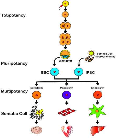
- In animals totipotent cells undergo a process of specialization called Differentiation. In general, the cells in an organism can be grouped under three major categories on the basis of the levels of differentiation:
- Undifferentiated cells: These cells are capable of undergoing division and development, for example, the stem cells (animals) and meristematic cells (plants).
- Differentiated cells: These are post mitotic cells which have undergone specialization or/and exhibit the division of labour. Therefore, these cells acquire a distinct character and perform a definite function. For example, RBCs carry out transportation of oxygen and carbon dioxide, the muscle cells perform kinetic functions or movement, and mesophyll cells carry out photosynthesis.
- Dedifferentiated cells: Some differentiated cells are capable of reverting back to the undifferentiated meristematic state, when required. These cells are important for wound healing, regeneration, and secondary growth. The process by which they lose their specialization is referred to as dedifferentiation.
Regulation of cell function
- The function of the cell are regulated by the flow of information. The flow of information in a cell or in a living system takes two routes
- Flow of intrinsic or genetic information
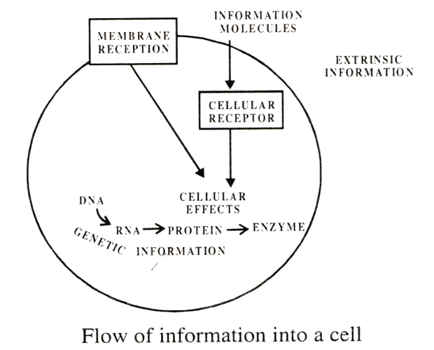
DNA molecules contain genetic information and control intrinsic activities like metabolism and expression of characters by synthesizing protein molecules. DNA transcribes the stored information into mRNA molecules which translate the genetic information into specific proteins. The proteins then perform and regulate all the activities of the cell. The flow of intrinsic information from DNA to mRNA & then to polypeptide is called central dogma of molecular biology or cell biology.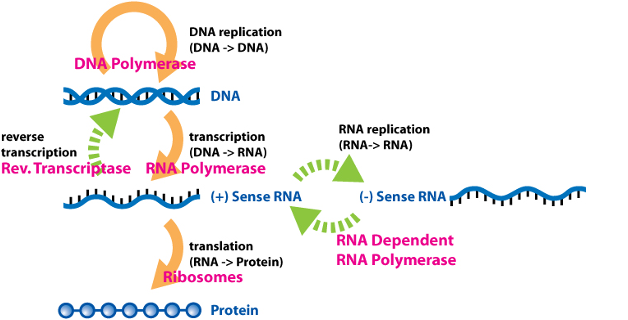
- Extrinsic information
It is brought to the cells in the form of specific stimuli and information molecules. Stimuli are commonly brought by nerves. The nerves secrete specific chemicals that stimulate endocrine glands to release hormones
(i)Flow of intrinsic or genetic information and
(ii)Flow of extrinsic information.
- Hormones are of two types :
- Some of them are able to pass into the cells. Here, they combine with specific intracellular receptors, which carry the hormones to the nucleus. The hormone functions as an inducer. The activated genes transcribe specific mRNAs for synthesis of particular proteins and enzymes inside the cytoplasm
- Other category of hormones combine with membrane receptors. The hormone-receptor complex can function in two ways. It may change the membrane permeability to facilitate passage of particular types of materials. The second method is through the formation of second chemical messenger known as cAMP. cAMP activates an enzyme system already present in the cell.
Flow of information
THE CELL THEORY
- The cell theory was jointly put forward by Schleiden and Schwann (1839) in their paper “Microscopic investigations on the similarity of structure and growth in animals and plants : As follows,
- All organisms are made of cell and their products.
- All cells are fundamentally similar and made up of similar kind of chemicals.
- All cells are structurally similar and similar kind of reactions occur in them.
- All cells arise from pre existing cells by division.
In 1855, a German medical doctor named Rudolph Virchow observed, under the microscope, cells dividing. He reasoned that all cells come from other pre-existing cells by cell division or Omnis cellule ‘e’ cellule.
Haeckel (1866) established that nucleus stores and transmits hereditary material. Cell theory was modified accordingly and was renamed as cell doctrine or cell principle.

SIZE OF CELL
- Generally cell size ranges between 0.2 µm-20 µm Smallest cell: PPLO (Pleuropneumonia Like Organism) : Mycoplasma gallisepticum: 0.1 µm
- Bacteria: Unicellular : About 10 times smaller than Animal cell
- Escherichia coli : Rod shaped Bacteria : 1-2 micron long
- Protozoa: Amoeba proteus : 220–760 µm
- Generally plant cells are larger than animal cell
- Generally Animal cell range from 10 µm to 30 µm & plant cell 10 µm
- Exceptions: Ostrich egg cell: 15.2 cm in diameter.
- Acetabularia a unicellular green alga is about 10 cms in length
- Nerve cells are the longest cell. The longest of nerve cells in any species are be found in the Colossal squid, with some nerve cells estimated to be over 10 meters in length.
Cell size

CELL SHAPE
- There is great variability in cell shape i.e. spherical, polygonal, disc like, cuboidal, columnar, spindle like or fiber like. Generally cells are spherical but in multicellular forms due to pressure become polyhedral. In plants as cells have cell wall appear hexagonal or polygonal . Some cells variable shape or they keep on changing shape such as Amoeba and leucocytes. Generally shape of animals cells are various depending upon their function while plants cells are rectangular or cube shaped.


Nucleo-cytoplasmic Ratio or Kernplasm ratio:
- The ratio in between Nucleus and Cytoplasm is called Nucleocytoplasmic ratio or Kernplasmic ratio or N/C ratio. It is very important property of a cell and tells about physiological nature of the cell, If N/C ratio is equal to 1 or N/C = 1 then cell is normal
- If N/C ratio is greater than 1 or N/C > 1 then cell is metabolically more active or young cell and will divide.
- If N/C ratio is lesser than 1 or N/C < 1 then cell is metabolically less active or dying.

Surface area to volume ratio or SA/V ratio:
- Surface area to volume ratio is an important property of a cell and actually tell us how cell interacts with the environment. Surface area to volume ration could have three values,
- Sa/V ratio is greater than 1 or Sa/V > 1, It is usually present in smaller cells, which have larger surface area.
- Sa/V ratio is lesser than 1 or Sa/V < 1, It is usually present in larger cells which have smaller surface area.
Sa/V ratio is equal to 1 or Sa/V = 1, It is usually present in normal size cells.
Factors which determine the size of cell:
- The factors which set the limit of cell size or volume are,
⁕
- Nucleocytoplasmic ratio which determines the range of control of metabolic activities by nucleus.
- ⁕Rate of metabolic activity .
- ⁕Ratio of surface area to the volume of the cell.
Metabolically active cells are usually smaller due to higher N/C ratio and higher surface volume ratio. The bigger sized nucleus will allow the nucleus to have better control of metabolic activities. The high surface area volume ratio will allow quicker exchange of materials between the cell and its outside environment. Surface volume ratio decreases with the increase in cell size or volume. Therefore larger cells tend to become less efficient. In order to overcome this deficiency, larger cells are either cylindrical in shape or possess several extensions of the cell membrane such as microvilli, axons, dendrons, pseudopodia, etc. are such extensions. They are found in all those cells which are active in absorption. Membrane infoldings also occur in transfer cells found in plants in the region of absorption or secretion of nutrients.

Cellular totipotency
- Cellular totipotency refers that vegetative cells of a plant body have got all the potentialities to grow into a new plant. Cultured plant cells, are used to obtain new information on the mechanism of gene function during cell reproduction, cell growth and differentiation and when the cells start to synthesize secondary substances. Isolated protoplasts and cultured cells are regarded as the basis of genetic engineering in plants.
History
- Gottlieb Haberlandt (1902), a German botanist, was first to grow on culture, cells isolated from plant. Although he was unsuccessful in his attempts to culture cells of leaves, he gave the concept of totipotency.
- Robbins and Kotte in (1922) were partially successful in growing isolated cells.
- White (1932) raised the ovule culture for a long period by changing the medium regularly with vitamin B. Gautheret, Nobecourt and White (1939) got success in raising the callus cultures of differentiated cells excised from mesophyll, pith, endosperm and secondary phloem.
Concept of totipotency
- Skoog & Miller - differentiation of callus into tissues.
- Steward's experiment : Steward et al. showed the phenomenon of cellular totipotency in carrot cultures. Small fragments (phloem) of mature carrot roots were placed in liquid medium, in special containers and growth factors like 'coconut milk' was added. F. C. Steward developed complete carrot plant.
- Steward (1963), Halperin and Wetharell (1964) put callus on high auxin medium and found out they develop into embryo like structures which undergoes somatic embryogenesis. These embryos were called as Embryoids or Artificial seeds.
- Vasiland Hildebrandt developed a special technique in 1965 to raise whole plants of tobacco from a single cell. Guha and Maheshwari (1965) cultured the anthers of Datura and obtained androgenic haploids called as Androids. Cellular totipotency was Confirmed by using different plant tissues obtained from stem, root, leaf, bud, floral bud, anther, endosperm, embryo, etc. This method has been used in rapid multiplication of desired characters in plants, to shorten the breeding cycle, to produce haploids: for genetic improvement of plants, for fusion of cell by Somatic hybridization. Cellular totipotency is not fully determined in higher animals because their fully differentiated cells do not undergo all the tissue and organ formation. Only the early blastomeres like the zygote are totipotent and have the potential to develop into complete embryos and form all the tissues of an adult organism. The cells of an early embryo lose the potential for the expression of all the genes contained in a fertilized egg. The more the specialized is the cell, the fewer the genes utilized.
- Experiments of Robert Briggs and Thomas Kind indicate that when the nuclei of fertilized eggs of frog were removed and substituted by the nuclei of early blastomeres of frog they developed into full fledged adult frogs. But when the nuclei of fertilized eggs of frog were substituted by the nuclei of cells of intestines of other tissue cells whose fate was fully determined did not develop into adult frogs and hence were not totipotent. However in 1996 Keith Campbell, Ian Wilmut and colleagues at the Roslin Institute with the help of Nuclear Transfer technique cloned Dolly sheep from udder cells and proved that totipotency is present in all eukaryotic cells but it is limited in animal cells.
The first evidence of cellular totipotency was given by F.C. Steward et al. in 1950 at Cornwell University of USA.

- In animals totipotent cells undergo a process of specialization called Differentiation. In general, the cells in an organism can be grouped under three major categories on the basis of the levels of differentiation:
- Undifferentiated cells: These cells are capable of undergoing division and development, for example, the stem cells (animals) and meristematic cells (plants).
- Differentiated cells: These are post mitotic cells which have undergone specialization or/and exhibit the division of labour. Therefore, these cells acquire a distinct character and perform a definite function. For example, RBCs carry out transportation of oxygen and carbon dioxide, the muscle cells perform kinetic functions or movement, and mesophyll cells carry out photosynthesis.
- Dedifferentiated cells: Some differentiated cells are capable of reverting back to the undifferentiated meristematic state, when required. These cells are important for wound healing, regeneration, and secondary growth. The process by which they lose their specialization is referred to as dedifferentiation.
Regulation of cell function
- The function of the cell are regulated by the flow of information. The flow of information in a cell or in a living system takes two routes
- Flow of intrinsic or genetic information
DNA molecules contain genetic information and control intrinsic activities like metabolism and expression of characters by synthesizing protein molecules. DNA transcribes the stored information into mRNA molecules which translate the genetic information into specific proteins. The proteins then perform and regulate all the activities of the cell. The flow of intrinsic information from DNA to mRNA & then to polypeptide is called central dogma of molecular biology or cell biology.
- Extrinsic information
It is brought to the cells in the form of specific stimuli and information molecules. Stimuli are commonly brought by nerves. The nerves secrete specific chemicals that stimulate endocrine glands to release hormones
(i)Flow of intrinsic or genetic information and
(ii)Flow of extrinsic information.
- Hormones are of two types :
- Some of them are able to pass into the cells. Here, they combine with specific intracellular receptors, which carry the hormones to the nucleus. The hormone functions as an inducer. The activated genes transcribe specific mRNAs for synthesis of particular proteins and enzymes inside the cytoplasm
- Other category of hormones combine with membrane receptors. The hormone-receptor complex can function in two ways. It may change the membrane permeability to facilitate passage of particular types of materials. The second method is through the formation of second chemical messenger known as cAMP. cAMP activates an enzyme system already present in the cell.
Flow of information

Differences in prokaryotic & eukaryotic cells
- Cytoplasm lacks organelles (endoplasmic reticulum, mitochondria, Golgi apparatus, centrosome, microfilaments, microtubules, intermediate fibres, microbodies), except ribosomes. Cytoplasm contains organelles, viz., endoplasmic reticulum, mitochondria, Golgi apparatus, lysosomes, centrosome, microfilaments, intermediate fibres, microtubules, microbodies, besides ribosomes.
- A prokaryotic cell is a single membrane system. Cell membrane bears respiratory enzymes. Photosynthetic lamellae i.e., (if present) thylakoids occur free in the cytoplasm.
A eukaryotic cell is a double membrane system. Cell membrane lacks respiratory enzymes. Photosynthetic lamellae if present, occur within the chloroplasts. - Ribosomes are 70S, lie free in cytoplasm or joined to cell membrane.
There are no streaming movements of cytoplasm. Sap vacuoles are lacking. Gas vacuoles may occur. Transcription and translation occur in the cytoplasm. Mitotic spindle is not formed in cell division. Mesosomes are formed by infolding of cell membrane. Protein synthesis takes place in cytoplasm only.
Ribosomes are 80S, may lie free or bound to E.R. and nuclear envelope. Cytoplasm shows streaming movements (cyclosis). Sap vacuoles are common. Transcription and translation occur in nucleus and cytoplasm respectively. Mitotic spindle is formed in cell division. Mesosomes are absent. Protein synthesis occurs in the cytoplasm, mitochondria and plastids. - Nuclear material is not enclosed by nuclear envelope and lies directly in cytoplasm. It is called Nucleoid or Nuclein or Genophore. There is no nucleolus. DNA is circular and without a protein coat.
Nuclear material is enclosed by nuclear envelope to form a nucleus distinct from cytoplasm. One or more nucleoli occur within the nucleus.
Nuclear DNA is linear with a protein coat, extra nuclear DNA is circular and naked. - DNA occurs in the cytoplasm only. Plasmids and pill occur in many prokaryotic cells. Flagella, if present, are single stranded and are formed of a protein flagellin. DNA occurs in the nucleus as well as in mitochondria and chloroplasts. There are no plasmids and pill in eukaryotic cells. Flagella, if present, are complex, have 9 + 2 pattern of microtubules formed of a protein tubulin.
Differences in Animals cells & plant cells
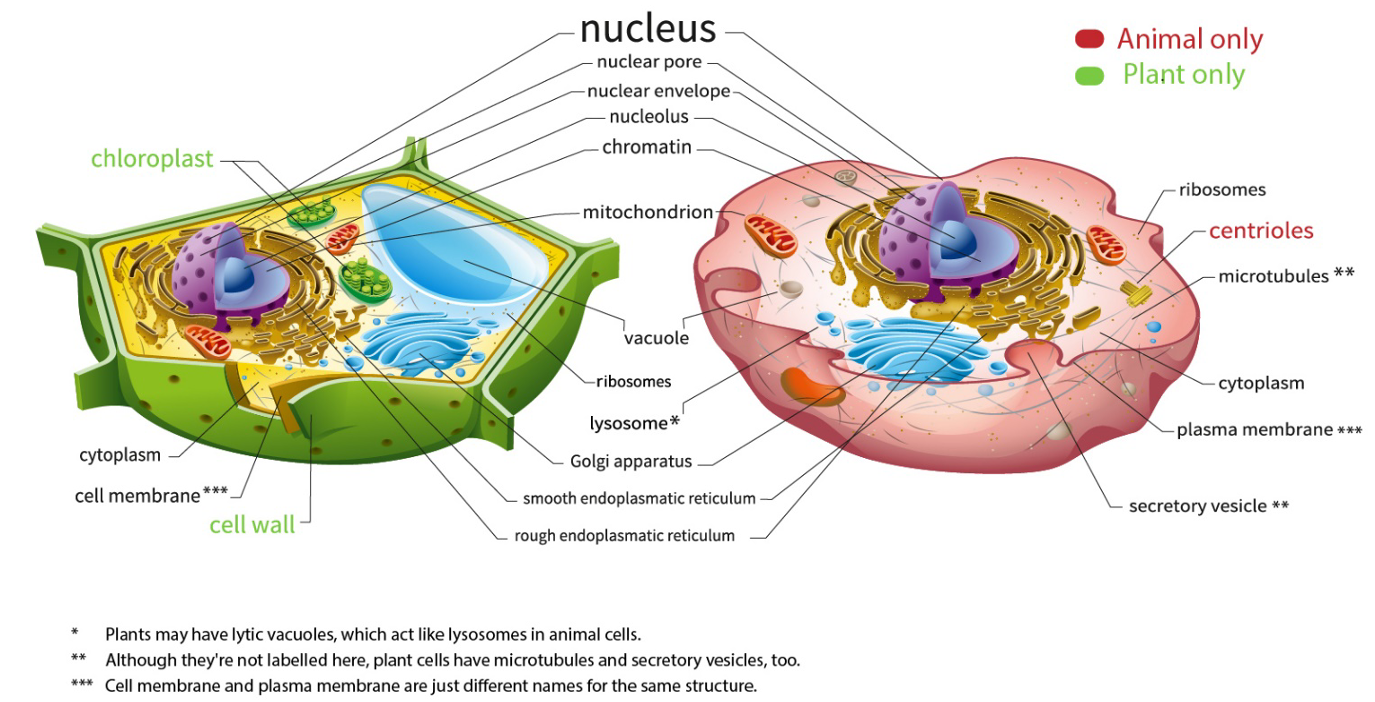
- Cell wall is absent in all animal cells. Cells are covered by a thin flexible living cell membrane. Cells are covered by thick rigid, cellulosic cell wall in addition to cell membrane.
- Animal cells have centrioles and centrosome in their cytoplasm. During cell division it forms amphiastral spindle. Small sized few food vacuoles and contractile vacuoles may be present in cytoplasm.
Centrioles and centrosome are absent in plant cells. During cell division, it forms ancestral spindle. Vacuoles are large in size and more in number. It has a large central sap vacuole. Food vacuoles and contractile vacuoles are absent. - In animals cell cytokinesis occurs by constriction or furrowing. Chloroplasts are absent and other plastids are generally absent.
In Plant cells cytokinesis occurs by cell plate method. Choloroplasts and other plastids are present in plant cells. - In animals cells Golgi complex is distinct, well developed, lies near the nucleus.
However in plant cells Golgi complex is indistinct in the form of unconnected units called Dictyosomes.
- Glyoxisomes are absent in animal cells. Many animal cells can change their shape and move about.
Glyoxisomes may be present in plant cells. Plant cells due to rigid cell wall cannot change their shape and cannot move about. - In animal cells, nucleus is generally centrally located in the cytoplasm. The lysosomes are present in animal cells.
In plant cells the large sap vacuole pushes the nucleus to peripheral position in cytoplasm. Lysosomes are generally absent in plant cells. Their activity is performed by pecialized vacuoles.

PROKARYOTIC CELL AND ITS ORGANISATION
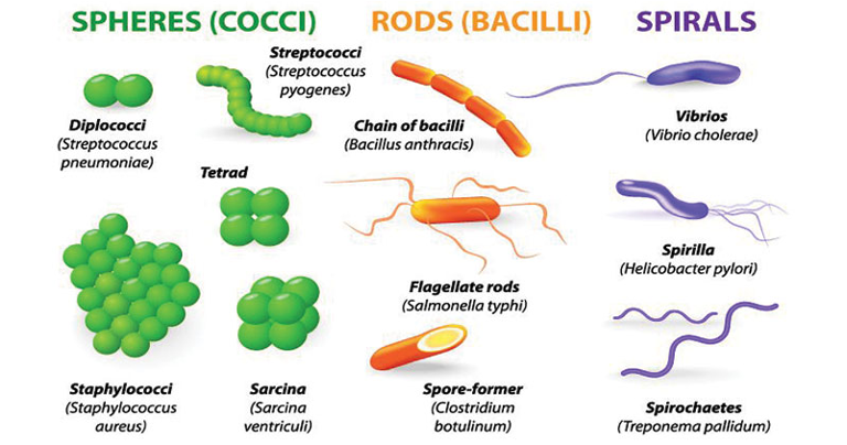
- The prokaryotic cells are represented by Bacteria, Blue-green algae, Mycoplasma or PPLO (pleuro pneumonia-like organism), Spirochete, and Rickettsiae. Generally, prokaryotic cells are supposed to be far smaller than the eukaryotic cells. The prokaryotic cells, however, multiply more rapidly as compared to most of the eukaryotic cells. The prokaryotic cells have remarkable amount of variation in shape and size. There are four basic shapes of bacteria,
- Bacillus: The most common shape is rod-like. The bacilli differ considerably in their shape of rod as it may be flat or round or cigar-shaped. The cells remain single but these may remain together after division to form pairs (Diplobacillus) or in groups of more than two (Streptobacillus) such as E.coli
- Coccus or Spherical: The spherical cells may be present singly (Monococcus) or in groups of two (Diplococcus) or in long chain (Streptococcus).
- Vibrios: A few rod-shaped bacteria may be slightly curved, or comma-shaped; V. cholera
- Spirilla: Many bacteria are shaped like along, twisted spiral; these may resemble acomma. Other bacteria may be highly coiled and resemble a cork screw (Spirochetes) such as Treponema pallidum.
Shapes of bacteria
- Prokaryotic cells vary in size. The smallest bacterial cells are about 100–200 nm in diameter and approach a size very close to that of large viruses. However, most eubacterial cells are 1.1–1.5 µm wide and 2.0–2.6 µm long. A few bacteria and blue-green algae become very long in length (upto 500 µm) e.g. Spirochetes and Oscillatoria. A huge bacterium, Epulopscium fishelsoni, which was discovered in the intestine of the brown surgeon fish, is as large as 600 µm and as wide as 80 µm. It is now well established that a few prokaryotic cells are much larger than the normal eukaryotic cell. Every prokaryotic cell is invariably bounded by a complex cell wall. Inside the cell wall is present Periplasmic space, Beneath this lies the plasma membrane. The plasma membrane can be invaginated to form internal membrane structures called Mesosomes. The prokaryotic cells do not have membrane-bound organelles. Hence, the interior appears very simple. The genetic material is localised within a discrete region, called nucleoid. The nucleoid is not separated from the surrounding cytoplasm by any delimiting membrane. The ribosomes and the inclusion bodies are randomly scattered in the cytoplasmic matrix.
Cell Envelope of Bacteria
- Most of the prokaryotic cells, particularly bacterial cells, have a chemically complex cell envelope. The layers of cell envelope stack upon one another and are often bonded together tightly. In electron
micrograph, three basic layers can be identified:
- The outermost glycocalyx,
- Followed by cell wall
- and cell membrane (plasma membrane).
Although each layer of the envelope performs a distinct function, together they act as a single protective unit.
Glycocalyx is the outermost layer comprising a coating of macromolecules, which protects cells and also helps in adhesion. This layer differs in thickness and chemical composition in different bacteria. Some have a loose sheath called Slime layer, which protects the cells from loss of water and nutrients. Others may have a thick and tough covering known as Capsule. The capsule and slime layer are made up of polysaccharides, but may sometimes contain proteins also. The capsule is responsible for giving gummy and sticky character to the cell. This layer is not absolutely essential for bacterial survival but can at times be highly specific and immunogenic.
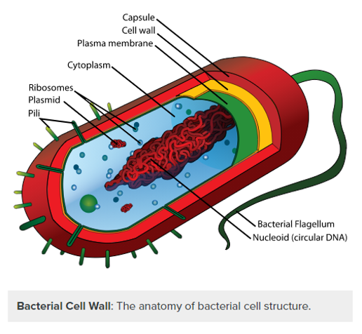
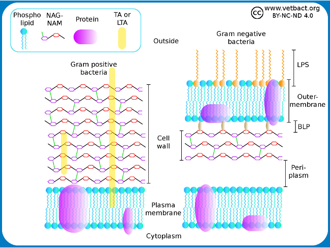
Cell wall is the second layer of the cell envelope below the glycocalyx. This layer determines the shape of the cell and provides a kind of strong structural support to prevent a bacterium from bursting or collapsing in a hypotonic solution. This layer is rigid due to a special macromolecule called Peptidoglycan (murein or mucopeptide). It is composed of repeating framework of long glycan strands (N-acetyl muramic acid and N-acetyl glucosamine), which are cross-linked by short peptide chains to provide a strong but flexible support framework. A number of antibiotics (e.g., penicillin and cephalosporins) inhibit cross linking of peptidoglycan strands. Therefore, cells undergo lysis in the presence of these antibiotics. Lysozyme, a naturally occurring enzyme in saliva and tears, also provides defense against certain bacteria by hydrolyzing the peptidoglycan.
Gram stain or Gram staining, also called Gram's method, is a method of staining used to distinguish and classify bacterial species into two large groups: gram-positive bacteria and gram-negative bacteria. The name comes from the Danish bacteriologist Hans Christian Gram, who developed the technique.
Gram staining
- Gram staining is a special technique, which is used to classify bacteria into two groups, viz. Gram-positive and Gram-negative bacteria. The bacteria are stained with weakly alkaline solution of crystal violet or gentian violet. The stained slide of bacteria is then treated with 0.5 per cent iodine solution. This is followed by washing with water and then with alcohol or acetone. There is difference between the cell envelopes of the two groups. In Gram-positive bacteria, the cell wall is thick and is primarily made up of peptidoglycan. In Gram-negative bacteria, the cell envelope shows three layers. The cell envelope is composed of outer membrane, and a thin layer of peptidoglycan followed by the plasma membrane. The Gram- positive cell wall is 20 – 80 nm in thickness and also contains tightly bound Teichoic acids. The outer face of the outer membrane of Gram-negative bacteria contains Lipopolysaccharides, a part of which is integrated into the membrane lipids. The inner face has a number of proteins which are anchored into peptidoglycan. The outer membrane of Gram-negative bacteria contains proteins called porins, and these proteins function as channels for the entry and exit of hydrophilic low molecular weight substances. In Mycobacterium and Noccardia, the wall is that of Gram-positive type but a part of their cell wall is made up of a very long chain of the fatty acid, called Mycoic acid.
Inclusion Bodies
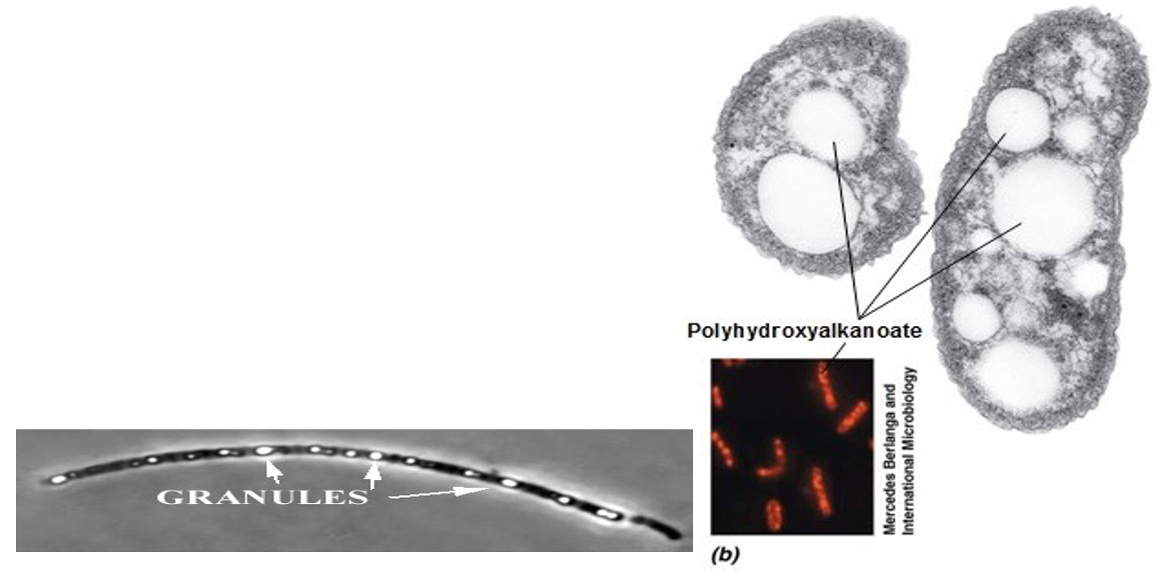
- The reserve materials of bacteria are stored in the cytoplasm as inclusion bodies or storage granules. These are not bounded by any membrane system and lie free in the cytoplasm, e.g., Phosphate granules, Cyanophycean granules and Glycogen granules. Some other inclusion bodies may be surrounded by a single layer non-unit membrane, which is 2–4 nm thick, e.g., Poly-β-hydroxybutyrate granules, Sulphur granules, and gas vacuoles.
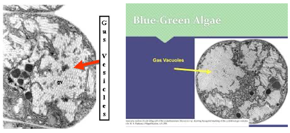
Other remarkable organic inclusion bodies are the gas vacuoles, which usually occur in cyanobacteria, purple and green photosynthetic bacteria and a few other aquatic forms that are free-floating (planktonic). These are basically aggregates of a number of small, hollow cylindrical gas vesicles. Gas vesicles are not permeable to water but are permeable to atmospheric gases. Because of gas vacuoles, these bacteria keep floating on or near the surface of water. These vacuoles help the organisms in positioning themselves in the water column for trapping sunlight for photosynthesis, or for avoiding intense sunlight.
The two major inorganic inclusions are the Polyphosphate granules or the Volutin granules and Sulphur granules. These granules take different colours with basic dyes, therefore, they are also termed as Metachromatic granules. The volutin granules are phosphate polymers and function as a storage reservoir for phosphate. Some bacteria also store sulphur temporarily as sulphur granules. These may be formed when bacteria use hydrogen sulphide as electron donor during photosynthesis. These granules may accumulate in either periplasmic space or in special cytoplasmic globules.

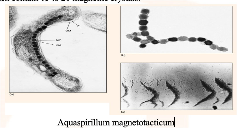
Some bacteria store Magnetosomes, these are membranous structures present in magnetotactic bacteria (MTB). They contain iron-rich magnetic particles that are enclosed within a lipid bilayer membrane. Each magnetosome can often contain 15 to 20 magnetite crystals.
Flagellum
- The electron microscopic studies have revealed that the bacterial flagellum is composed of three parts - Filament, Hook and Basal body. The filament is the longest and the most obvious portion of the flagellum. It extends from the cell surface to the top. It is a hollow rigid cylindrical structure made of the protein called Flagellin. The protein molecules are arranged in a spiral manner in the filament. The filament measures about 20 nm in diameter and 1-70 nm in length. It is inserted into a curved tubular hook which is anchored to the cell by basal body. The hook is quite different from the filament and is made up of different protein subunits.
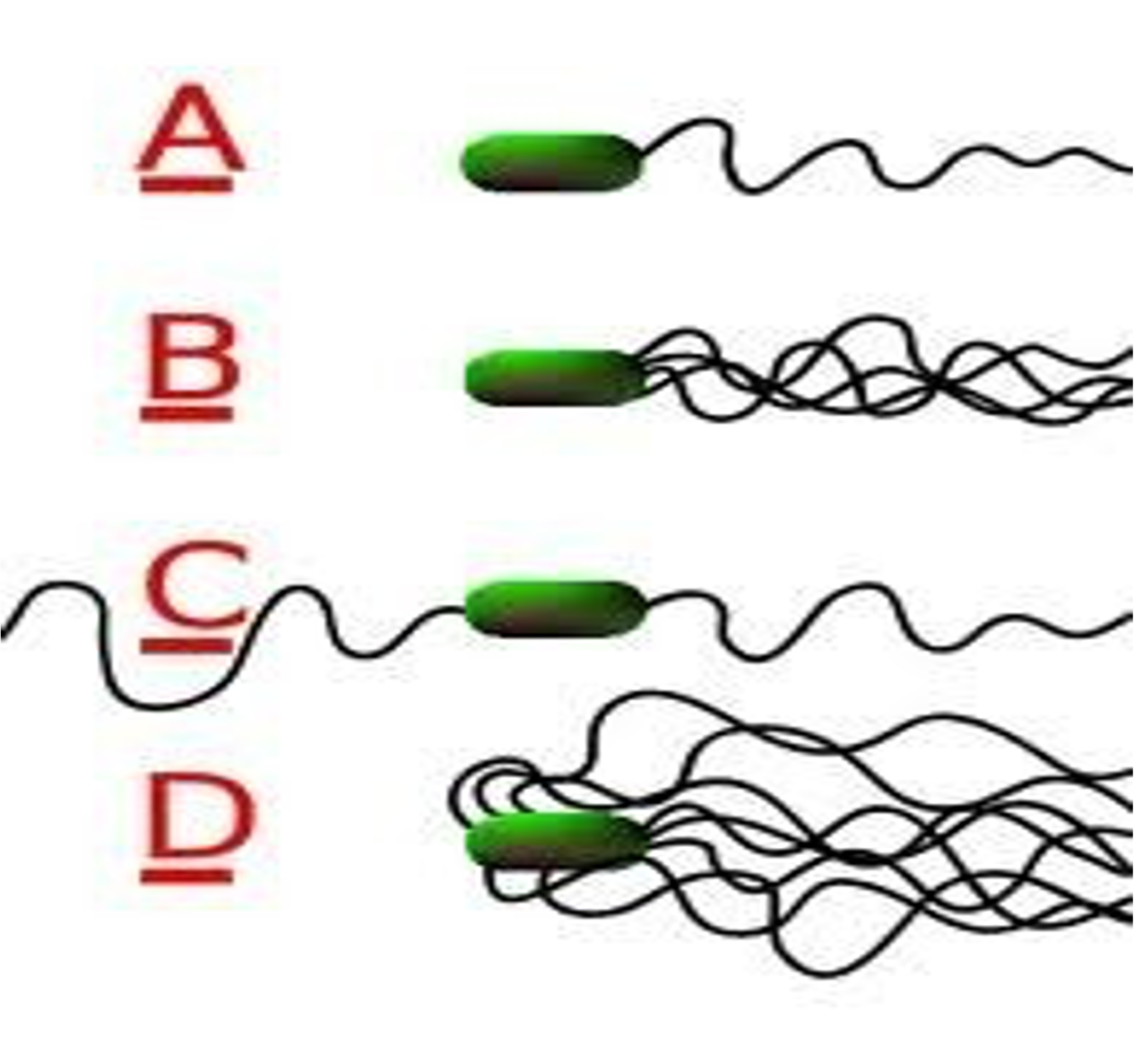
- Monotrichous- One flagellum
- Lophotrichous- A cluster of flagella at one or both ends.
- Amphitrichous- Single flagellum at each pole
- Peritrichous- Flagella are spread over the whole Cell.
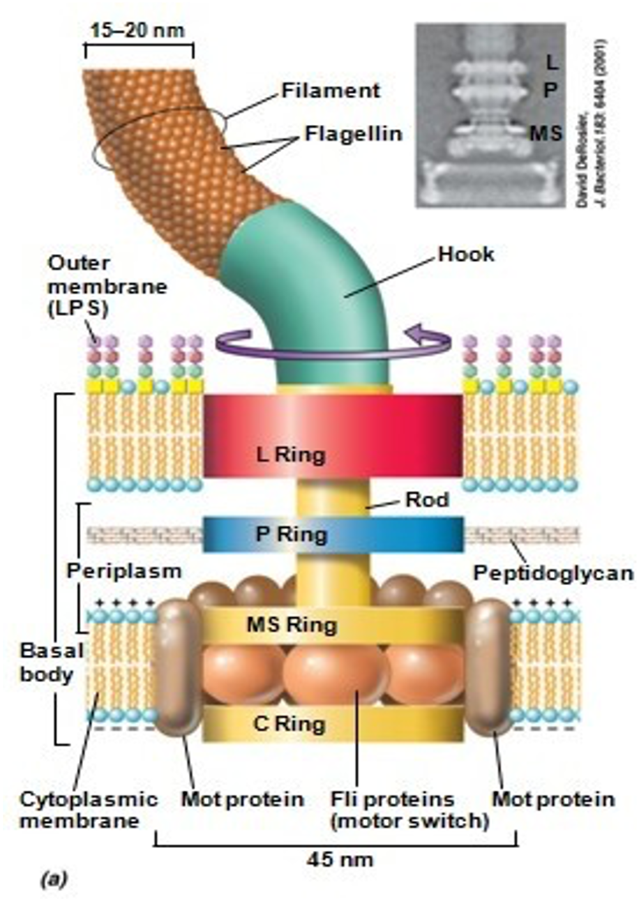
The basal body is the most complex part of the flagellum and consists of four rings connected to a central rod in Gram-negative bacteria. The Gram-positive cells have only two basal body rings. The inner ring is connected to plasma membrane, while the outer ring is connected to peptidoglycan membrane. The filament, hook and basal body are arranged in such a manner that the structure permits the filament to rotate by 360° rather than undulating back and forth like a whip. As the flagellum rotates, it spins the body in the opposite direction and pushes the bacterium in forward direction.
Depending on the arrangement of Flagella, they are of four types.
Pilus and Fimbriae
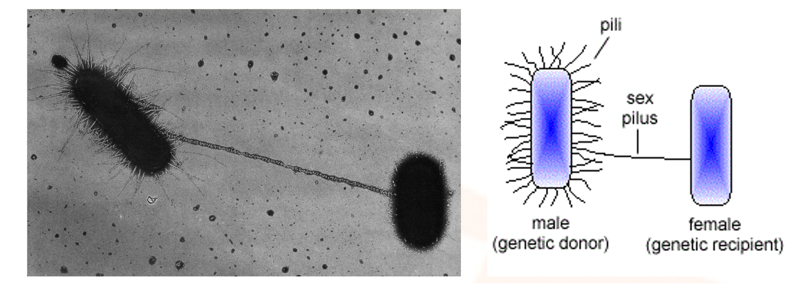
- Fimbriae and Pili are thin, protein tubes originating from the cytoplasmic membrane of many bacteria. Both are able to stick bacteria to surfaces, but pili are typically longer and fewer in number than fimbriae. They are found in virtually all Gram-negative bacteria but not in many Gram-positive bacteria. The fimbriae and pili have a shaft composed of a protein called Pilin. At the end of the shaft is the adhesive tip structure having a shape corresponding to that of specific glycoprotein or glycolipid receptors on a host cell. There are two basic types of pili: Short attachment pili and Long conjugation pili.

- Short attachment pili, also known as Fimbriae, are usually short and quite numerous and enable bacteria to colonize environmental surfaces or cells and resist flushing. Long conjugation pili, also called "F" or sex pili, that are longer and very few in number. The conjugation pilus enables conjugation. As will be seen later in this unit, conjugation is the transfer of DNA from one bacterium to another by cell-to-cell contact. In gram-negative bacteria it is typically the transfer of DNA from a donor or “Male bacterium" with a sex pilus to a recipient or “Female bacterium" to enable genetic recombination.
PROKARYOTIC CELL AND ITS ORGANISATION
- The prokaryotic cells are represented by Bacteria, Blue-green algae, Mycoplasma or PPLO (pleuro pneumonia-like organism), Spirochete, and Rickettsiae. Generally, prokaryotic cells are supposed to be far smaller than the eukaryotic cells. The prokaryotic cells, however, multiply more rapidly as compared to most of the eukaryotic cells. The prokaryotic cells have remarkable amount of variation in shape and size. There are four basic shapes of bacteria,
- Bacillus: The most common shape is rod-like. The bacilli differ considerably in their shape of rod as it may be flat or round or cigar-shaped. The cells remain single but these may remain together after division to form pairs (Diplobacillus) or in groups of more than two (Streptobacillus) such as E.coli
- Coccus or Spherical: The spherical cells may be present singly (Monococcus) or in groups of two (Diplococcus) or in long chain (Streptococcus).
- Vibrios: A few rod-shaped bacteria may be slightly curved, or comma-shaped; V. cholera
- Spirilla: Many bacteria are shaped like along, twisted spiral; these may resemble acomma. Other bacteria may be highly coiled and resemble a cork screw (Spirochetes) such as Treponema pallidum.
Shapes of bacteria

- Prokaryotic cells vary in size. The smallest bacterial cells are about 100–200 nm in diameter and approach a size very close to that of large viruses. However, most eubacterial cells are 1.1–1.5 µm wide and 2.0–2.6 µm long. A few bacteria and blue-green algae become very long in length (upto 500 µm) e.g. Spirochetes and Oscillatoria. A huge bacterium, Epulopscium fishelsoni, which was discovered in the intestine of the brown surgeon fish, is as large as 600 µm and as wide as 80 µm. It is now well established that a few prokaryotic cells are much larger than the normal eukaryotic cell. Every prokaryotic cell is invariably bounded by a complex cell wall. Inside the cell wall is present Periplasmic space, Beneath this lies the plasma membrane. The plasma membrane can be invaginated to form internal membrane structures called Mesosomes. The prokaryotic cells do not have membrane-bound organelles. Hence, the interior appears very simple. The genetic material is localised within a discrete region, called nucleoid. The nucleoid is not separated from the surrounding cytoplasm by any delimiting membrane. The ribosomes and the inclusion bodies are randomly scattered in the cytoplasmic matrix.
Cell Envelope of Bacteria
- Most of the prokaryotic cells, particularly bacterial cells, have a chemically complex cell envelope. The layers of cell envelope stack upon one another and are often bonded together tightly. In electron
micrograph, three basic layers can be identified:
- The outermost glycocalyx,
- Followed by cell wall
- and cell membrane (plasma membrane).
Although each layer of the envelope performs a distinct function, together they act as a single protective unit.
Glycocalyx is the outermost layer comprising a coating of macromolecules, which protects cells and also helps in adhesion. This layer differs in thickness and chemical composition in different bacteria. Some have a loose sheath called Slime layer, which protects the cells from loss of water and nutrients. Others may have a thick and tough covering known as Capsule. The capsule and slime layer are made up of polysaccharides, but may sometimes contain proteins also. The capsule is responsible for giving gummy and sticky character to the cell. This layer is not absolutely essential for bacterial survival but can at times be highly specific and immunogenic.

Cell wall is the second layer of the cell envelope below the glycocalyx. This layer determines the shape of the cell and provides a kind of strong structural support to prevent a bacterium from bursting or collapsing in a hypotonic solution. This layer is rigid due to a special macromolecule called Peptidoglycan (murein or mucopeptide). It is composed of repeating framework of long glycan strands (N-acetyl muramic acid and N-acetyl glucosamine), which are cross-linked by short peptide chains to provide a strong but flexible support framework. A number of antibiotics (e.g., penicillin and cephalosporins) inhibit cross linking of peptidoglycan strands. Therefore, cells undergo lysis in the presence of these antibiotics. Lysozyme, a naturally occurring enzyme in saliva and tears, also provides defense against certain bacteria by hydrolyzing the peptidoglycan.
Gram stain or Gram staining, also called Gram's method, is a method of staining used to distinguish and classify bacterial species into two large groups: gram-positive bacteria and gram-negative bacteria. The name comes from the Danish bacteriologist Hans Christian Gram, who developed the technique.
Gram staining
- Gram staining is a special technique, which is used to classify bacteria into two groups, viz. Gram-positive and Gram-negative bacteria. The bacteria are stained with weakly alkaline solution of crystal violet or gentian violet. The stained slide of bacteria is then treated with 0.5 per cent iodine solution. This is followed by washing with water and then with alcohol or acetone. There is difference between the cell envelopes of the two groups. In Gram-positive bacteria, the cell wall is thick and is primarily made up of peptidoglycan. In Gram-negative bacteria, the cell envelope shows three layers. The cell envelope is composed of outer membrane, and a thin layer of peptidoglycan followed by the plasma membrane. The Gram- positive cell wall is 20 – 80 nm in thickness and also contains tightly bound Teichoic acids. The outer face of the outer membrane of Gram-negative bacteria contains Lipopolysaccharides, a part of which is integrated into the membrane lipids. The inner face has a number of proteins which are anchored into peptidoglycan. The outer membrane of Gram-negative bacteria contains proteins called porins, and these proteins function as channels for the entry and exit of hydrophilic low molecular weight substances. In Mycobacterium and Noccardia, the wall is that of Gram-positive type but a part of their cell wall is made up of a very long chain of the fatty acid, called Mycoic acid.

Inclusion Bodies
- The reserve materials of bacteria are stored in the cytoplasm as inclusion bodies or storage granules. These are not bounded by any membrane system and lie free in the cytoplasm, e.g., Phosphate granules, Cyanophycean granules and Glycogen granules. Some other inclusion bodies may be surrounded by a single layer non-unit membrane, which is 2–4 nm thick, e.g., Poly-β-hydroxybutyrate granules, Sulphur granules, and gas vacuoles.

Other remarkable organic inclusion bodies are the gas vacuoles, which usually occur in cyanobacteria, purple and green photosynthetic bacteria and a few other aquatic forms that are free-floating (planktonic). These are basically aggregates of a number of small, hollow cylindrical gas vesicles. Gas vesicles are not permeable to water but are permeable to atmospheric gases. Because of gas vacuoles, these bacteria keep floating on or near the surface of water. These vacuoles help the organisms in positioning themselves in the water column for trapping sunlight for photosynthesis, or for avoiding intense sunlight.

The two major inorganic inclusions are the Polyphosphate granules or the Volutin granules and Sulphur granules. These granules take different colours with basic dyes, therefore, they are also termed as Metachromatic granules. The volutin granules are phosphate polymers and function as a storage reservoir for phosphate. Some bacteria also store sulphur temporarily as sulphur granules. These may be formed when bacteria use hydrogen sulphide as electron donor during photosynthesis. These granules may accumulate in either periplasmic space or in special cytoplasmic globules.

Some bacteria store Magnetosomes, these are membranous structures present in magnetotactic bacteria (MTB). They contain iron-rich magnetic particles that are enclosed within a lipid bilayer membrane. Each magnetosome can often contain 15 to 20 magnetite crystals.

Flagellum
- The electron microscopic studies have revealed that the bacterial flagellum is composed of three parts - Filament, Hook and Basal body. The filament is the longest and the most obvious portion of the flagellum. It extends from the cell surface to the top. It is a hollow rigid cylindrical structure made of the protein called Flagellin. The protein molecules are arranged in a spiral manner in the filament. The filament measures about 20 nm in diameter and 1-70 nm in length. It is inserted into a curved tubular hook which is anchored to the cell by basal body. The hook is quite different from the filament and is made up of different protein subunits.
- Monotrichous- One flagellum
- Lophotrichous- A cluster of flagella at one or both ends.
- Amphitrichous- Single flagellum at each pole
- Peritrichous- Flagella are spread over the whole Cell.

The basal body is the most complex part of the flagellum and consists of four rings connected to a central rod in Gram-negative bacteria. The Gram-positive cells have only two basal body rings. The inner ring is connected to plasma membrane, while the outer ring is connected to peptidoglycan membrane. The filament, hook and basal body are arranged in such a manner that the structure permits the filament to rotate by 360° rather than undulating back and forth like a whip. As the flagellum rotates, it spins the body in the opposite direction and pushes the bacterium in forward direction.
Depending on the arrangement of Flagella, they are of four types.

Pilus and Fimbriae
- Fimbriae and Pili are thin, protein tubes originating from the cytoplasmic membrane of many bacteria. Both are able to stick bacteria to surfaces, but pili are typically longer and fewer in number than fimbriae. They are found in virtually all Gram-negative bacteria but not in many Gram-positive bacteria. The fimbriae and pili have a shaft composed of a protein called Pilin. At the end of the shaft is the adhesive tip structure having a shape corresponding to that of specific glycoprotein or glycolipid receptors on a host cell. There are two basic types of pili: Short attachment pili and Long conjugation pili.

- Short attachment pili, also known as Fimbriae, are usually short and quite numerous and enable bacteria to colonize environmental surfaces or cells and resist flushing. Long conjugation pili, also called "F" or sex pili, that are longer and very few in number. The conjugation pilus enables conjugation. As will be seen later in this unit, conjugation is the transfer of DNA from one bacterium to another by cell-to-cell contact. In gram-negative bacteria it is typically the transfer of DNA from a donor or “Male bacterium" with a sex pilus to a recipient or “Female bacterium" to enable genetic recombination.

EUKARYOTIC CELL AND ITS ORGANISATION
- Eukaryotic cells, by definition, have a nucleus, which contains nuclear material enclosed by adouble layered membrane. All plant and animal cells fall under this category. In general, the eukaryotic cells have the following components:
- Cell wall (absent in animal cells and some protists),
- Plasma membrane,
- Cytoplasm and
- Organelles.
Until the discovery of electron microscope, the cytoplasm was thought to have a simple organisation. It is now known that cytoplasm has a highly complex organization consisting of cytoplasmic matrix and several organelles. The motile cells have additional appendages for locomotion. The plant cells have a defined, rigid cell wall, which animal cells do not posseses; therefore, animal cells generally have an irregular shape.
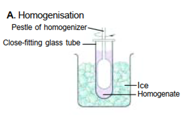
- With a view to study the components of the cell, the cell has to be homogenised or broken and components separated individually by different methods. The whole exercise is termed as cell fractionation. This involves homogenisation of the cell which can be brought about by
⁕Simple grinding of cells with glass beads,
⁕Breaking the cell by the physical forces like pressure,
⁕Using osmotic shock, and
⁕Employing ultrasound waves.
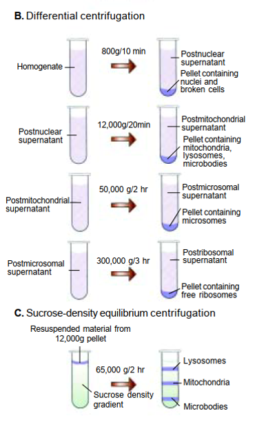
The prepared slurry is known as cell homogenate. Usually the intact cells or other debris may also be present along with the homogenate. These components can be removed from the homogenate by low speed centrifugation. The supernatant thus obtained is called cell free extract and is used to isolate further the different components like the various cell organelles, by different types of centrifugation.
Centrifugation
- For fractionation of cell components from the homogenate, usually differential Centrifugation technique is applied. Centrifuge is an instrument that is used to create a centrifugal force on different components of cell. As the components are put to centrifugal force, they tend to move outward depending upon their size and density. The components, therefore, move in the medium with different speeds. This character and principle is exploited for separation. There are different kinds of centrifuges. Normal clinical centrifuge is a motorized instrument which can rotate upto 5,000 rpm (revolutions per minute). The high speed centrifuge may have a speed upto 50,000 to 1,00,000 rpm. The high speed centrifuges are also called Ultracentrifuges.
- The plant cells have a fixed shape and size due to the presence of
a cell wall a specialised form of extra-cellular matrix, which is closely associated with the external surface of plant cell membrane. It is sufficiently thick, strong, and rigid and measures 0.1 nm to several μms. The cell wall was first by Robert Hooke in 1665. The composition and appearance of cell wall differs according to cell type and the function it performs. The fungal cell walls have a thick layer of polysaccharide fibres composed of either chitin or cellulose and a thin outer layer of mixed glycans. In the cell walls of algae, the substances commonly found are cellulose, galactans, mannans and minerals, such as silicon dioxide and calcium carbonate. In higher plants, the cell wall fibres are made up of the polysaccharide cellulose and are embedded in a highly cross-linked matrix of polysaccharides such as pectin, lignin and hemicellulose.
In higher plant cell wall is made up of layers.
1.Middle lamella.
2.Primary Cell Wall
3.Secondary cell wall
4.Tertiary cell wall
Structure of Cell Wall
The structure of cell wall usually consists of two parts, 1.Matrix 2.Depositions The cell wall in plant cells is composed of an intricate network of microfibrils in a gel-like matrix arranged in the following manner: (i) Cellulose microfibrils: The microfibrils are connected with the help of xyloglucan chains through hydrogen bonds. Thus a complete and continuous lattice is formed, which is embedded in a second network. (ii) Pectic polysaccharides: This constitutes second network, which is rich in galactouronic acid residue and forms cross links based on calcium bridges and other ionic interactions. (iii) Structural proteins: This is the third interlocking network, which consists of structural proteins that interweaves through the other two domains of network and forms wart and weft structure.
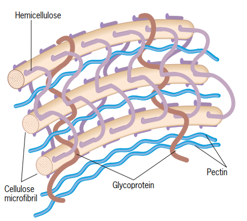
When a cell is young and small, the cellulose fibres are loosely packed and crosslinking of the cellulose fibrils is not complete. However, in mature cells due to crosslinking of fibres present in the wall, the cell is able to grow further., a new class of proteins called Expansin has been found to be responsible for wall loosening and cell expansion by addition of cellulose molecules to cellulose microfibrils.
2. Depositions Many chemicals are deposited in cell wall according to the requirement of the cell such as, 1.Silica (Si): It is deposited in the cell wall of Grasses where it protects the plant from fungal infection and also from grazers.
2.Iron (Fe): Iron is deposited in cell wall of Chara and helps in the floating.
3.Cutin: Cutin is a wax which covers plant surfaces and reduces the rate of transpiration. 4.Suberin: Suberin is also a wax which makes plants surface unwettable.
Structure of Cell Wall
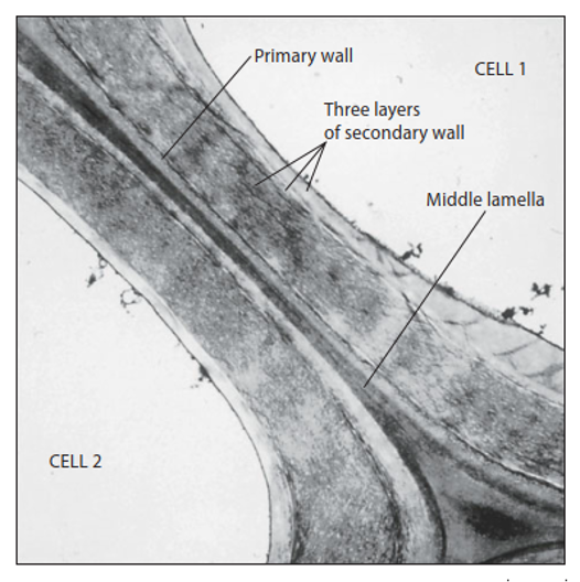
1.Middle lamella. Middle lamella is the outermost layer of a plant cell in fact it is the only layer in between two plant cells. It is made up of Ca2+ and Mg2+ salts of pectin. 2.Primary Cell Wall Primary cell wall is the most common type of cell wall present in the plant cell. It is made up of Pectin, Hemicellulose and Fibrils of cellulose.
Secondary Cell Wall
Inner to the primary wall the secondary wall is present, it is made up of Cellulose fibrils in parallel rows at an angle with the other fibre bundles. Sometimes, Hemicellulose, Pectin and Lignin may also be deposited in the secondary wall. The thick walls which have lignin deposits may also have pits, which are unthickened areas present in otherwise thick walls. Generally, the pits are present in pairs (on both the sides of wall). Therefore, these are called as pit pairs. Secondary cell wall is made up layers which are formed in different times. Due to presence of Lignin it became impermeable to water.
Tertiary Cell Wall
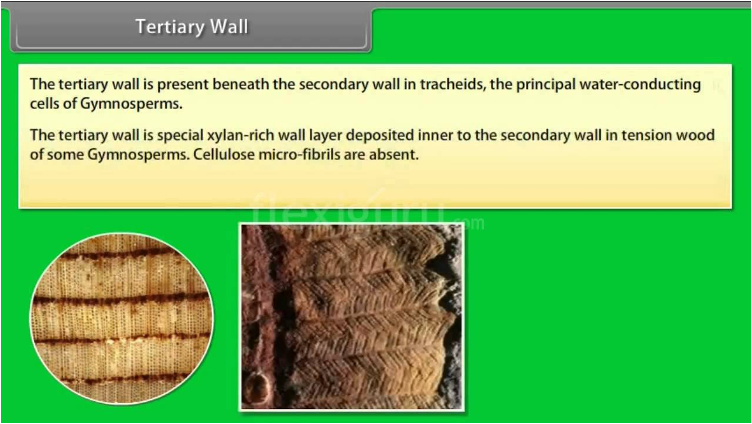
In Tracheid elements of Gymnosperm beneath secondary cell wall Tertiary cell wall is present. It is made up of Lignin.
 For the transport and communication, there are cytoplasmic bridges present between adjacent plant cells which are called Plasmodesmata. It is a fine cytoplasmic canal, which is lined by plasma membrane and often has ER (endoplasmic reticulum) tubule called Desmotubule.
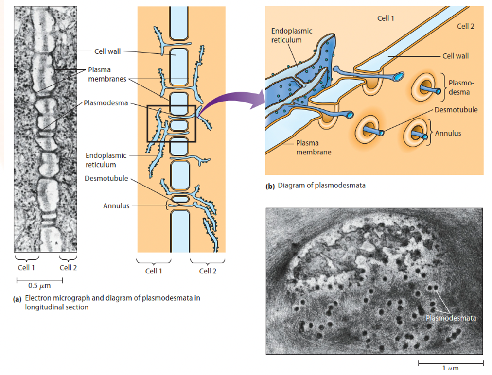
The plasmodesmata, help in maintaining continuity of living matter and the cytoplasm in such condition is often called Symplasm. In contrast to this, the intercellular space containing non-living matter constituents is called Apoplasm.
Plasma membrane
Its presence was first recognised by Nageli & Cramer (1855) who gave the term ‘Plasma membrane’. It was experimentally confirmed by E. Overton (1899) suggesting that it is made of lipid. In 1906 Friske reported that conductivity of intact cell is very low or it is made up of hydrophobic molecules. J.O. Plowe (1931) called it Plasmalemma.
Plasma membrane is a semi permeable and selectively permeable membrane which constitute the limiting membrane of the cell. It regulate all the activity of the cell and also called the soul of the cell.
Structure of Plasma membrane
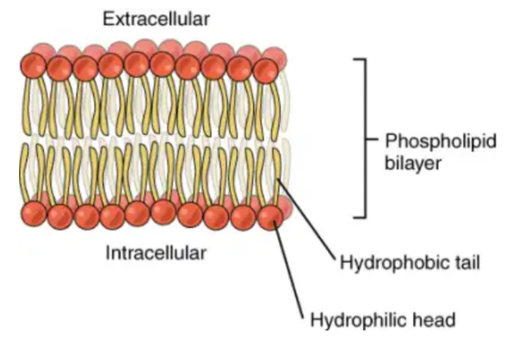
To find out the structure of Plasma membrane many models were given. In 1925 Gorter and Grendel gave Lipid membrane Model or it is made up of “Bimolecular lipid layer”. Evert Gorter and François Grendel (Dutch physiologists) approached the discovery of our present model of the plasma membrane structure as a lipid bi-layer. They simply hypothesized that if the plasma membrane is a bi-layer, then the surface area of the mono-layer of lipids measured would be double the surface area of the plasma membrane.
They measured the total surface area of the plasma membrane of red blood cells, and using Langmuir's method, they measured the area of the mono-layer of lipids. In comparing the two, they calculated an estimated ratio of 2:1 Mono-layer of lipids: Plasma membrane. This supported their hypothesis, which led to the conclusion that cell membranes are composed of two apposing molecular layers.
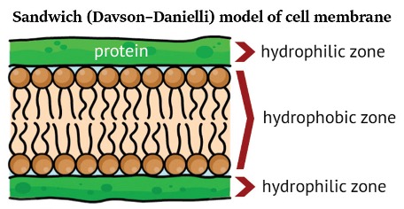
In 1935, Davson and Danielli proposed that biological membranes are made up of lipid bi-layers that are coated on both sides with thin sheets of protein and they simplified their model into the "pauci-molecular" theory. This theory declared that all biological membranes have a "lipoid" center surrounded by mono-layers of lipid that are covered by protein mono-layers. In short, their model was illustrated as a "sandwich" of protein-lipid-protein. The Davson-Danielli model threw new light on the understanding of cell membranes, by stressing the important role played by proteins in biological membranes.
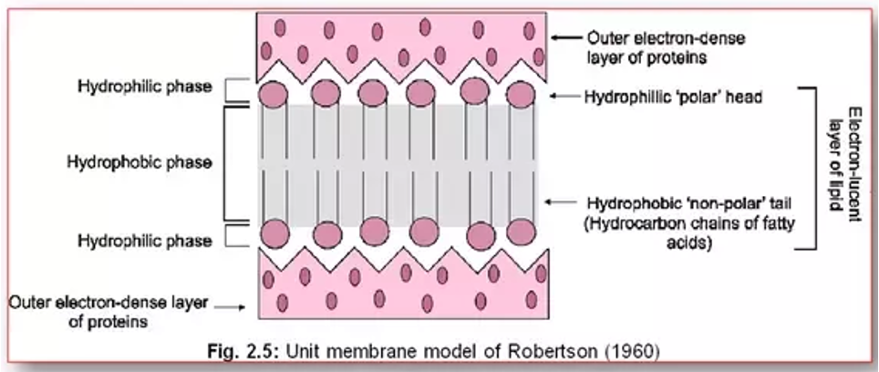
J. David Robertson used electron microscopy in 1950 to propose the Unit membrane model. Basically, he suggested that all cellular membranes share a similar underlying structure, the unit membrane. Using heavy metal staining, Robertson's proposal also seemed to agree instantaneously with the Davson-Danielli model. According to the trilaminar pattern of the cellular membrane viewed by Robertson, he suggested that the membranes consist of a lipid bi-layer covered on both surfaces with thin sheets of proteins. He proposed that membrane is a single unit of 75 Å having of Bilayer of phospholipids of 35 Å which is surrounded by two layers of proteins each is 20 Å.
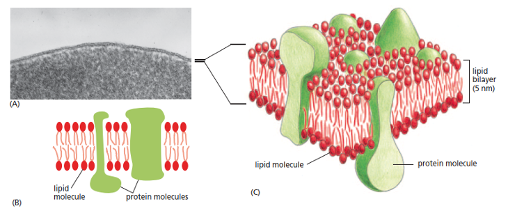
In 1972, Jonathan Singer and Garth Nicolson proposed the fluid mosaic model of membrane structure, in which membranes are viewed as two-dimensional fluids with proteins inserted into lipid bilayers. One of the central features of the fluid mosaic model is that, because they are inserted into a fluid lipid bilayer, both proteins and lipids are able to diffuse laterally through the membrane. According to this model proteins are like the icebergs which are floating in sea of phospholipids. The structure and arrangement of membrane proteins in the fluid-mosaic model differ from those of previous models in that they occur as a “mosaic” of discontinuous particles that penetrate the lipid sheet. Most importantly, the fluid-mosaic model presents cellular membranes as dynamic structures in which the components are mobile and capable of coming together to engage in various types of transient or semi permanent interactions.
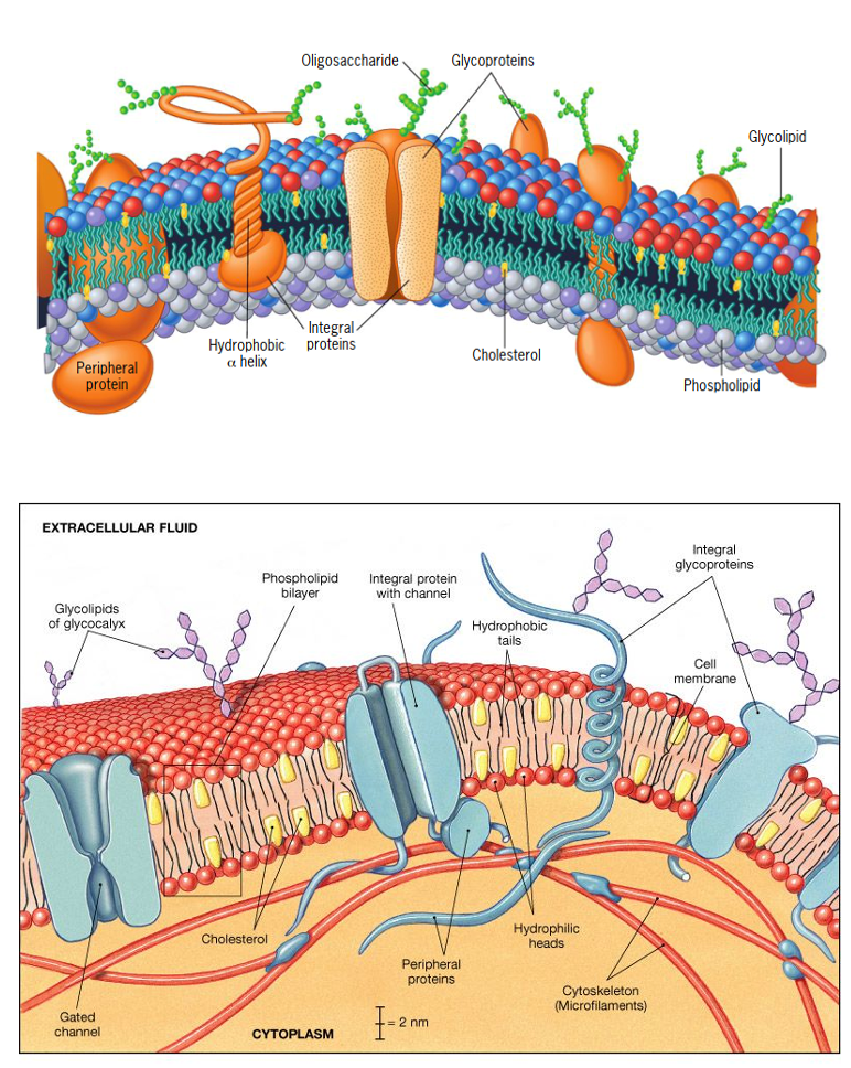
Biochemical investigation clearly revealed that the cell membranes also possess protein and carbohydrate. The ratio of protein and lipid varies considerably in different cell types. In human beings, the membrane of the erythrocyte has approximately 52 per cent Protein, 40% Phospholipids and 8% Oligosaccharides.
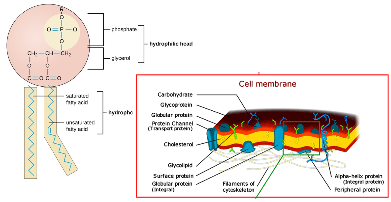
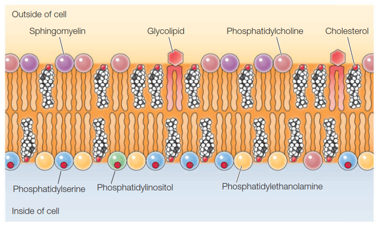
Phospholipids: The lipids in cell membranes combine two very different properties in a single molecule: each lipid has a Hydrophilic (“Water-loving”) head and a Hydrophobic (“Water-fearing”) tail. The most abundant lipids in cell membranes are the Phospholipids, which have a phosphate-containing, hydrophilic head linked to a pair of hydrophobic tails.
Phosphatidylcholine, for example, has the small molecule choline attached to a phosphate group as its hydrophilic head.
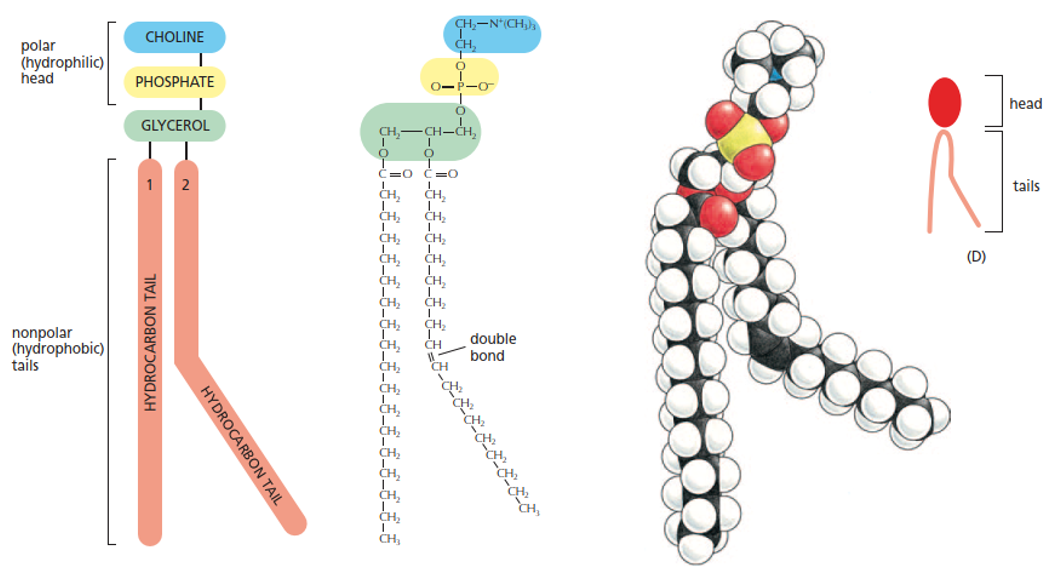
Molecules with both hydrophilic and hydrophobic parts are termed amphipathic, a property shared by other types of membrane lipids, including the cholesterol, which is found in animal cell membranes and the glycolipids, which have sugars as part of their hydrophilic head. Having both hydrophilic and hydrophobic parts plays a crucial part in driving these lipid molecules to assemble into bilayers in an aqueous environment. The aqueous environment inside and outside a cell prevents membrane lipids from escaping from the bilayer, but nothing stops these molecules from moving about and changing places with one another within the plane of the bilayer. The membrane therefore behaves as a two-dimensional fluid, a fact that is crucial for membrane function and integrity. Inside membrane phospholipids exhibit two type of movements,
1.Criss-cross : These are lateral movements within the same monolayer, they are frequent and does not require energy or enzymes.
2.Flip-flop : These type of movements are rare and require energy or enzymes.
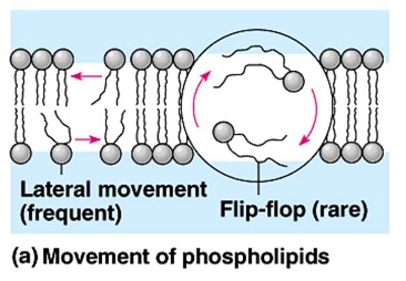
Besides this to maintain fluidity and stability in phospholipids mixed fatty acids are used or one saturated and one fatty acids are present in Membrane Phospholipids.
At the site of double bond in one unsaturated fatty acid pie π electrons are present which attract carbon atoms from saturated chain of adjacent phospholipids and thereby a weak interaction is formed in between two phospholipids which stabilize the membrane but to maintain fluidity in between two phospholipids Cholesterol is also present. Presence of Cholesterol increases the fluidity or provide membrane ‘Quasi Fluid’ nature means Neither pure solid nor pure Liquid. Besides this presence of cholesterol also makes membrane cold tolerant.
Most cell membranes are asymmetrical: the two halves of the bilayer often include strikingly different sets of phospholipids. But if membranes emerge from the ER with an evenly scrambled set of phospholipids. It begins in the Golgi apparatus. The Golgi membrane contains another family of phospholipid-handling enzyme, called flippases. These enzymes remove specific phospholipids from the side of the bilayer facing the exterior space and flip them into the monolayer that faces the cytosol. The action of these flippases—and similar enzymes in the plasma Membrane initiates and maintains the asymmetric arrangement of phospholipids that is characteristic of the membranes of animal cells. This asymmetry is preserved as membranes bud from one organelle and fuse with another—or with the plasma membrane.
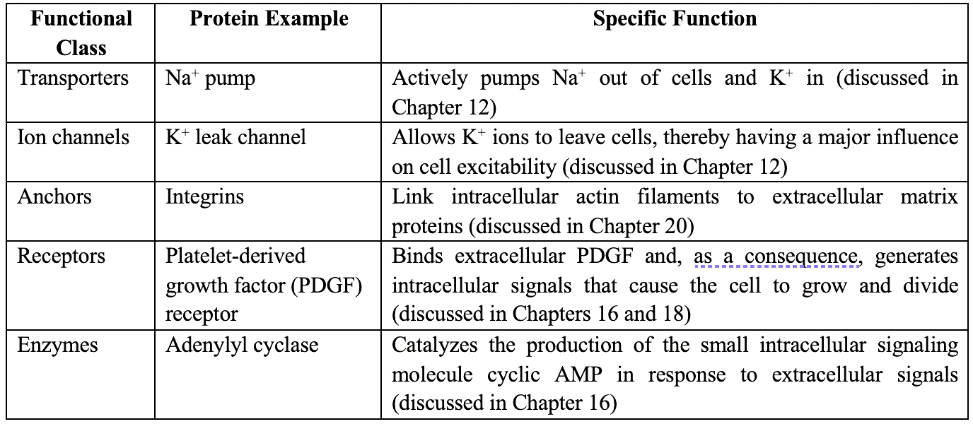
Most membrane functions are carried out by Membrane proteins. In animals, proteins constitute about 50% of the mass of most plasma membranes, the remainder being lipid plus the relatively small amounts of carbohydrate found on some of the lipids (glycolipids) and many of the proteins (glycoproteins). Because lipid molecules are much smaller than proteins, however, a cell membrane typically contains about 50 times more lipid molecules than protein molecules.
Membrane proteins serve many functions. Some transport particular nutrients, metabolites, and ions across the lipid bilayer. Others anchor the membrane to macromolecules on either side. Still others function as receptors that detect chemical signals in the cell’s environment and relay them into the cell interior, or work as enzymes to catalyze specific reactions at the membrane. Each type of cell membrane contains a different set of proteins.
Membrane proteins are of two types,

These proteins are easily isolated from the membrane.
They are either absorbed or adsorbed on the membrane.
They are not covalently attached to the membrane.
Eg. Spectrin and Actin in RBC and Protein kinase C.
Intrinsic Proteins
These proteins are not easily isolated from the membrane.
They are covalently attached to the membrane.
Many of them cross the membrane and present on both the side.
Eg. All transport channels, pumps and permeases.
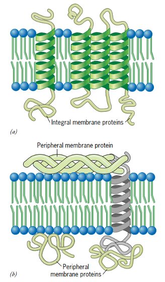
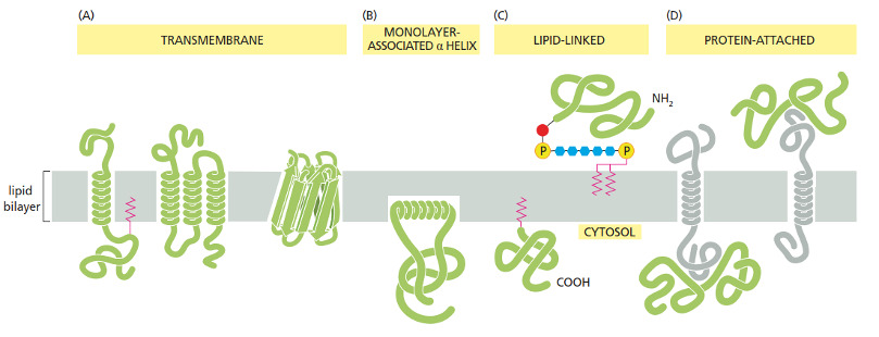
Membrane proteins are attached to the membrane by different methods,
Membrane proteins are not static they are in constant state of motion. Particularly convincing evidence for the mobility of at least somemembrane proteins has come from cell fusion experiments. In these studies, David Frye and Michael Edidin took advantage of two powerful techniques, one that enabled them to fuse cells from two different species and another that made it possible for them to label specific proteins on the surfaces of cells with antibodies containing fluorescent dye.
Antibodies are immune system proteins that recognize and bind to specific molecular antigens such as cell surface proteins. Frye and Edidin prepared two fluorescent antibodies, each one having a differently-colored dye linked to it, so that the human and mouse proteins could be distinguished. The anti-mouse antibodies were linked to a green fluorescent dye called fluorescein, whereas the anti-human antibodies were linked to a red fluorescent dye, Rhodamine. Frye and Edidin fused mouse and human cells using Sendai virus, exposed them to the red and green fluorescent antibodies, and observed the fused cells by fluorescence microscopy.
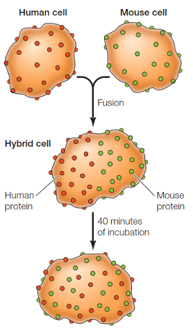
At first, the green fluorescent membrane proteins from the mouse cell were localized on one-half of the hybrid cell surface, and the red fluorescent membrane proteins derived from the human cell were restricted to the other half. In a few minutes, however, the proteins from the two parent cells began to intermix. After 40 minutes, the separate regions of green and red fluorescence were completely intermingled.
Membrane transport
- To survive and grow, cells must be able to exchange molecules with their environment. They must import nutrients such as sugars and amino acids and eliminate metabolic waste products. They must also regulate the concentrations of a variety of inorganic ions in their cytosol and organelles. A few molecules, such as CO2 and O2, can simply diffuse across the lipid bilayer of the plasma membrane. But the vast majority cannot. Instead, their transfer depends on specialized membrane transport proteins that span the lipid bilayer, providing private passageways across the membrane for select substances. Given enough time, virtually any molecule will diffuse across a lipid bilayer. The rate at which it diffuses, however, varies enormously depending on the size of the molecule and its solubility properties. In general, the smaller the molecule and the more hydrophobic, or nonpolar, it is, the more rapidly it will diffuse across the membrane.
The principle behind the membrane transport is Diffusion and the driving force is concentration gradient of the molecule.

Fats and oils can pass directly through:
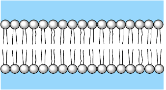
The Diffusion of Water through Membranes Water molecules move much more rapidly through a cell membrane than do dissolved ions or small polar organic solutes, which are essentially non penetrating. Because of this difference in the penetrability of water versus solutes, membranes are said to be semipermeable. Water moves readily through a semipermeable membrane from a region of lower solute concentration to a region of higher solute concentration. This process is called Osmosis, and it is readily demonstrated by placing a cell into a solution containing a non penetrating solute at a concentration different than that present within the cell itself.
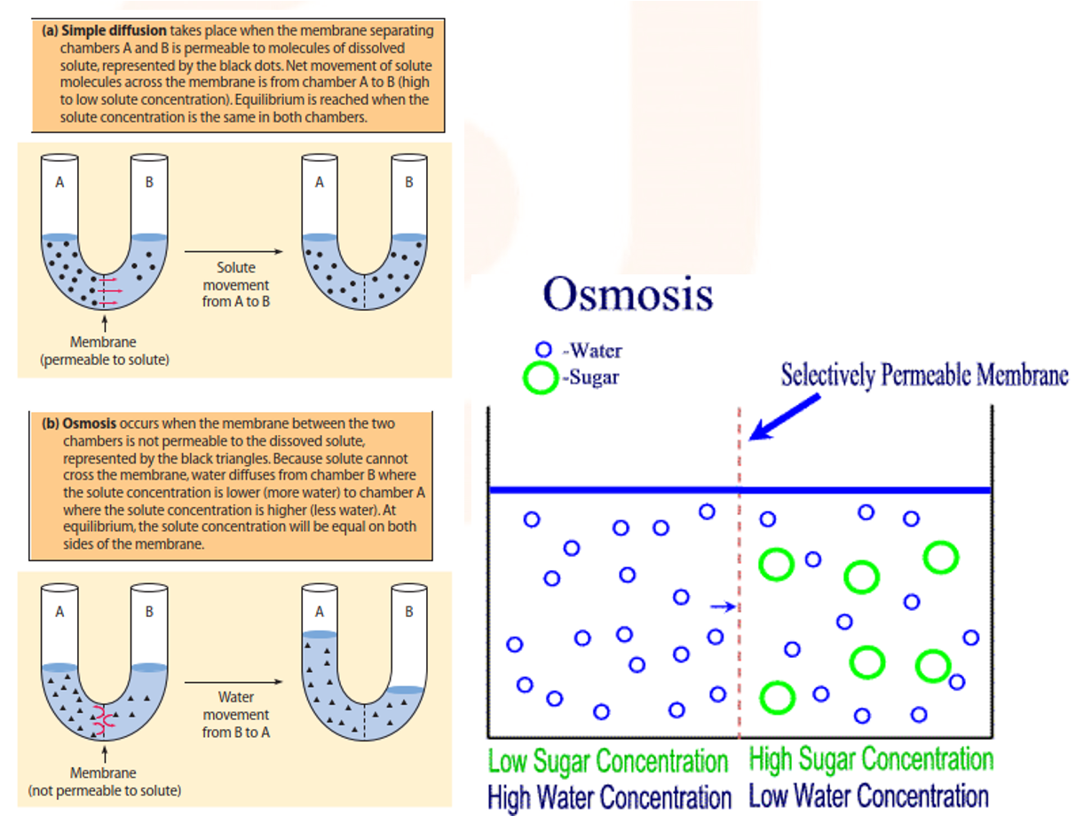
In 1882 Sydney Ringer discovered that 0.9% salt solution is isotonic to our body fluids. Depending upon this concept solutions are of three types,

Because most solutes cannot cross cell membranes, water tends to move across membranes in response to differences in solute concentrations on the two sides of the membrane. Specifically, water tends to diffuse from the side of the membrane with the lower solute concentration to the side with the higher solute concentration. Osmosis, is readily observed when a selectively permeable membrane separates two compartments, one of them containing a solute that cannot cross the membrane. Water will move across the membrane to equalize the concentration of solutes on both sides of the membrane.
An animal cell that starts out in an isotonic solution will shrink and shrivel if it is transferred to a hypertonic solution. On the other hand, the cell will swell if placed in a hypotonic solution. And it will actually lyse—or burst—if placed in a very hypotonic solution, such as pure water containing no solutes.
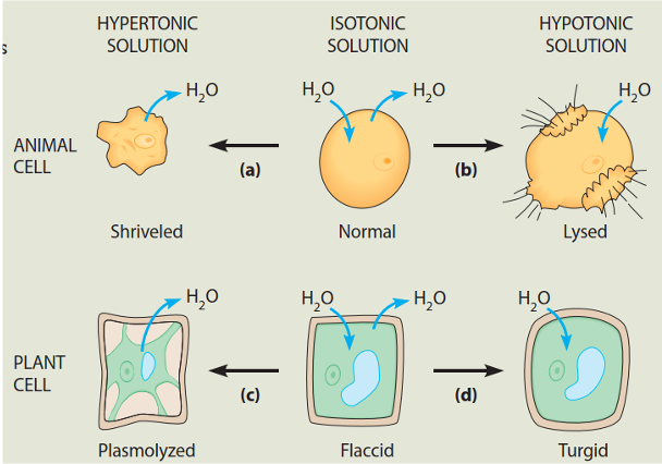
Cells of plants, algae, fungi, and many bacteria have rigid cell walls that keep the cells from swelling and bursting in a hypotonic solution. Instead, the cells become very firm from the turgor pressure that builds up due to the inward movement of water. The resulting turgidity accounts for the firmness, or turgor, of fully hydrated plant tissue. Without this turgidity, the tissue will wilt.
In a hypertonic solution, on the other hand, the outward movement of water causes the plasma membrane to pull away from the cell wall by a process called Plasmolysis. The wilting of a plant or a plant part during water deprivation is due to the plasmolysis of its cells.
Many cells are much more permeable to water than can be explained by simple diffusion through the lipid bilayer. In the early 1990s, Peter Agre and colleagues at Johns Hopkins University were attempting to isolate and purify the membrane proteins responsible for the Rh antigen on the surface of RBCs. During this pursuit, they identified a protein they thought might be the long-sought water channel of the erythrocyte membrane. To test their hypothesis, they engineered frog oocytes to incorporate the newly discovered protein into their plasma membranes and then placed the oocytes in a hypotonic medium.
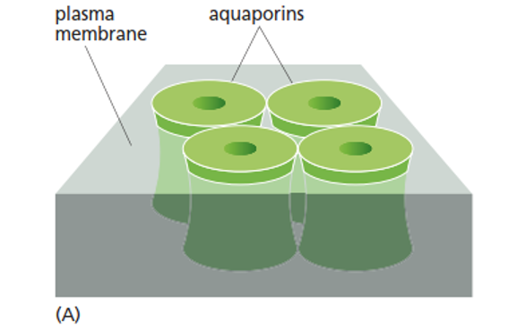
Just as predicted, the oocytes swelled due to the influx of water and eventually burst. The team had discovered a family of small integral proteins, called Aquaporins, that allow the passive movement of water from one side of the plasma membrane to the other.
Depending upon the concentration gradient of the solute membrane transport is of two types,

Passive transport occurs from higher concentration of solute to lower concentration of solute. The driving force is the concentration gradient and no energy is utilized. The transport of water soluble solute occurs with the help of membrane transport proteins.
Membrane transport proteins occur in many forms and are present in all cell membranes. Each provides a private portal across the membrane for a particular small, water-soluble molecule—an ion, sugar, or amino acid. There are two main classes of membrane transport proteins: Transporters and Channels. These proteins differ in the way they discriminate between solutes, transporting some but not others. Channels discriminate mainly on the basis of size and electric charge: when the channel is open, any ion or molecule that is small enough and carries the appropriate charge can pass through.

The principle behind Passive transport is Diffusion. Inside cell diffusion is of two type,

Simple Diffusion
Simple Diffusion is the simple transport of solute from higher concentration to low concentration. It occurs with the help of Protein channels, these channels are of two types,
1.Open channels
2.Closed channels
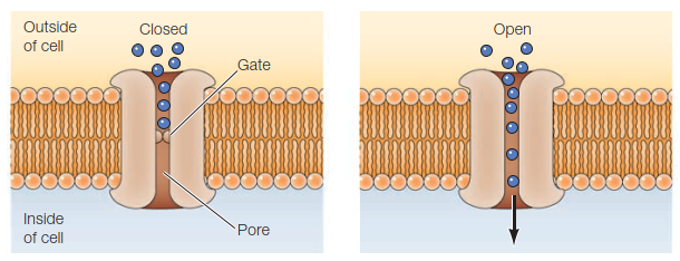
1.Open channels: These channels remain open and allow the transport of solute according to their concentration gradient. A. Porins: Porins are beta barrel proteins that cross a cellular membrane and act as a pore, through which molecules can diffuse. Unlike other membrane transport proteins, porins are large enough to allow passive diffusion, i.e., they act as channels that are specific to different types of molecules. They are present in the outer membrane of gram-negative bacteria and some gram-positive mycobacteria (mycolic acid-containing actinomycetes), the outer membrane of mitochondria, and the outer chloroplast membrane.
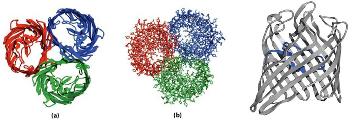 B. Potassium Leaking channels: Potassium channels are the most widely distributed type of ion channel and are found in virtually all living organisms. They form potassium-selective pores that span cell membranes. Potassium channels are found in most cell types and control a wide variety of cell functions. They participate in regulation of the cardiac action potential duration in cardiac muscle, malfunction of potassium channels may cause life-threatening arrhythmias. Potassium channels may also be involved in maintaining vascular tone.
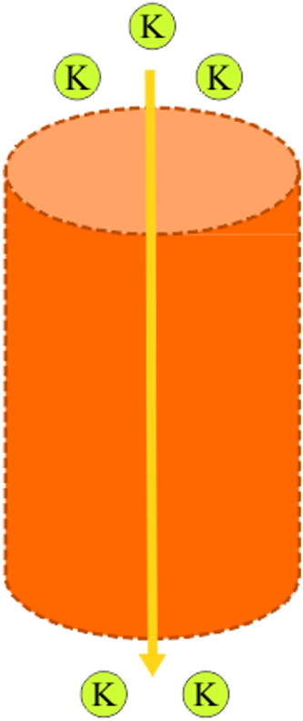 They also regulate cellular processes such as the secretion of hormones (e.g., insulin release from beta-cells in the pancreas) so their malfunction can lead to diseases (such as diabetes). Some toxins, such as Dendrotoxin, are potent because they block potassium channels.
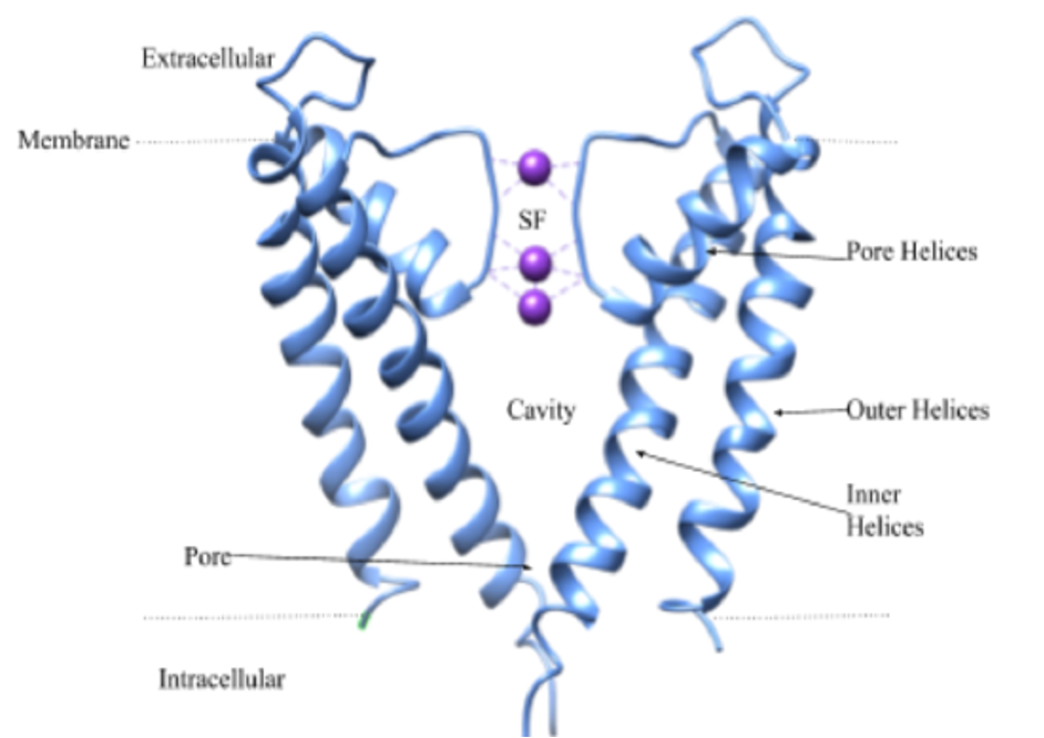 2.Closed channels
Closed channels remain closed unless and until chemical or electrical environment changes around them.
Depending upon the environment they are of two types,
1.Voltage operated
2.Ligand operated
1.Voltage operated
Most of ion channels are gated and many of them are voltage operated or whenever there is a potential change across the membrane these channels undergo conformation change and open or closes according to the stimulus.
E.g. Voltage operated Na+ channels and voltage operated K+ channels, both these channels participate in generation of action potential in Neurons and Muscle cells. Similarly Ca2+ channels are present in Presynaptic nerve terminal.
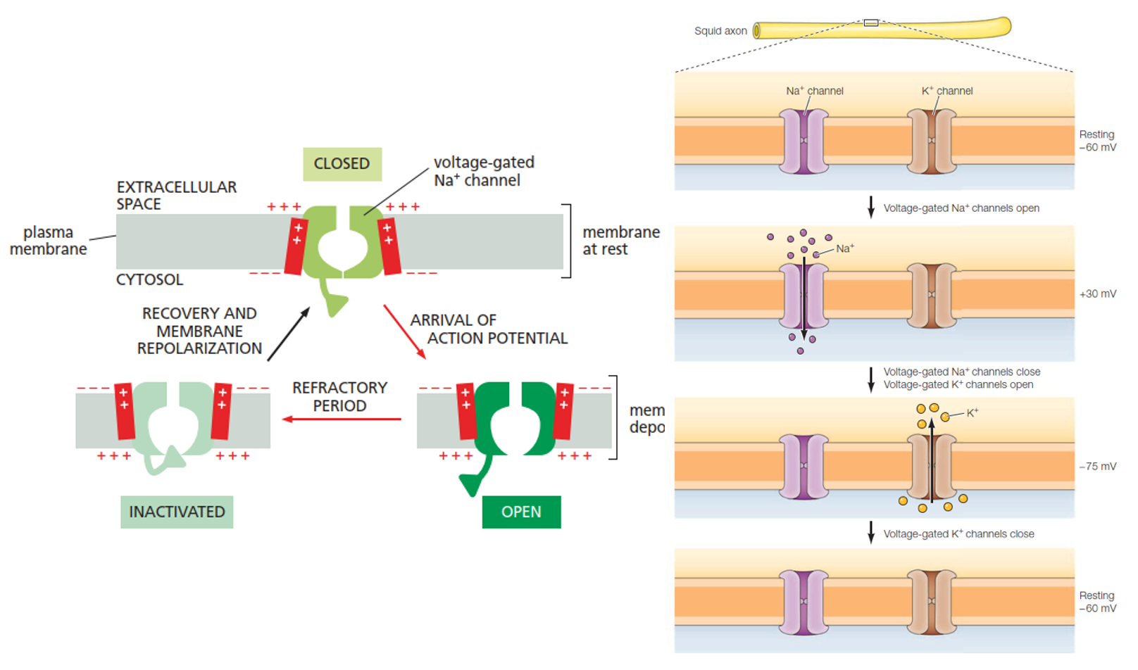
2.Ligand operated
Ligand are chemicals which bind to specific receptors. These chemicals bind to channels and brings about a change in their conformation which opens them, ●Acetylcholine dependent Na+ channels,
●GABA dependent Cl- channels
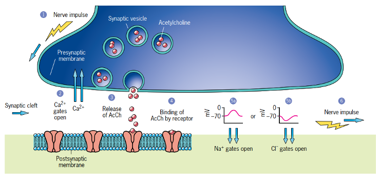
Faciliated Diffusion
- Substances always diffuse across a membrane from a region of higher concentration on one side to a region of lower concentration on the other side, but they do not always diffuse through the lipid bilayer or through a channel. In many cases, the diffusing substance first binds selectively to a membrane-spanning protein, called a facilitative transporter, that facilitates the diffusion process. The binding of the solute to the facilitative transporter on one side of the membrane is thought to trigger a conformational change in the protein, exposing the solute to the other surface of the membrane, from where it can diffuse down its concentration gradient.
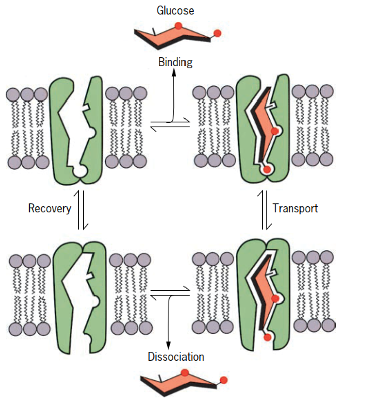
Because they operate passively, that is, without being coupled to an energy-releasing system, facilitated transporters can mediate the movement of solutes equally well in both directions. The direction of net flux depends on the relative concentration of the substance on the two sides of the membrane. Facilitated diffusion is highly specific and carried out with the help of specialized proteins called Permeases or Carrier Proteins.
Depending upon the number of solute molecule transported Facilitated Diffusion is of two types

Uniport
A uniport is transport protein which transport one solute molecule across the membrane according to concentration gradient. These proteins first bind to the solute molecule and this binding is highly specific.
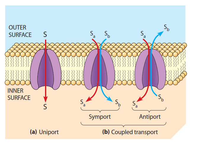
An excellent example of uniport transporter is Glucose transporters, Glucose transporters are a wide group of membrane proteins that facilitate the transport of glucose across the plasma membrane, a process known as facilitated diffusion. Because glucose is a vital source of energy for all life, these transporters are present in all phyla. The GLUT or SLC2A family are a protein family that is found in most mammalian cells. 14 GLUTS are encoded by human genome. GLUT is a type of uniporter transporter protein.
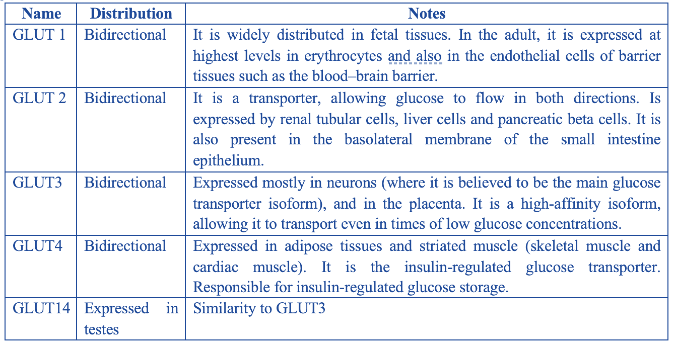
Biport A Biport is a carrier protein which transport two solute molecule at a time,

Symport
A Symport is a protein which transport two solute molecules in the same direction. SGLT1 in the intestinal epithelium transports sodium ions (Na+) and glucose across luminal membrane of the epithelial cells so that it can be absorbed into the bloodstream. This is the basis of oral rehydration therapy. If this symporter did not exist, individual sodium channels and glucose uniporters would not be able to transfer glucose against the concentration gradient and into the bloodstream. Na+/K+/2Cl− symporter in the loop of Henle in the renal tubules of the kidney transports 4 molecules of 3 different types; a sodium ion (Na+), a potassium ion (K+) and two chloride ions (2Cl−). Loop diuretics such as furosemide (Lasix) act on this protein. Marine invertebrates use symporters to transport against strong chemical gradients. Amino acids and sugars are taken up from sea water in the presence of extracellular sodium and is driven by the NA+/K+-ATPase pump.
Six examples are shown in this figure. The Na+/glucose cotransporter (SGLT1) is found in the apical membrane of kidney proximal tubule epithelial cells, where it functions along with SGLT2 to reabsorb glucose from the ultrafiltrate, as well as in the apical membrane of small intestine epithelial cells, where it functions to absorb glucose from the lumen of the small intestine. The Na+/phosphate cotransporter (NaPi) is found in the apical membrane of epithelial cells of the small intestine, where it serves to absorb dietary phosphate, and renal proximal tubules, where it serves to reabsorb filtered phosphate. The Na+/I− symporter (NIS) is located in the basolateral membrane of thyroid follicular cells, and is responsible for the accumulation of iodide into the thyroid gland. The Na+/K+/2Cl− cotransporter (NKCC), Na+/Cl− cotransporter (NCC), and K+/Cl− cotransporter (KCC) are found in many different cell types, where they perform essential roles in ion homeostasis. Na+, sodium; K+, potassium; Cl−, chloride; I−, iodide; Pi, inorganic phosphate.
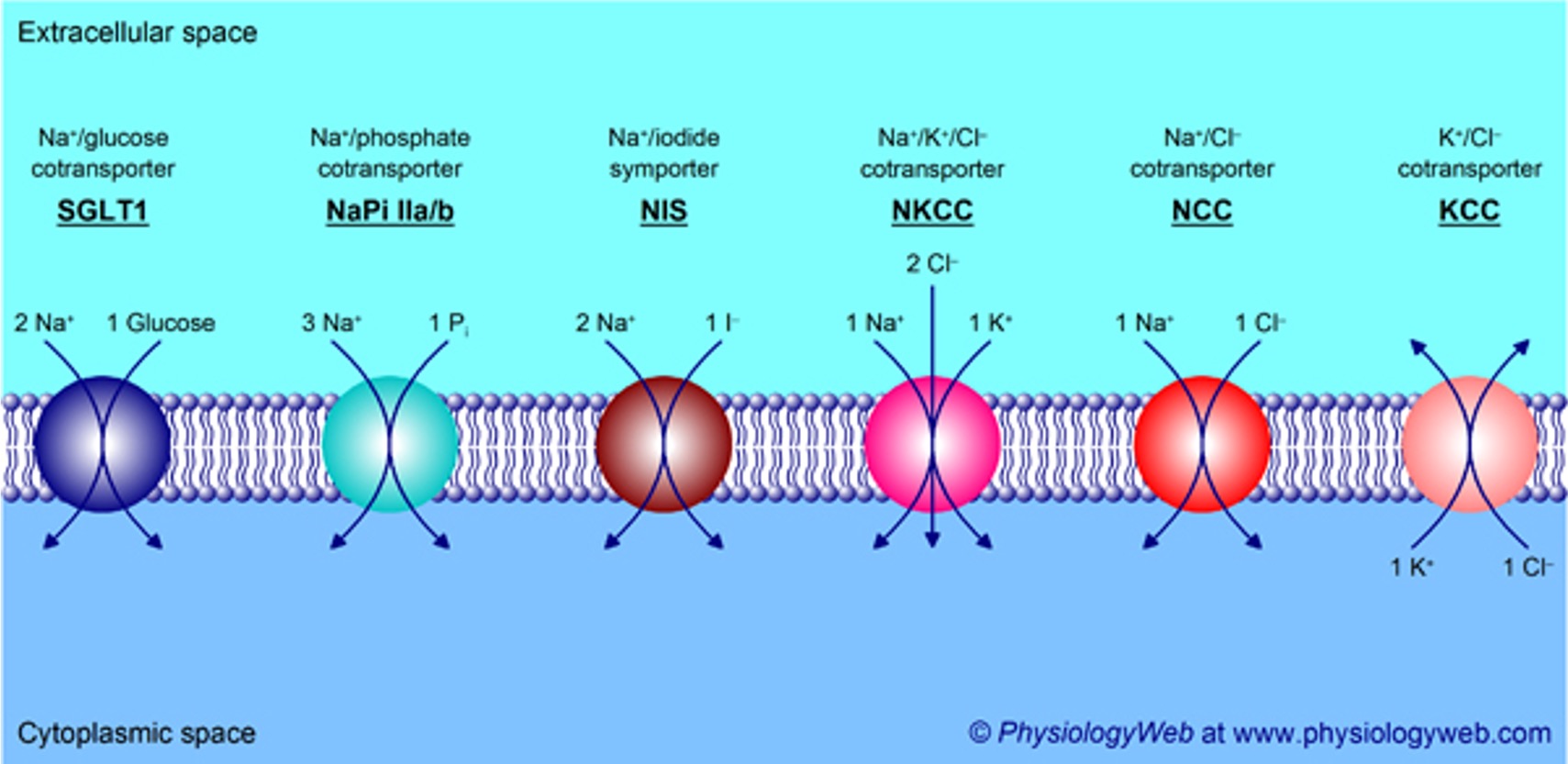
Antiport
An antiport protein is a carrier protein which transport two solute molecules in opposite direction.
Eg., 1.Transport of Chloride ions and Bicarbonate ion in RBC: To maintain the transport of CO2 in RBC carbonic anhydrase enzyme converts C02 into HCO3- ions which are transported to plasma by Chloride Exchange Protein which is present in RBC membrane.
2.Transport of ATP/ADP in mitochondria.
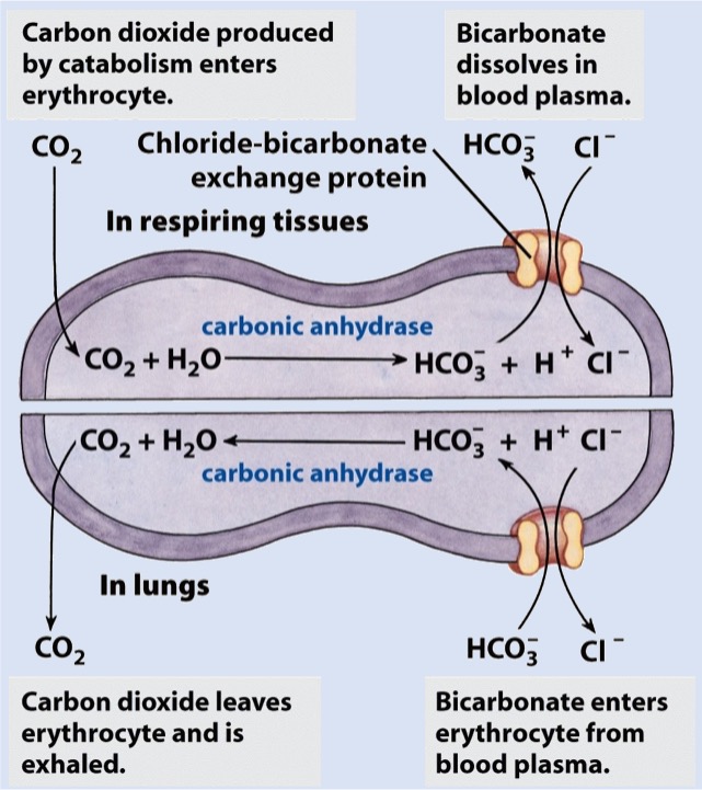
Active Transport
- Active transport is the movement of molecules across a cell membrane from a region of lower concentration to a region of higher concentration against the concentration gradient. Active transport requires cellular energy to achieve this movement.
There are two types of active transport: ●Primary active transport that uses adenosine triphosphate (ATP), and
●Secondary active transport that uses an electrochemical gradient.
Primary active transport:
Primary active transport, also called direct active transport, directly uses metabolic energy to transport molecules across a membrane. Substances that are transported across the cell membrane by primary active transport include metal ions, such as Na+, K+, Mg2+, and Ca2+. Most of the enzymes that perform this type of transport are transmembrane ATPases. Active transport performs three major functions in cells and organelles. First, it makes possible the uptake of essential nutrients from the environment or surrounding fluid, even when their concentrations in the cell are already higher.
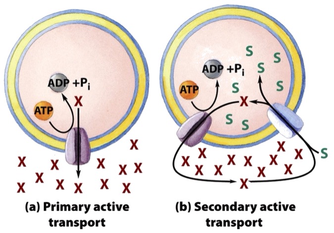
Second, it allows various substances, such as secretory products and waste materials, to be removed from the cell or organelle, even when the concentration outside is greater than the concentration inside. Third, it enables the cell to maintain constant, non equilibrium intracellular concentrations of specific inorganic ions.
Examples of primary active transport: 1.
Proton Pump in Lysosomes. Lysosomes are membrane vesicles having Acid Hydrolases which help in various activities of the cell because these enzymes functions at acidic pH therefore in Lysosomes pH is maintained in between 4 to 5 with the help of Proton pump present in the Lysosomal Membrane. 2.
Calcium Pump in Sarcoplasmic Reticulum: The calcium pump allows muscles to relax after this frenzied wave of calcium-induced contraction. The pump is found in the membrane of the sarcoplasmic reticulum. In some cases, it is so plentiful that it may make up 90% of the protein there. Powered by ATP, it pumps calcium ions back into the sarcoplasmic reticulum, reducing the calcium level around the actin and myosin filaments and allowing the muscle to relax.
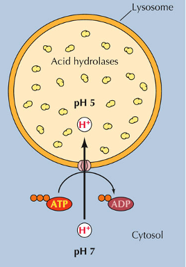
The H+/K+ ATPase
The parietal cells of your stomach use this pump to secrete gastric juice. These cells transport protons (H+) from a concentration of about 4 x 10⁻⁸ M within the cell to a concentration of about 0.15 M in the gastric juice (giving it a pH close to 1). Small wonder that parietal cells are stuffed with mitochondria and uses huge amounts of ATP as they carry out this three-million fold concentration of protons.
Na+/K+-ATPase
Na⁺/K⁺-ATPase (sodium–potassium adenosine triphosphatase, also known as the Na⁺/K⁺ pump or sodium–potassium pump) is an enzyme (an electrogenic transmembrane ATPase) found in the membrane of all animal cells. The Na⁺/K⁺-ATPase enzyme is active (i.e. it uses energy from ATP). For every ATP molecule that the pump uses, three sodium ions are exported and two potassium ions are imported; there is hence a net export of a single positive charge per pump cycle. The Na⁺/K⁺-ATPase helps maintain resting potential, affects transport, and regulates cellular volume. In fact, all cells expend a large fraction of the ATP they produce (typically 30% and up to 70% in nerve cells) to maintain their required cytosolic Na and K concentrations.
Secondary active transport:
In secondary active transport, also known as coupled transport or cotransport, energy is used to transport molecules across a membrane; however, in contrast to primary active transport, there is no direct coupling of ATP. Instead, it relies upon the electrochemical potential difference created by pumping ions in/out of the cell. Permitting one ion or molecule to move down an electrochemical gradient, but possibly against the concentration gradient where it is more concentrated to that where it is less concentrated, increases entropy and can serve as a source of energy for metabolism (e.g. in ATP synthase). The energy derived from the pumping of protons across a cell membrane is frequently used as the energy source in secondary active transport. Usually along with pump carrier proteins are present which functions in a coupled reaction. These pumps are of two types
1.Symport pumps
2.Anitport pumps
Symport pumps
In this type of indirect active transport, the driving ion (Na+) and the pumped molecules pass through the membrane pump in the same direction. Examples:
The Na+/glucose transporter. ●This transmembrane protein allows sodium ions and glucose to enter the cell together. The Na+/glucose transporter is used to actively transport glucose out of the intestine and also out of the kidney tubules and back into the blood.
●All the amino acids can be actively transported, for example out of the kidney tubules and into the blood, by sodium-driven symport pumps.
●Sodium-driven symport pumps also return neurotransmitters to the presynaptic neuron .
●The Na+/iodide transporter. This symporter pumps iodide ions into the cells of the thyroid gland (for the manufacture of thyroxine) and also into the cells of the mammary gland (to supply the baby's need for iodide).
●The permease encoded by the lac operon of E. coli that transports lactose into the cell.
The Na+/glucose transporter
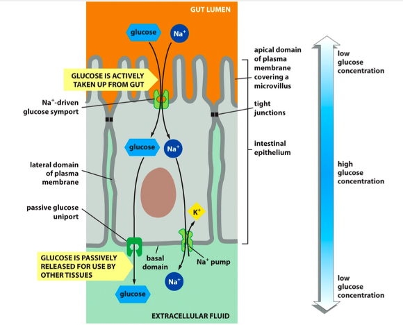
Antiport Pumps
In antiport pumps, the driving ion (again, usually sodium) diffuses through the pump in one direction providing the energy for the active transport of some other molecule or ion in the opposite direction. Example: Ca2+ ions are pumped out of cells by sodium-driven antiport pumps. Antiport pumps in the vacuole of some plants harness the outward facilitated diffusion of protons (themselves pumped into the vacuole by a H+ ATPase) to the active inward transport of sodium ions. This sodium/proton antiport pump enables the plant to sequester sodium ions in its vacuole. Transgenic tomato plants that overexpress this sodium/proton antiport pump are able to thrive in saline soils too salty for conventional tomatoes. to the active inward transport of nitrate ions (NO3−). Cell membranes are semi-permeable, meaning they allow certain small molecules and ions to passively diffuse through them. Other small molecules are able to make their way into or out of the cell through carrier proteins or channels.
But there are materials that are too large to pass through the cell membrane using these methods. There are times when a cell will need to engulf a bacterium or release a hormone. It is during these instances that bulk transport mechanisms are needed. Bulk transport is of two types, Endocytosis and exocytosis are the bulk transport mechanisms used in eukaryotes. As these transport processes require energy, they are known as active transport processes.

During bulk transport, larger substances or large packages of small molecules are transported through the cell membrane, also known as the plasma membrane, by way of membrane vesicles.
EUKARYOTIC CELL AND ITS ORGANISATION
- Eukaryotic cells, by definition, have a nucleus, which contains nuclear material enclosed by adouble layered membrane. All plant and animal cells fall under this category. In general, the eukaryotic cells have the following components:
- Cell wall (absent in animal cells and some protists),
- Plasma membrane,
- Cytoplasm and
- Organelles.
Until the discovery of electron microscope, the cytoplasm was thought to have a simple organisation. It is now known that cytoplasm has a highly complex organization consisting of cytoplasmic matrix and several organelles. The motile cells have additional appendages for locomotion. The plant cells have a defined, rigid cell wall, which animal cells do not posseses; therefore, animal cells generally have an irregular shape.
- With a view to study the components of the cell, the cell has to be homogenised or broken and components separated individually by different methods. The whole exercise is termed as cell fractionation. This involves homogenisation of the cell which can be brought about by
⁕Simple grinding of cells with glass beads,
⁕Breaking the cell by the physical forces like pressure,
⁕Using osmotic shock, and
⁕Employing ultrasound waves.
The prepared slurry is known as cell homogenate. Usually the intact cells or other debris may also be present along with the homogenate. These components can be removed from the homogenate by low speed centrifugation. The supernatant thus obtained is called cell free extract and is used to isolate further the different components like the various cell organelles, by different types of centrifugation.

Centrifugation
- For fractionation of cell components from the homogenate, usually differential Centrifugation technique is applied. Centrifuge is an instrument that is used to create a centrifugal force on different components of cell. As the components are put to centrifugal force, they tend to move outward depending upon their size and density. The components, therefore, move in the medium with different speeds. This character and principle is exploited for separation. There are different kinds of centrifuges. Normal clinical centrifuge is a motorized instrument which can rotate upto 5,000 rpm (revolutions per minute). The high speed centrifuge may have a speed upto 50,000 to 1,00,000 rpm. The high speed centrifuges are also called Ultracentrifuges.

- The plant cells have a fixed shape and size due to the presence of
a cell wall a specialised form of extra-cellular matrix, which is closely associated with the external surface of plant cell membrane. It is sufficiently thick, strong, and rigid and measures 0.1 nm to several μms. The cell wall was first by Robert Hooke in 1665. The composition and appearance of cell wall differs according to cell type and the function it performs. The fungal cell walls have a thick layer of polysaccharide fibres composed of either chitin or cellulose and a thin outer layer of mixed glycans. In the cell walls of algae, the substances commonly found are cellulose, galactans, mannans and minerals, such as silicon dioxide and calcium carbonate. In higher plants, the cell wall fibres are made up of the polysaccharide cellulose and are embedded in a highly cross-linked matrix of polysaccharides such as pectin, lignin and hemicellulose.
In higher plant cell wall is made up of layers.
1.Middle lamella.
2.Primary Cell Wall
3.Secondary cell wall
4.Tertiary cell wall
Structure of Cell Wall
The structure of cell wall usually consists of two parts, 1.Matrix 2.Depositions The cell wall in plant cells is composed of an intricate network of microfibrils in a gel-like matrix arranged in the following manner: (i) Cellulose microfibrils: The microfibrils are connected with the help of xyloglucan chains through hydrogen bonds. Thus a complete and continuous lattice is formed, which is embedded in a second network. (ii) Pectic polysaccharides: This constitutes second network, which is rich in galactouronic acid residue and forms cross links based on calcium bridges and other ionic interactions. (iii) Structural proteins: This is the third interlocking network, which consists of structural proteins that interweaves through the other two domains of network and forms wart and weft structure.

When a cell is young and small, the cellulose fibres are loosely packed and crosslinking of the cellulose fibrils is not complete. However, in mature cells due to crosslinking of fibres present in the wall, the cell is able to grow further., a new class of proteins called Expansin has been found to be responsible for wall loosening and cell expansion by addition of cellulose molecules to cellulose microfibrils.
2. Depositions Many chemicals are deposited in cell wall according to the requirement of the cell such as, 1.Silica (Si): It is deposited in the cell wall of Grasses where it protects the plant from fungal infection and also from grazers.
2.Iron (Fe): Iron is deposited in cell wall of Chara and helps in the floating.
3.Cutin: Cutin is a wax which covers plant surfaces and reduces the rate of transpiration. 4.Suberin: Suberin is also a wax which makes plants surface unwettable.
Structure of Cell Wall
1.Middle lamella. Middle lamella is the outermost layer of a plant cell in fact it is the only layer in between two plant cells. It is made up of Ca2+ and Mg2+ salts of pectin. 2.Primary Cell Wall Primary cell wall is the most common type of cell wall present in the plant cell. It is made up of Pectin, Hemicellulose and Fibrils of cellulose.

Secondary Cell Wall
Inner to the primary wall the secondary wall is present, it is made up of Cellulose fibrils in parallel rows at an angle with the other fibre bundles. Sometimes, Hemicellulose, Pectin and Lignin may also be deposited in the secondary wall. The thick walls which have lignin deposits may also have pits, which are unthickened areas present in otherwise thick walls. Generally, the pits are present in pairs (on both the sides of wall). Therefore, these are called as pit pairs. Secondary cell wall is made up layers which are formed in different times. Due to presence of Lignin it became impermeable to water.
Tertiary Cell Wall
In Tracheid elements of Gymnosperm beneath secondary cell wall Tertiary cell wall is present. It is made up of Lignin.
 For the transport and communication, there are cytoplasmic bridges present between adjacent plant cells which are called Plasmodesmata. It is a fine cytoplasmic canal, which is lined by plasma membrane and often has ER (endoplasmic reticulum) tubule called Desmotubule.
The plasmodesmata, help in maintaining continuity of living matter and the cytoplasm in such condition is often called Symplasm. In contrast to this, the intercellular space containing non-living matter constituents is called Apoplasm.
Plasma membrane
Its presence was first recognised by Nageli & Cramer (1855) who gave the term ‘Plasma membrane’. It was experimentally confirmed by E. Overton (1899) suggesting that it is made of lipid. In 1906 Friske reported that conductivity of intact cell is very low or it is made up of hydrophobic molecules. J.O. Plowe (1931) called it Plasmalemma.
Plasma membrane is a semi permeable and selectively permeable membrane which constitute the limiting membrane of the cell. It regulate all the activity of the cell and also called the soul of the cell.

Structure of Plasma membrane
To find out the structure of Plasma membrane many models were given. In 1925 Gorter and Grendel gave Lipid membrane Model or it is made up of “Bimolecular lipid layer”. Evert Gorter and François Grendel (Dutch physiologists) approached the discovery of our present model of the plasma membrane structure as a lipid bi-layer. They simply hypothesized that if the plasma membrane is a bi-layer, then the surface area of the mono-layer of lipids measured would be double the surface area of the plasma membrane.
They measured the total surface area of the plasma membrane of red blood cells, and using Langmuir's method, they measured the area of the mono-layer of lipids. In comparing the two, they calculated an estimated ratio of 2:1 Mono-layer of lipids: Plasma membrane. This supported their hypothesis, which led to the conclusion that cell membranes are composed of two apposing molecular layers.

In 1935, Davson and Danielli proposed that biological membranes are made up of lipid bi-layers that are coated on both sides with thin sheets of protein and they simplified their model into the "pauci-molecular" theory. This theory declared that all biological membranes have a "lipoid" center surrounded by mono-layers of lipid that are covered by protein mono-layers. In short, their model was illustrated as a "sandwich" of protein-lipid-protein. The Davson-Danielli model threw new light on the understanding of cell membranes, by stressing the important role played by proteins in biological membranes.

J. David Robertson used electron microscopy in 1950 to propose the Unit membrane model. Basically, he suggested that all cellular membranes share a similar underlying structure, the unit membrane. Using heavy metal staining, Robertson's proposal also seemed to agree instantaneously with the Davson-Danielli model. According to the trilaminar pattern of the cellular membrane viewed by Robertson, he suggested that the membranes consist of a lipid bi-layer covered on both surfaces with thin sheets of proteins. He proposed that membrane is a single unit of 75 Å having of Bilayer of phospholipids of 35 Å which is surrounded by two layers of proteins each is 20 Å.

In 1972, Jonathan Singer and Garth Nicolson proposed the fluid mosaic model of membrane structure, in which membranes are viewed as two-dimensional fluids with proteins inserted into lipid bilayers. One of the central features of the fluid mosaic model is that, because they are inserted into a fluid lipid bilayer, both proteins and lipids are able to diffuse laterally through the membrane. According to this model proteins are like the icebergs which are floating in sea of phospholipids. The structure and arrangement of membrane proteins in the fluid-mosaic model differ from those of previous models in that they occur as a “mosaic” of discontinuous particles that penetrate the lipid sheet. Most importantly, the fluid-mosaic model presents cellular membranes as dynamic structures in which the components are mobile and capable of coming together to engage in various types of transient or semi permanent interactions.

Biochemical investigation clearly revealed that the cell membranes also possess protein and carbohydrate. The ratio of protein and lipid varies considerably in different cell types. In human beings, the membrane of the erythrocyte has approximately 52 per cent Protein, 40% Phospholipids and 8% Oligosaccharides.
Phospholipids: The lipids in cell membranes combine two very different properties in a single molecule: each lipid has a Hydrophilic (“Water-loving”) head and a Hydrophobic (“Water-fearing”) tail. The most abundant lipids in cell membranes are the Phospholipids, which have a phosphate-containing, hydrophilic head linked to a pair of hydrophobic tails.
Phosphatidylcholine, for example, has the small molecule choline attached to a phosphate group as its hydrophilic head.

Molecules with both hydrophilic and hydrophobic parts are termed amphipathic, a property shared by other types of membrane lipids, including the cholesterol, which is found in animal cell membranes and the glycolipids, which have sugars as part of their hydrophilic head. Having both hydrophilic and hydrophobic parts plays a crucial part in driving these lipid molecules to assemble into bilayers in an aqueous environment. The aqueous environment inside and outside a cell prevents membrane lipids from escaping from the bilayer, but nothing stops these molecules from moving about and changing places with one another within the plane of the bilayer. The membrane therefore behaves as a two-dimensional fluid, a fact that is crucial for membrane function and integrity. Inside membrane phospholipids exhibit two type of movements,
1.Criss-cross : These are lateral movements within the same monolayer, they are frequent and does not require energy or enzymes.
2.Flip-flop : These type of movements are rare and require energy or enzymes.

Besides this to maintain fluidity and stability in phospholipids mixed fatty acids are used or one saturated and one fatty acids are present in Membrane Phospholipids.
At the site of double bond in one unsaturated fatty acid pie π electrons are present which attract carbon atoms from saturated chain of adjacent phospholipids and thereby a weak interaction is formed in between two phospholipids which stabilize the membrane but to maintain fluidity in between two phospholipids Cholesterol is also present. Presence of Cholesterol increases the fluidity or provide membrane ‘Quasi Fluid’ nature means Neither pure solid nor pure Liquid. Besides this presence of cholesterol also makes membrane cold tolerant.

Most cell membranes are asymmetrical: the two halves of the bilayer often include strikingly different sets of phospholipids. But if membranes emerge from the ER with an evenly scrambled set of phospholipids. It begins in the Golgi apparatus. The Golgi membrane contains another family of phospholipid-handling enzyme, called flippases. These enzymes remove specific phospholipids from the side of the bilayer facing the exterior space and flip them into the monolayer that faces the cytosol. The action of these flippases—and similar enzymes in the plasma Membrane initiates and maintains the asymmetric arrangement of phospholipids that is characteristic of the membranes of animal cells. This asymmetry is preserved as membranes bud from one organelle and fuse with another—or with the plasma membrane.

Most membrane functions are carried out by Membrane proteins. In animals, proteins constitute about 50% of the mass of most plasma membranes, the remainder being lipid plus the relatively small amounts of carbohydrate found on some of the lipids (glycolipids) and many of the proteins (glycoproteins). Because lipid molecules are much smaller than proteins, however, a cell membrane typically contains about 50 times more lipid molecules than protein molecules.
Membrane proteins serve many functions. Some transport particular nutrients, metabolites, and ions across the lipid bilayer. Others anchor the membrane to macromolecules on either side. Still others function as receptors that detect chemical signals in the cell’s environment and relay them into the cell interior, or work as enzymes to catalyze specific reactions at the membrane. Each type of cell membrane contains a different set of proteins.

Membrane proteins are of two types,

These proteins are easily isolated from the membrane.
They are either absorbed or adsorbed on the membrane.
They are not covalently attached to the membrane.
Eg. Spectrin and Actin in RBC and Protein kinase C.
Intrinsic Proteins
These proteins are not easily isolated from the membrane.
They are covalently attached to the membrane.
Many of them cross the membrane and present on both the side.
Eg. All transport channels, pumps and permeases.

Membrane proteins are attached to the membrane by different methods,

Membrane proteins are not static they are in constant state of motion. Particularly convincing evidence for the mobility of at least somemembrane proteins has come from cell fusion experiments. In these studies, David Frye and Michael Edidin took advantage of two powerful techniques, one that enabled them to fuse cells from two different species and another that made it possible for them to label specific proteins on the surfaces of cells with antibodies containing fluorescent dye.
Antibodies are immune system proteins that recognize and bind to specific molecular antigens such as cell surface proteins. Frye and Edidin prepared two fluorescent antibodies, each one having a differently-colored dye linked to it, so that the human and mouse proteins could be distinguished. The anti-mouse antibodies were linked to a green fluorescent dye called fluorescein, whereas the anti-human antibodies were linked to a red fluorescent dye, Rhodamine. Frye and Edidin fused mouse and human cells using Sendai virus, exposed them to the red and green fluorescent antibodies, and observed the fused cells by fluorescence microscopy.

At first, the green fluorescent membrane proteins from the mouse cell were localized on one-half of the hybrid cell surface, and the red fluorescent membrane proteins derived from the human cell were restricted to the other half. In a few minutes, however, the proteins from the two parent cells began to intermix. After 40 minutes, the separate regions of green and red fluorescence were completely intermingled.
Membrane transport
- To survive and grow, cells must be able to exchange molecules with their environment. They must import nutrients such as sugars and amino acids and eliminate metabolic waste products. They must also regulate the concentrations of a variety of inorganic ions in their cytosol and organelles. A few molecules, such as CO2 and O2, can simply diffuse across the lipid bilayer of the plasma membrane. But the vast majority cannot. Instead, their transfer depends on specialized membrane transport proteins that span the lipid bilayer, providing private passageways across the membrane for select substances. Given enough time, virtually any molecule will diffuse across a lipid bilayer. The rate at which it diffuses, however, varies enormously depending on the size of the molecule and its solubility properties. In general, the smaller the molecule and the more hydrophobic, or nonpolar, it is, the more rapidly it will diffuse across the membrane.
The principle behind the membrane transport is Diffusion and the driving force is concentration gradient of the molecule.

Fats and oils can pass directly through:

The Diffusion of Water through Membranes Water molecules move much more rapidly through a cell membrane than do dissolved ions or small polar organic solutes, which are essentially non penetrating. Because of this difference in the penetrability of water versus solutes, membranes are said to be semipermeable. Water moves readily through a semipermeable membrane from a region of lower solute concentration to a region of higher solute concentration. This process is called Osmosis, and it is readily demonstrated by placing a cell into a solution containing a non penetrating solute at a concentration different than that present within the cell itself.

In 1882 Sydney Ringer discovered that 0.9% salt solution is isotonic to our body fluids. Depending upon this concept solutions are of three types,

Because most solutes cannot cross cell membranes, water tends to move across membranes in response to differences in solute concentrations on the two sides of the membrane. Specifically, water tends to diffuse from the side of the membrane with the lower solute concentration to the side with the higher solute concentration. Osmosis, is readily observed when a selectively permeable membrane separates two compartments, one of them containing a solute that cannot cross the membrane. Water will move across the membrane to equalize the concentration of solutes on both sides of the membrane.
An animal cell that starts out in an isotonic solution will shrink and shrivel if it is transferred to a hypertonic solution. On the other hand, the cell will swell if placed in a hypotonic solution. And it will actually lyse—or burst—if placed in a very hypotonic solution, such as pure water containing no solutes.

Cells of plants, algae, fungi, and many bacteria have rigid cell walls that keep the cells from swelling and bursting in a hypotonic solution. Instead, the cells become very firm from the turgor pressure that builds up due to the inward movement of water. The resulting turgidity accounts for the firmness, or turgor, of fully hydrated plant tissue. Without this turgidity, the tissue will wilt.
In a hypertonic solution, on the other hand, the outward movement of water causes the plasma membrane to pull away from the cell wall by a process called Plasmolysis. The wilting of a plant or a plant part during water deprivation is due to the plasmolysis of its cells.
Many cells are much more permeable to water than can be explained by simple diffusion through the lipid bilayer. In the early 1990s, Peter Agre and colleagues at Johns Hopkins University were attempting to isolate and purify the membrane proteins responsible for the Rh antigen on the surface of RBCs. During this pursuit, they identified a protein they thought might be the long-sought water channel of the erythrocyte membrane. To test their hypothesis, they engineered frog oocytes to incorporate the newly discovered protein into their plasma membranes and then placed the oocytes in a hypotonic medium.

Just as predicted, the oocytes swelled due to the influx of water and eventually burst. The team had discovered a family of small integral proteins, called Aquaporins, that allow the passive movement of water from one side of the plasma membrane to the other.
Depending upon the concentration gradient of the solute membrane transport is of two types,

Passive transport occurs from higher concentration of solute to lower concentration of solute. The driving force is the concentration gradient and no energy is utilized. The transport of water soluble solute occurs with the help of membrane transport proteins.
Membrane transport proteins occur in many forms and are present in all cell membranes. Each provides a private portal across the membrane for a particular small, water-soluble molecule—an ion, sugar, or amino acid. There are two main classes of membrane transport proteins: Transporters and Channels. These proteins differ in the way they discriminate between solutes, transporting some but not others. Channels discriminate mainly on the basis of size and electric charge: when the channel is open, any ion or molecule that is small enough and carries the appropriate charge can pass through.

The principle behind Passive transport is Diffusion. Inside cell diffusion is of two type,

Simple Diffusion
Simple Diffusion is the simple transport of solute from higher concentration to low concentration. It occurs with the help of Protein channels, these channels are of two types,
1.Open channels
2.Closed channels

1.Open channels: These channels remain open and allow the transport of solute according to their concentration gradient. A. Porins: Porins are beta barrel proteins that cross a cellular membrane and act as a pore, through which molecules can diffuse. Unlike other membrane transport proteins, porins are large enough to allow passive diffusion, i.e., they act as channels that are specific to different types of molecules. They are present in the outer membrane of gram-negative bacteria and some gram-positive mycobacteria (mycolic acid-containing actinomycetes), the outer membrane of mitochondria, and the outer chloroplast membrane.
 B. Potassium Leaking channels: Potassium channels are the most widely distributed type of ion channel and are found in virtually all living organisms. They form potassium-selective pores that span cell membranes. Potassium channels are found in most cell types and control a wide variety of cell functions. They participate in regulation of the cardiac action potential duration in cardiac muscle, malfunction of potassium channels may cause life-threatening arrhythmias. Potassium channels may also be involved in maintaining vascular tone.
 They also regulate cellular processes such as the secretion of hormones (e.g., insulin release from beta-cells in the pancreas) so their malfunction can lead to diseases (such as diabetes). Some toxins, such as Dendrotoxin, are potent because they block potassium channels.
 2.Closed channels
Closed channels remain closed unless and until chemical or electrical environment changes around them.
Depending upon the environment they are of two types,
1.Voltage operated
2.Ligand operated
1.Voltage operated
Most of ion channels are gated and many of them are voltage operated or whenever there is a potential change across the membrane these channels undergo conformation change and open or closes according to the stimulus.
E.g. Voltage operated Na+ channels and voltage operated K+ channels, both these channels participate in generation of action potential in Neurons and Muscle cells. Similarly Ca2+ channels are present in Presynaptic nerve terminal.

2.Ligand operated
Ligand are chemicals which bind to specific receptors. These chemicals bind to channels and brings about a change in their conformation which opens them, ●Acetylcholine dependent Na+ channels,
●GABA dependent Cl- channels

Faciliated Diffusion
- Substances always diffuse across a membrane from a region of higher concentration on one side to a region of lower concentration on the other side, but they do not always diffuse through the lipid bilayer or through a channel. In many cases, the diffusing substance first binds selectively to a membrane-spanning protein, called a facilitative transporter, that facilitates the diffusion process. The binding of the solute to the facilitative transporter on one side of the membrane is thought to trigger a conformational change in the protein, exposing the solute to the other surface of the membrane, from where it can diffuse down its concentration gradient.

Because they operate passively, that is, without being coupled to an energy-releasing system, facilitated transporters can mediate the movement of solutes equally well in both directions. The direction of net flux depends on the relative concentration of the substance on the two sides of the membrane. Facilitated diffusion is highly specific and carried out with the help of specialized proteins called Permeases or Carrier Proteins.
Depending upon the number of solute molecule transported Facilitated Diffusion is of two types

Uniport
A uniport is transport protein which transport one solute molecule across the membrane according to concentration gradient. These proteins first bind to the solute molecule and this binding is highly specific.

An excellent example of uniport transporter is Glucose transporters, Glucose transporters are a wide group of membrane proteins that facilitate the transport of glucose across the plasma membrane, a process known as facilitated diffusion. Because glucose is a vital source of energy for all life, these transporters are present in all phyla. The GLUT or SLC2A family are a protein family that is found in most mammalian cells. 14 GLUTS are encoded by human genome. GLUT is a type of uniporter transporter protein.

Biport A Biport is a carrier protein which transport two solute molecule at a time,

Symport
A Symport is a protein which transport two solute molecules in the same direction. SGLT1 in the intestinal epithelium transports sodium ions (Na+) and glucose across luminal membrane of the epithelial cells so that it can be absorbed into the bloodstream. This is the basis of oral rehydration therapy. If this symporter did not exist, individual sodium channels and glucose uniporters would not be able to transfer glucose against the concentration gradient and into the bloodstream. Na+/K+/2Cl− symporter in the loop of Henle in the renal tubules of the kidney transports 4 molecules of 3 different types; a sodium ion (Na+), a potassium ion (K+) and two chloride ions (2Cl−). Loop diuretics such as furosemide (Lasix) act on this protein. Marine invertebrates use symporters to transport against strong chemical gradients. Amino acids and sugars are taken up from sea water in the presence of extracellular sodium and is driven by the NA+/K+-ATPase pump.
Six examples are shown in this figure. The Na+/glucose cotransporter (SGLT1) is found in the apical membrane of kidney proximal tubule epithelial cells, where it functions along with SGLT2 to reabsorb glucose from the ultrafiltrate, as well as in the apical membrane of small intestine epithelial cells, where it functions to absorb glucose from the lumen of the small intestine. The Na+/phosphate cotransporter (NaPi) is found in the apical membrane of epithelial cells of the small intestine, where it serves to absorb dietary phosphate, and renal proximal tubules, where it serves to reabsorb filtered phosphate. The Na+/I− symporter (NIS) is located in the basolateral membrane of thyroid follicular cells, and is responsible for the accumulation of iodide into the thyroid gland. The Na+/K+/2Cl− cotransporter (NKCC), Na+/Cl− cotransporter (NCC), and K+/Cl− cotransporter (KCC) are found in many different cell types, where they perform essential roles in ion homeostasis. Na+, sodium; K+, potassium; Cl−, chloride; I−, iodide; Pi, inorganic phosphate.

Antiport
An antiport protein is a carrier protein which transport two solute molecules in opposite direction.
Eg., 1.Transport of Chloride ions and Bicarbonate ion in RBC: To maintain the transport of CO2 in RBC carbonic anhydrase enzyme converts C02 into HCO3- ions which are transported to plasma by Chloride Exchange Protein which is present in RBC membrane.
2.Transport of ATP/ADP in mitochondria.

Active Transport
- Active transport is the movement of molecules across a cell membrane from a region of lower concentration to a region of higher concentration against the concentration gradient. Active transport requires cellular energy to achieve this movement.
There are two types of active transport: ●Primary active transport that uses adenosine triphosphate (ATP), and
●Secondary active transport that uses an electrochemical gradient.
Primary active transport:
Primary active transport, also called direct active transport, directly uses metabolic energy to transport molecules across a membrane. Substances that are transported across the cell membrane by primary active transport include metal ions, such as Na+, K+, Mg2+, and Ca2+. Most of the enzymes that perform this type of transport are transmembrane ATPases. Active transport performs three major functions in cells and organelles. First, it makes possible the uptake of essential nutrients from the environment or surrounding fluid, even when their concentrations in the cell are already higher.

Second, it allows various substances, such as secretory products and waste materials, to be removed from the cell or organelle, even when the concentration outside is greater than the concentration inside. Third, it enables the cell to maintain constant, non equilibrium intracellular concentrations of specific inorganic ions.
Examples of primary active transport: 1.
Proton Pump in Lysosomes. Lysosomes are membrane vesicles having Acid Hydrolases which help in various activities of the cell because these enzymes functions at acidic pH therefore in Lysosomes pH is maintained in between 4 to 5 with the help of Proton pump present in the Lysosomal Membrane. 2.
Calcium Pump in Sarcoplasmic Reticulum: The calcium pump allows muscles to relax after this frenzied wave of calcium-induced contraction. The pump is found in the membrane of the sarcoplasmic reticulum. In some cases, it is so plentiful that it may make up 90% of the protein there. Powered by ATP, it pumps calcium ions back into the sarcoplasmic reticulum, reducing the calcium level around the actin and myosin filaments and allowing the muscle to relax.

The H+/K+ ATPase
The parietal cells of your stomach use this pump to secrete gastric juice. These cells transport protons (H+) from a concentration of about 4 x 10⁻⁸ M within the cell to a concentration of about 0.15 M in the gastric juice (giving it a pH close to 1). Small wonder that parietal cells are stuffed with mitochondria and uses huge amounts of ATP as they carry out this three-million fold concentration of protons.
Na+/K+-ATPase
Na⁺/K⁺-ATPase (sodium–potassium adenosine triphosphatase, also known as the Na⁺/K⁺ pump or sodium–potassium pump) is an enzyme (an electrogenic transmembrane ATPase) found in the membrane of all animal cells. The Na⁺/K⁺-ATPase enzyme is active (i.e. it uses energy from ATP). For every ATP molecule that the pump uses, three sodium ions are exported and two potassium ions are imported; there is hence a net export of a single positive charge per pump cycle. The Na⁺/K⁺-ATPase helps maintain resting potential, affects transport, and regulates cellular volume. In fact, all cells expend a large fraction of the ATP they produce (typically 30% and up to 70% in nerve cells) to maintain their required cytosolic Na and K concentrations.
Secondary active transport:
In secondary active transport, also known as coupled transport or cotransport, energy is used to transport molecules across a membrane; however, in contrast to primary active transport, there is no direct coupling of ATP. Instead, it relies upon the electrochemical potential difference created by pumping ions in/out of the cell. Permitting one ion or molecule to move down an electrochemical gradient, but possibly against the concentration gradient where it is more concentrated to that where it is less concentrated, increases entropy and can serve as a source of energy for metabolism (e.g. in ATP synthase). The energy derived from the pumping of protons across a cell membrane is frequently used as the energy source in secondary active transport. Usually along with pump carrier proteins are present which functions in a coupled reaction. These pumps are of two types
1.Symport pumps
2.Anitport pumps
Symport pumps
In this type of indirect active transport, the driving ion (Na+) and the pumped molecules pass through the membrane pump in the same direction. Examples:
The Na+/glucose transporter. ●This transmembrane protein allows sodium ions and glucose to enter the cell together. The Na+/glucose transporter is used to actively transport glucose out of the intestine and also out of the kidney tubules and back into the blood.
●All the amino acids can be actively transported, for example out of the kidney tubules and into the blood, by sodium-driven symport pumps.
●Sodium-driven symport pumps also return neurotransmitters to the presynaptic neuron .
●The Na+/iodide transporter. This symporter pumps iodide ions into the cells of the thyroid gland (for the manufacture of thyroxine) and also into the cells of the mammary gland (to supply the baby's need for iodide).
●The permease encoded by the lac operon of E. coli that transports lactose into the cell.
The Na+/glucose transporter

Antiport Pumps
In antiport pumps, the driving ion (again, usually sodium) diffuses through the pump in one direction providing the energy for the active transport of some other molecule or ion in the opposite direction. Example: Ca2+ ions are pumped out of cells by sodium-driven antiport pumps. Antiport pumps in the vacuole of some plants harness the outward facilitated diffusion of protons (themselves pumped into the vacuole by a H+ ATPase) to the active inward transport of sodium ions. This sodium/proton antiport pump enables the plant to sequester sodium ions in its vacuole. Transgenic tomato plants that overexpress this sodium/proton antiport pump are able to thrive in saline soils too salty for conventional tomatoes. to the active inward transport of nitrate ions (NO3−). Cell membranes are semi-permeable, meaning they allow certain small molecules and ions to passively diffuse through them. Other small molecules are able to make their way into or out of the cell through carrier proteins or channels.
But there are materials that are too large to pass through the cell membrane using these methods. There are times when a cell will need to engulf a bacterium or release a hormone. It is during these instances that bulk transport mechanisms are needed. Bulk transport is of two types, Endocytosis and exocytosis are the bulk transport mechanisms used in eukaryotes. As these transport processes require energy, they are known as active transport processes.

During bulk transport, larger substances or large packages of small molecules are transported through the cell membrane, also known as the plasma membrane, by way of membrane vesicles.
ENDOCYTOSIS
- Endocytosis is the process by which cells take in substances from outside of the cell by engulfing them in a vesicle. These can include things like nutrients to support the cell or pathogens that immune cells engulf and destroy.
Endocytosis occurs when a portion of the cell membrane folds in on itself, encircling extracellular fluid and various molecules or microorganisms. The resulting vesicle breaks off and is transported within the cell. These vesicles are called Endosomes.
Endocytosis serves many purposes, including:
⁕Taking in nutrients for cellular growth, function and repair: Cells need materials like proteins and lipids to function.
⁕Capturing pathogens or other unknown substances that may endanger the organism: When pathogens like bacteria are identified by the immune system, they are engulfed by immune cells to be destroyed.
⁕Disposing of old or damaged cells: Cells must be safely disposed of when they stop functioning properly to prevent damage to other cells. These cells are eliminated through endocytosis.
Types of endocytosis:
There are two types of endocytosis: Phagocytosis and Pinocytosis.
 Phagocytosis
 (from Ancient Greek Phagein 'to eat', and Kytos= 'cell') is the process by which a cell uses its plasma membrane to engulf a large particle (≥ 0.5 μm), giving rise to an internal compartment called the Phagosome. It is one type of endocytosis. A cell that performs phagocytosis is called a phagocyte. It is also called Cellular Eating. In a multicellular organism's immune system, phagocytosis is a major mechanism used to remove pathogens and cell debris. The ingested material is then digested in the phagosome. Bacteria, dead tissue cells, and small mineral particles are all examples of objects that may be phagocytized. Some protozoa use phagocytosis as means to obtain nutrients. Phagocytosis was first noted by Canadian physician William Osler (1876), and later studied and named by Élie Metchnikoff in1880.
Phagocytosis
Phagocytosis (from Ancient Greek Phagein 'to eat', and Kytos= 'cell') is the process by which a cell uses its plasma membrane to engulf a large particle (≥ 0.5 μm), giving rise to an internal compartment called the Phagosome. It is one type of endocytosis. A cell that performs phagocytosis is called a phagocyte. It is also called Cellular Eating. In a multicellular organism's immune system, phagocytosis is a major mechanism used to remove pathogens and cell debris. The ingested material is then digested in the phagosome. Bacteria, dead tissue cells, and small mineral particles are all examples of objects that may be phagocytized. Some protozoa use phagocytosis as means to obtain nutrients. Phagocytosis was first noted by Canadian physician William Osler (1876), and later studied and named by Élie Metchnikoff in1880.
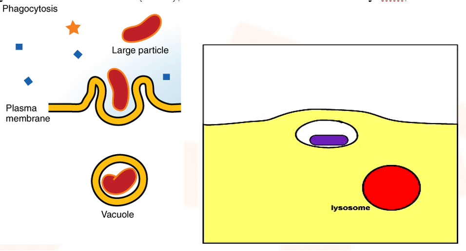 Phagocytosis occurs with the help of receptors.
Pinocytosis
Pinocytosis, also known as cell drinking, is common in plant and animal cells. During pinocytosis, the cell takes in substances from the extracellular fluid that it needs to function. These include things like water and nutrients.
Receptor-mediated endocytosis is a specialized type of pinocytosis. During receptor-mediated endocytosis, macromolecules bind to receptors along the surface of the cell’s plasma membrane. Cholesterol uptake is an example of receptor-mediated endocytosis.
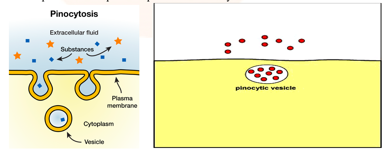 Exocytosis is the process by which cells move materials from within the cell into the extracellular fluid. Exocytosis occurs when a vesicle fuses with the plasma membrane, allowing its contents to be released outside the cell.
Exocytosis serves the following purposes:
●Removing toxins or waste products from the cell’s interior: Cells create waste or toxins that must be removed from the cell to maintain homeostasis. The membrane vesicles formed during the Exocytosis are called Exosomes.
●Facilitating cellular communication: Cells create signaling molecules like hormones and neurotransmitters. They are delivered to other cells following their release from the cell through exocytosis.
●Facilitating cellular membrane growth, repair, signaling and migration: When cells absorb materials from outside the cell during endocytosis, they use lipids and proteins from the plasma membrane to create vesicles. When certain exocytotic vesicles fuse with the cellular membrane, they replenish the cell membrane with these materials.
One of the example of Exocytosis is Ephagy or Cellular Defecation.


ENDOCYTOSIS
- Endocytosis is the process by which cells take in substances from outside of the cell by engulfing them in a vesicle. These can include things like nutrients to support the cell or pathogens that immune cells engulf and destroy.
Endocytosis occurs when a portion of the cell membrane folds in on itself, encircling extracellular fluid and various molecules or microorganisms. The resulting vesicle breaks off and is transported within the cell. These vesicles are called Endosomes.
Endocytosis serves many purposes, including:
⁕Taking in nutrients for cellular growth, function and repair: Cells need materials like proteins and lipids to function.
⁕Capturing pathogens or other unknown substances that may endanger the organism: When pathogens like bacteria are identified by the immune system, they are engulfed by immune cells to be destroyed.
⁕Disposing of old or damaged cells: Cells must be safely disposed of when they stop functioning properly to prevent damage to other cells. These cells are eliminated through endocytosis.
Types of endocytosis:
There are two types of endocytosis: Phagocytosis and Pinocytosis.
 Phagocytosis
 (from Ancient Greek Phagein 'to eat', and Kytos= 'cell') is the process by which a cell uses its plasma membrane to engulf a large particle (≥ 0.5 μm), giving rise to an internal compartment called the Phagosome. It is one type of endocytosis. A cell that performs phagocytosis is called a phagocyte. It is also called Cellular Eating. In a multicellular organism's immune system, phagocytosis is a major mechanism used to remove pathogens and cell debris. The ingested material is then digested in the phagosome. Bacteria, dead tissue cells, and small mineral particles are all examples of objects that may be phagocytized. Some protozoa use phagocytosis as means to obtain nutrients. Phagocytosis was first noted by Canadian physician William Osler (1876), and later studied and named by Élie Metchnikoff in1880.
Phagocytosis
Phagocytosis (from Ancient Greek Phagein 'to eat', and Kytos= 'cell') is the process by which a cell uses its plasma membrane to engulf a large particle (≥ 0.5 μm), giving rise to an internal compartment called the Phagosome. It is one type of endocytosis. A cell that performs phagocytosis is called a phagocyte. It is also called Cellular Eating. In a multicellular organism's immune system, phagocytosis is a major mechanism used to remove pathogens and cell debris. The ingested material is then digested in the phagosome. Bacteria, dead tissue cells, and small mineral particles are all examples of objects that may be phagocytized. Some protozoa use phagocytosis as means to obtain nutrients. Phagocytosis was first noted by Canadian physician William Osler (1876), and later studied and named by Élie Metchnikoff in1880.
 Phagocytosis occurs with the help of receptors.
Pinocytosis
Pinocytosis, also known as cell drinking, is common in plant and animal cells. During pinocytosis, the cell takes in substances from the extracellular fluid that it needs to function. These include things like water and nutrients.
Receptor-mediated endocytosis is a specialized type of pinocytosis. During receptor-mediated endocytosis, macromolecules bind to receptors along the surface of the cell’s plasma membrane. Cholesterol uptake is an example of receptor-mediated endocytosis.
 Exocytosis is the process by which cells move materials from within the cell into the extracellular fluid. Exocytosis occurs when a vesicle fuses with the plasma membrane, allowing its contents to be released outside the cell.
Exocytosis serves the following purposes:
●Removing toxins or waste products from the cell’s interior: Cells create waste or toxins that must be removed from the cell to maintain homeostasis. The membrane vesicles formed during the Exocytosis are called Exosomes.
●Facilitating cellular communication: Cells create signaling molecules like hormones and neurotransmitters. They are delivered to other cells following their release from the cell through exocytosis.
●Facilitating cellular membrane growth, repair, signaling and migration: When cells absorb materials from outside the cell during endocytosis, they use lipids and proteins from the plasma membrane to create vesicles. When certain exocytotic vesicles fuse with the cellular membrane, they replenish the cell membrane with these materials.
One of the example of Exocytosis is Ephagy or Cellular Defecation.


PROTOPLASM/h2>
- Protoplasm is the living part of the cell, which comprises of different cellular organelles. It is a jelly-like, colourless, transparent and viscous living substances present within the cell wall. The term protoplasm was proposed in the year 1835 and is known as the primary substance, as it is responsible for various living processes. In 1840s Huxley proposed that Protoplasm is a Physical Basis of Life. In eukaryotic cell protoplasm divides into two parts,
The Cytoplasm
The cellular material filled between the nucleus and plasma membrane is called cytoplasm, a term coined by Polisk german botanist Eduard Strasburger in 1882. It is not just a structure less (amorphous) mass of "living matter" (protoplasm) but a highly organized and compartmentalized "miniature chemical factory" in which "life" is produced.
For producing life, numerous chemical reactions of several thousand types occur every moment in the cytoplasm, so that the cellular materials are continuously metabolized (transformed). The sum total of these reactions is therefore, called cell metabolism. The different types of metabolic processes mainly occur in different types of membrane-bound compartments called Cell organelles or Organoids. All organelles are embedded in a semifluid “Intracellular milieu", or cytoplasmic matrix. Besides membrane-bound organelles, the matrix also contains certain types of non membranous organelles. Certain metabolically inactive structures containing secretory, excretory or storage materials formed as metabolic products are also usually found in this matrix. These are called Deutoplasts (metaplasts or simply cell inclusions). Thus, the cytoplasm consists of
 Cytosol
This is also called the continuous fluid phase which fills all spaces between plasma membrane, nucleus and the formed structures. It is called Cytosol or Hyaloplasm. It constitutes about half of the total volume of a cell. It is the life substance or living matter for which Felix Dujardin (1835) used the term “Sarcode", while J.E. Purkinje (1840) and Von Mohl (1846) called it “Protoplasm". Max Schultze recognized it as “Physical basis of life".
Chemical organization of cytosol: The cytosol is a complex organized mixture of mineral ions and small and large organic molecules dissolved in water (solvent) as solutes. Water constitutes about 70 to 85% by weight of the cell. Some water remains bound to proteins and other macromolecules but bulk of it, remains free to act as osmotically active solvent. Ions and molecules smaller than 0.001 μ, such as those of sugars, amino acids, nucleotides (tRNAs), enzymes, vitamins, etc occur as crystalloid solutes, whereas larger molecules such as those of proteins and polysaccharides occur as Colloidal solutes. Or Cytosol is a Crystallo-colloidal solution in which cell organelles and cell inclusions are suspended.
Cytosolic constituents remain in a continuous movement called Cytoplasmic Streaming (Amici, 1818). It is an autonomic vital movement that occurs continually in the cytoplasmic matrix of eucaryotic cells. Cytoplasm streaming is also called Protoplasmic streaming or Cyclosis. It is of two types-
Rotation and circulation.
In rotation, the cytoplasmic matrix continuously moves in one direction around a central vacuole, e.g., young leaves of Hydrilla,
In circulation, the cytoplasmic matrix moves in different directions around different vacuoles of the cell, e.g., staminal hair of Rhoeo or Tradescantia.
Cytoplasmic streaming takes part in
Movement of organelles inside the cell like chloroplasts in relation to light intensity.
Distribution of various substances in the cell.
Distribution of food vacuoles as in Amoeba and Paramecium.
Formation of pseudopodia in white blood corpuscles and Amoeba.
Quick repair of membrane and other constituents.
Distribution of heat inside the cell.
Cytosol
This is also called the continuous fluid phase which fills all spaces between plasma membrane, nucleus and the formed structures. It is called Cytosol or Hyaloplasm. It constitutes about half of the total volume of a cell. It is the life substance or living matter for which Felix Dujardin (1835) used the term “Sarcode", while J.E. Purkinje (1840) and Von Mohl (1846) called it “Protoplasm". Max Schultze recognized it as “Physical basis of life".
Chemical organization of cytosol: The cytosol is a complex organized mixture of mineral ions and small and large organic molecules dissolved in water (solvent) as solutes. Water constitutes about 70 to 85% by weight of the cell. Some water remains bound to proteins and other macromolecules but bulk of it, remains free to act as osmotically active solvent. Ions and molecules smaller than 0.001 μ, such as those of sugars, amino acids, nucleotides (tRNAs), enzymes, vitamins, etc occur as crystalloid solutes, whereas larger molecules such as those of proteins and polysaccharides occur as Colloidal solutes. Or Cytosol is a Crystallo-colloidal solution in which cell organelles and cell inclusions are suspended.
Cytosolic constituents remain in a continuous movement called Cytoplasmic Streaming (Amici, 1818). It is an autonomic vital movement that occurs continually in the cytoplasmic matrix of eucaryotic cells. Cytoplasm streaming is also called Protoplasmic streaming or Cyclosis. It is of two types-
Rotation and circulation.
In rotation, the cytoplasmic matrix continuously moves in one direction around a central vacuole, e.g., young leaves of Hydrilla,
In circulation, the cytoplasmic matrix moves in different directions around different vacuoles of the cell, e.g., staminal hair of Rhoeo or Tradescantia.
Cytoplasmic streaming takes part in
Movement of organelles inside the cell like chloroplasts in relation to light intensity.
Distribution of various substances in the cell.
Distribution of food vacuoles as in Amoeba and Paramecium.
Formation of pseudopodia in white blood corpuscles and Amoeba.
Quick repair of membrane and other constituents.
Distribution of heat inside the cell.
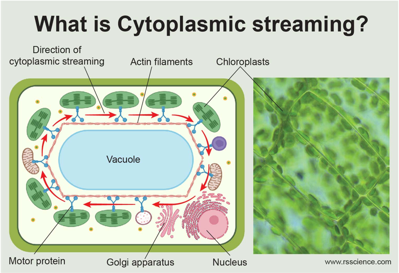 There are two possibilities,
(a)Sol-gel changes
(b)Movement of microfilaments
There are two possibilities,
(a)Sol-gel changes
(b)Movement of microfilamentsPROTOPLASM/h2>
- Protoplasm is the living part of the cell, which comprises of different cellular organelles. It is a jelly-like, colourless, transparent and viscous living substances present within the cell wall. The term protoplasm was proposed in the year 1835 and is known as the primary substance, as it is responsible for various living processes. In 1840s Huxley proposed that Protoplasm is a Physical Basis of Life. In eukaryotic cell protoplasm divides into two parts,
The Cytoplasm
The cellular material filled between the nucleus and plasma membrane is called cytoplasm, a term coined by Polisk german botanist Eduard Strasburger in 1882. It is not just a structure less (amorphous) mass of "living matter" (protoplasm) but a highly organized and compartmentalized "miniature chemical factory" in which "life" is produced.
For producing life, numerous chemical reactions of several thousand types occur every moment in the cytoplasm, so that the cellular materials are continuously metabolized (transformed). The sum total of these reactions is therefore, called cell metabolism. The different types of metabolic processes mainly occur in different types of membrane-bound compartments called Cell organelles or Organoids. All organelles are embedded in a semifluid “Intracellular milieu", or cytoplasmic matrix. Besides membrane-bound organelles, the matrix also contains certain types of non membranous organelles. Certain metabolically inactive structures containing secretory, excretory or storage materials formed as metabolic products are also usually found in this matrix. These are called Deutoplasts (metaplasts or simply cell inclusions). Thus, the cytoplasm consists of
 Cytosol
This is also called the continuous fluid phase which fills all spaces between plasma membrane, nucleus and the formed structures. It is called Cytosol or Hyaloplasm. It constitutes about half of the total volume of a cell. It is the life substance or living matter for which Felix Dujardin (1835) used the term “Sarcode", while J.E. Purkinje (1840) and Von Mohl (1846) called it “Protoplasm". Max Schultze recognized it as “Physical basis of life".
Chemical organization of cytosol: The cytosol is a complex organized mixture of mineral ions and small and large organic molecules dissolved in water (solvent) as solutes. Water constitutes about 70 to 85% by weight of the cell. Some water remains bound to proteins and other macromolecules but bulk of it, remains free to act as osmotically active solvent. Ions and molecules smaller than 0.001 μ, such as those of sugars, amino acids, nucleotides (tRNAs), enzymes, vitamins, etc occur as crystalloid solutes, whereas larger molecules such as those of proteins and polysaccharides occur as Colloidal solutes. Or Cytosol is a Crystallo-colloidal solution in which cell organelles and cell inclusions are suspended.
Cytosolic constituents remain in a continuous movement called Cytoplasmic Streaming (Amici, 1818). It is an autonomic vital movement that occurs continually in the cytoplasmic matrix of eucaryotic cells. Cytoplasm streaming is also called Protoplasmic streaming or Cyclosis. It is of two types-
Rotation and circulation.
In rotation, the cytoplasmic matrix continuously moves in one direction around a central vacuole, e.g., young leaves of Hydrilla,
In circulation, the cytoplasmic matrix moves in different directions around different vacuoles of the cell, e.g., staminal hair of Rhoeo or Tradescantia.
Cytoplasmic streaming takes part in
Movement of organelles inside the cell like chloroplasts in relation to light intensity.
Distribution of various substances in the cell.
Distribution of food vacuoles as in Amoeba and Paramecium.
Formation of pseudopodia in white blood corpuscles and Amoeba.
Quick repair of membrane and other constituents.
Distribution of heat inside the cell.
Cytosol
This is also called the continuous fluid phase which fills all spaces between plasma membrane, nucleus and the formed structures. It is called Cytosol or Hyaloplasm. It constitutes about half of the total volume of a cell. It is the life substance or living matter for which Felix Dujardin (1835) used the term “Sarcode", while J.E. Purkinje (1840) and Von Mohl (1846) called it “Protoplasm". Max Schultze recognized it as “Physical basis of life".
Chemical organization of cytosol: The cytosol is a complex organized mixture of mineral ions and small and large organic molecules dissolved in water (solvent) as solutes. Water constitutes about 70 to 85% by weight of the cell. Some water remains bound to proteins and other macromolecules but bulk of it, remains free to act as osmotically active solvent. Ions and molecules smaller than 0.001 μ, such as those of sugars, amino acids, nucleotides (tRNAs), enzymes, vitamins, etc occur as crystalloid solutes, whereas larger molecules such as those of proteins and polysaccharides occur as Colloidal solutes. Or Cytosol is a Crystallo-colloidal solution in which cell organelles and cell inclusions are suspended.
Cytosolic constituents remain in a continuous movement called Cytoplasmic Streaming (Amici, 1818). It is an autonomic vital movement that occurs continually in the cytoplasmic matrix of eucaryotic cells. Cytoplasm streaming is also called Protoplasmic streaming or Cyclosis. It is of two types-
Rotation and circulation.
In rotation, the cytoplasmic matrix continuously moves in one direction around a central vacuole, e.g., young leaves of Hydrilla,
In circulation, the cytoplasmic matrix moves in different directions around different vacuoles of the cell, e.g., staminal hair of Rhoeo or Tradescantia.
Cytoplasmic streaming takes part in
Movement of organelles inside the cell like chloroplasts in relation to light intensity.
Distribution of various substances in the cell.
Distribution of food vacuoles as in Amoeba and Paramecium.
Formation of pseudopodia in white blood corpuscles and Amoeba.
Quick repair of membrane and other constituents.
Distribution of heat inside the cell.
 There are two possibilities,
(a)Sol-gel changes
(b)Movement of microfilaments
There are two possibilities,
(a)Sol-gel changes
(b)Movement of microfilamentsENDOMEMBRANOUS SYSTEM/h2>
- While each of the membranous organelles is distinct in terms of its structure and function, many of these are considered together as an Endomembrane system because their functions are coordinated. The endomembrane system include Endoplasmic reticulum (ER), Golgi complex, Lysosomes and Vacuoles.
Endoplasmic Reticulum
It was discovered independently by Porter and Thompson (1945). The term endoplasmic reticulum was used by Keith Porter and Kallman in 1953 to identify a fine reticulum (network) in endoplasm of the cell. In 1953 Watson proposed that ER is continuous with Nuclear membrane.
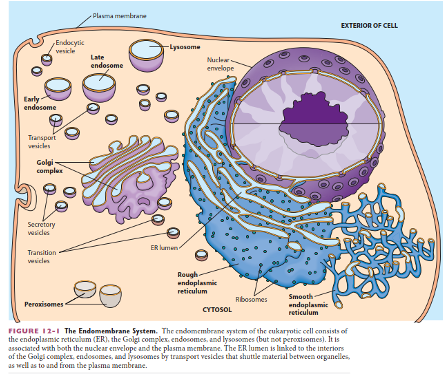 Endoplasmic reticulum
It is an irregular 3-D network of membranous tubules, forming a continuous sheet enclosing a single internal space. The extent of endoplasmic reticulum varies from cell to cell. Normally it forms 30-60% of membrane system of the cell which increases the internal surface 30-40 times as compared to external surface.
Structure of ER
Endoplasmic reticulum consists of membrane lined channels or spaces. The channels or spaces contain a fluid called Endoplasmic matrix, which is quite different from cytoplasmic matrix present outside the reticulum. The membranes of endoplasmic reticulum are 50-60 Å thick.
Endoplasmic reticulum
It is an irregular 3-D network of membranous tubules, forming a continuous sheet enclosing a single internal space. The extent of endoplasmic reticulum varies from cell to cell. Normally it forms 30-60% of membrane system of the cell which increases the internal surface 30-40 times as compared to external surface.
Structure of ER
Endoplasmic reticulum consists of membrane lined channels or spaces. The channels or spaces contain a fluid called Endoplasmic matrix, which is quite different from cytoplasmic matrix present outside the reticulum. The membranes of endoplasmic reticulum are 50-60 Å thick.
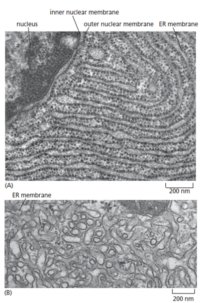 Endoplasmic reticulum consists of three parts,
 1.Cisternae: They are flat interconnected sac-like parts of the endoplasmic reticulum which are 40-50 nm in diameter. The cisternae are found in bundles where they lie parallel to one another. They occur in the cells actively involved in synthetic activity.
2.Tubules: They are tube-like extensions which may be connected with cisternae or vesicles to form a reticular system. The tubules can be irregular or regular, branched or unbranched with a diameter of 50-100 nm.
3.Vesicles: They are oval or rounded sacs of 25 - 500 nm in diameter. The vesicles appear as small vacuoles. They remain isolated in the cytoplasm. The vesicles are also called Microsomes.
4.
 Endoplasmic reticulum is quite extensive in metabolically active cells (e.g., cells of pancreas, liver), simple in storage cells (in the form of tubules in adipose cells), reduced in spermatocytes (in the form of a few vesicles), and absent in eggs, mature erythrocytes, embryonic cells, resting cells, prokaryotic cells, etc.
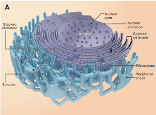 In the cells that are involved in active secretion, the Endoplasmic reticulum (ER) is studded on its outer surface with ribosomes and, because of its appearance such ER is referred to as Rough endoplasmic reticulum (RER). Whereas the cells, which produce large quantities of lipid, have ER that is not studded with ribosomes. Such ER is known as Smooth ER (SER). RER is greatly developed in those cells which are involved in the synthesis of Secretory Proteins such as Pancreatic cells, while SER is developed in those cells which participate in steroid biosynthesis and detoxification.
 The endoplasmic reticulum, apart from giving mechanical support to cytoplasmic matrix, also performs a number of functions:
⁕Synthesis of secretory (serum proteins), lysosomal or membrane proteins on the ER membrane and its transport through ER lumen,
⁕Synthesis of lipids,
⁕Detoxification of drugs and toxins (Xenobiotics)
However in Muscle cells the SER gets modified and store Ca2+ ions and called Sarcoplasmic Reticulum. Most proteins that are synthesised at ER, are glycoproteins which, after being transported to Golgi apparatus, are modified therein. Depending upon the target site, and the different fates, specific groups are added to the proteins here and then sent to their proper location enclosed in the vesicles that bud off from Golgi apparatus.
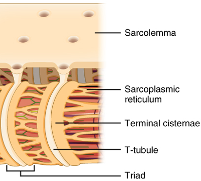 Membrane vesicles
Sphaerosomes
Sphaerosomes are single membrane bound spherical structures present in cytoplasmic matrix. They are associated with synthesis and storage of lipids. They develop from endoplasmic reticulum. In certain plants, the sphaerosomes may also posses lysosomal activity.
Lomasomes
Lomasomes are also single membrane bound spherical structures present in between plasma membrane and cell wall of Fungus. They are filled by the enzymes which help in digestion of food. They are synthesized by the ER.
Golgi Apparatus:
Golgi complex is a complex cytoplasmic structure made up of smooth membrane saccules or cisternae, a network of tubules with vesicles and vacuoles, which takes part in membrane transformation, secretion and production of complex biochemicals. It is surrounded by an organelle free cytoplasm called zone of exclusion or Golgi ground substance. They are present in all eukaryotic cells except RBC, sieve tube elements, sperms of Bryophytes and Pteridophytes.
It was first seen by George (1867) but is named after Italian scientist Camillo Golgi, who in 1898 recognised the apparatus in the nerve cells of barn owl and cat by means of metallic impregnation method, he called them as Apparato Reticular Inferno (Internal branching apparatus). In 1910 Perroncito coined Dictyosomes for Golgi Bodies. In 1914 Cajal called them as Golgi bodies after great Italian cytologist Camillo Golgi. They are also known as Baker’s Bodies, Dalton’s complexes and Terrific police man of the cell.
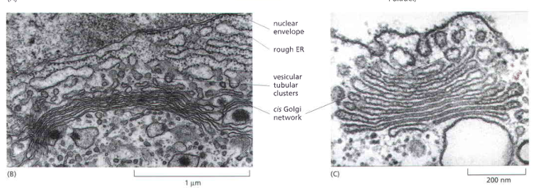 It is a membranous organelle composed of flattened sac-like cisternae stacked on one another. These cisternae resemble smooth ER. There are usually 3-8 cisternae in a stack, though the number and size of Golgi stacks vary with the cell type and with the metabolic activity of the cell. Some cells have one large stack, whereas others—especially active secretory cells—have hundreds or even thousands of Golgi stacks. Besides cisternae, tubules, vesicles and vacuoles also constitute Golgi Bodies.
On the outer edges of the cisternae, a complex network of tubules and vesicles exists; this structure appears to have a definite polarity.
The two ends or faces are quite different from one another. The sacs on the forming face (cis – face) are associated with ER and differ from the sacs present on maturing face (trans - face) in thickness, its contents and the degree of vesicle formation.
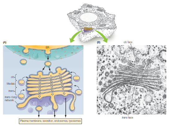 It appears that materials are transported from cis to trans face by vesicles that keep budding off from the cisternal edge to the next sac and so on.
While most of the eukaryotic cells possess Golgi apparatus, many fungi and ciliated protozoans have well-formed Golgi bodies. The cisternae in these organisms may have one stack or many. These stacks are also called Dictyosomes.
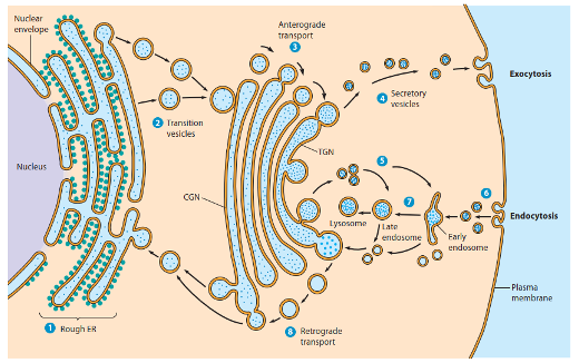 The important function of Golgi apparatus is to package the material and prepare for secretions. The material to be secreted moves from ER to the Golgi apparatus, during which vesicles are budded off from the ER. These vesicles travel to Golgi apparatus and fuse with cis – cisternae. Therefore, Golgi apparatus is closely associated with ER in structural as well.
1.Secretion. All glandular cells depend upon Golgi complex for concentrating and pouring their secretion to the outside. Secretion vesicles of the glands usually contain Zymogen Granules. They are sent out of the cells through exocytosis or reverse pinocytosis.
2.Transformation of Membranes. Golgi complex brings about membrane transformation, that is, converting one type of membrane (e.g., that of ER) into other types (e.g., selectively permeable plasma membrane, differentiated membrane of lysosome). The complex also takes part in the recycling of plasma membrane.
3.Glycoprotein. Proteins synthesised by the rough endoplasmic reticulum reach the cisternae of the Golgi apparatus. Here, they combine with carbohydrates to form glycoproteins.
4.Special Simple Carbohydrates. Sialic acid and galactose are synthesised inside Golgi complex.
5.Hormones. The production of hormones by endocrine glands is mediated through Golgi apparatus.
6.Complex Carbohydrates. Most of the complex carbohydrates, other than glycogen and starch, are synthesised inside the Golgi complex, e.g., pectic compounds, mucopolysaccharides, hyaluronic acid, chondroitin sulphate, hemicellulose, etc.
7.Matrix. Matrix of connective tissue is formed by Golgi complex of its cells.
8.Synthesis of Pigments. In Chick embryo the retinal pigment has been observed to be synthesised by Golgi complex.
9.Formation of Acrosome. Acrosome is an important constituent of the tip of animal sperms which helps in digesting away the covering sheath of the egg or ovum during fertilization
10.Vitellogenesis. In oocytes of animals, Golgi apparatus functions as the centre around which yolk is deposited. The process is called vitellogenesis.
11.Root Hair. The formation of root hair from their mother cells is believed to take place through the agency of Golgi apparatus.
12.Formation of Lysosomes. Some of the vesicles or vacuoles of the Golgi apparatus store digestive enzymes obtained through endoplasmic reticulum in the inactive state. They function as primary lysosomes.
13.Cell wall formation. In plant cells they help in the formation of cell wall by cell plate method.
Lysosomes
They were discovered accidently by a Belgian scientist, Christian de Duve, in 1955 through fractionation technique. The organelles were observed under electron microscope by Novikoff (1956). He also coined the term, Lysosomes. Lysosomes (Gk. lysis- digestive or loose, soma- body) are small vesicles which are bounded by a single membrane and contain hydrolytic enzymes in the form of minute crystalline or semi crystalline granules of 5-8 nm. They are also called Suicidal Begs of the cell. Lysosomes are known to contain more than 60 different enzymes, and have more than 50 membrane proteins. All the enzymes do not occur in the same lysosome but there are different sets of enzymes in different types of lysosomes.
The important enzymes are acid phosphatases, sulphatases, proteases, nucleases, lipases and glycosidases, They are also called acid hydrolases because these digestive enzymes usually function in acidic medium or pH less than 7. Acidic conditions are maintained inside the lysosomes by pumping of H+ or protons into them.
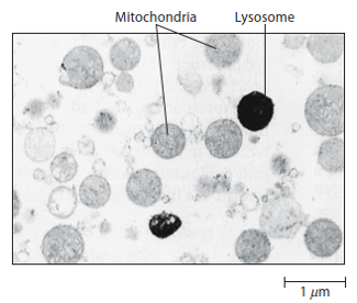 These are formed by budding off vesicles from Golgi apparatus. They are bound by a single membrane measuring about 500 nm. They are involved in intracellular digestion and contain the enzymes needed to digest all types of macromolecules. The digestive enzymes are synthesised on RER and packed into the lysosomes. The portion of SER near the Golgi apparatus may also bud off lysosomes. The covering membrane of lysosomes keeps the hydrolytic enzymes out of contact from the cellular contents.
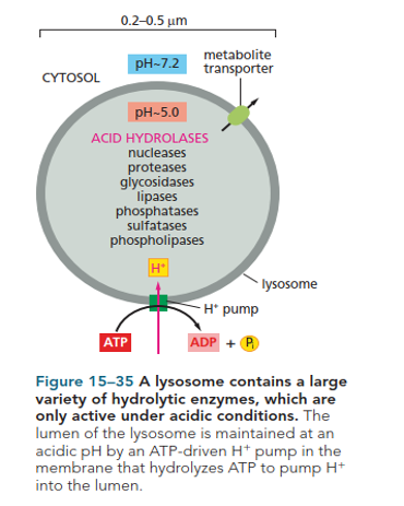 They have been recorded in fungi, Euglena, root tip cells of Maize, Cotton and Pea seeds. In other plants the function of lysosomes is shared by sphaerosomes, aleurone grains and vacuoles. In animals, lysosomes are abundant in leucocytes, macrophages, Kupffer's cells and similar cells with phagocytic activity. The diameter varies from 0.2-0.8 μm but sometimes it may grow to a very large size (upto 5 μm in leucocytes, kidney cells, etc.).
The Lysosomes are believed to be formed by the joint activity of ER and Golgi complex. The precursors of hydrolytic enzymes are mostly synthesised at the rough endoplasmic reticulum. The latter transfers them to the forming face of Golgi complex either directly or from smooth endoplasmic reticulum through its vesicles. In Golgi complex the precursors are changed to enzymes. The enzymes are then packed in larger vesicles which are pinched off as lysosomes from the maturing face. Portions of smooth endoplasmic reticulum lying near Golgi apparatus may also bud off lysosomes directly. Lysosomes do not normally burst in the cytoplasm. All materials which are to be acted by lysosome enzymes must enter them. Rather the materials are usually enclosed inside vesicles and the vacuoles fuse with the lysosomes for digestion of materials. Lysosomes take part in intracellular digestion of various types of materials of endogenous or exogenous origin. Extracellular digestion can be performed by them under certain conditions. They help in removing various toxic substances including carcinogens.
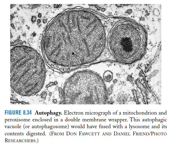 The covering membrane becomes fragile in the absence of the oxygen, or the presence of excess of vitamins A and E, male and female hormones, bile salts, X-rays and ultra-violet rays. These substances are called Membrane Labilisers. The membrane is protected from these agencies by Cortisone, Cortisol, Chloroquine and a type of cholesterol, which are termed as Membrane Stabilisers.
Lysosomes exhibit Polymorphism. Depending upon their morphology and function, there are two types of lysosomes- Primary and Secondary. Seconday Lysosomes further divide into Autophagosome, Hetrophagosome and Residual bodies.
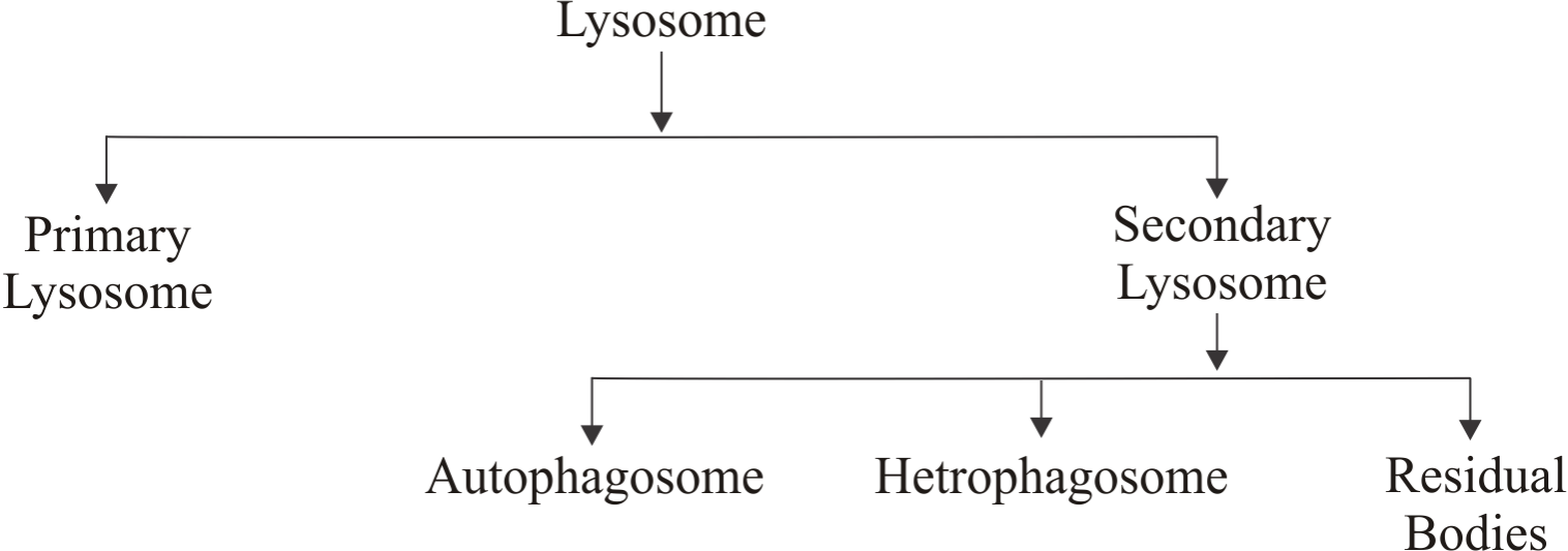 1. Primary Lysosomes: They are newly pinched off vesicles from the Golgi apparatus. The primary lysosomes are small in size. They contain hydrolytic enzymes in the form of granules.
2. Secondary Lysosomes: They are also called heterophagosomes or digestive vacuoles. A secondary lysosome is formed by the fusion of membrane vesicles with lysosome (having hydrolytic or digestive enzymes). Secondary Lysosomes are of three types,
Autophagosome
Autophagic Vacuoles (Autophagosomes, Autolysosomes). They are produced by the fusion of a number of primary lysosomes around worn out or degenerate intracellular organelles. The latter are digested. The phenomenon is also called autophagy or autodigestion. It helps in disposal of cell debris. Therefore, lysosomes are also called disposal bags or disposal units. The digested products are made available to the cell for new synthesis. Lysosomes are, therefore, also known as recycling centres. Besides removing worn out organelles, old or diseased cells, the autophagic vacuoles are also used in removing internal obstructions. Autophagic vacuoles provide nourishment during starvation (de Duve, 1967).
Hetrophagosome
They are also called heterophagosomes or digestive vacuoles. A Hetrophagosome is formed by the fusion of food containing phagosome with lysosome (having hydrolytic or digestive enzymes). Digestion occurs. The digested food passes out into the cytoplasm. Ultimately, the secondary lysosome is left with only undigested or indigestible food.
Residual Bodies
(Residual or Tertiary Lysosomes). They are those lysosomes in which only indigestible food materials have been left. The residual bodies or lysosomes pass outwardly and fuse with the plasma membrane to throw out the debris into external environment by exocytosis or ephagy. Sometimes, residual bodies remain inside the cells due to
(i)Failure of exocytosis and
(ii)Absence of some hydrolytic enzymes. This leads to pathological diseases called Storage Disorders like hepatitis and polynephritis. Ageing is also due to them.
Lysosomes storage disorders
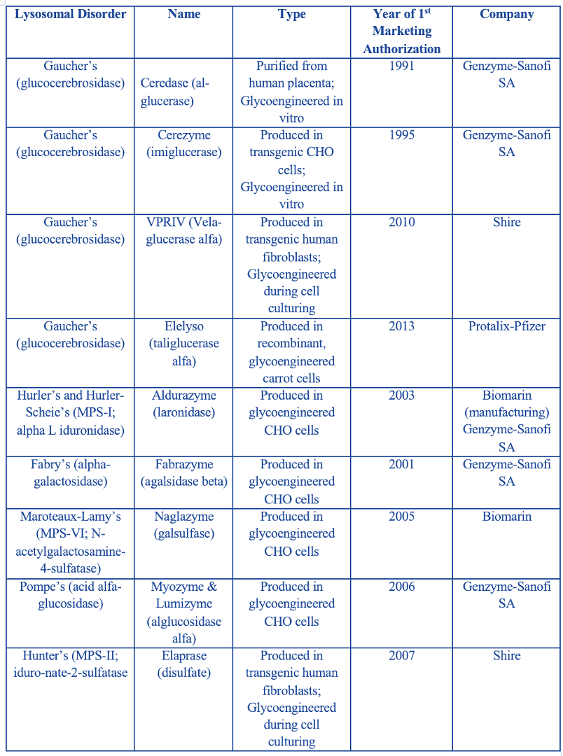 Microbodies
 are also single membrane bound organelles, associated with oxidation reaction other than those of respiration. These organelles often possesses a crystalloid core and granular matrix. There are two types of microbodies namely Peroxisomes and Glyoxysomes. The peroxisomes have enzymes for peroxide biosynthesis. These occur in most of the animals and plants, but are quite common in photosynthetic cells. Their number can be 70-100 per mesophyll cell wherein they interact with mitochondria and chloroplasts to take part in photorespiration. The glyoxysomes usually occur in fat-rich plant cells and are associated with triglyceride metabolism through glyoxylate cycle.
Microbodies
Microbodies are also single membrane bound organelles, associated with oxidation reaction other than those of respiration. These organelles often possesses a crystalloid core and granular matrix. There are two types of microbodies namely Peroxisomes and Glyoxysomes. The peroxisomes have enzymes for peroxide biosynthesis. These occur in most of the animals and plants, but are quite common in photosynthetic cells. Their number can be 70-100 per mesophyll cell wherein they interact with mitochondria and chloroplasts to take part in photorespiration. The glyoxysomes usually occur in fat-rich plant cells and are associated with triglyceride metabolism through glyoxylate cycle.
 Peroxisomes
A peroxisome is a membrane-bound organelle (formerly known as a microbody), found in the cytoplasm of virtually all eukaryotic cells. Peroxisomes are oxidative organelles. Peroxisomes owe their name to hydrogen peroxide generating and scavenging activities. They perform key roles in lipid metabolism and the conversion of reactive oxygen species. Peroxisomes (microbodies) were first described by a Swedish doctoral student, J. Rhodin in 1954. They were identified as organelles by the Belgian cytologist Christian de Duve in 1967. De Duve and co-workers discovered that peroxisomes contain several oxidases involved in the production of hydrogen peroxide (H2O2) as well as catalase involved in the decomposition of H2O2 to oxygen and water. Due to their role in peroxide metabolism, De Duve named them “Peroxisomes”, replacing the formerly used morphological term “microbodies”.
Structure:
Peroxisomes are small (0.1–1 µm diameter) subcellular compartments (organelles) with a fine, granular matrix and surrounded by a single biomembrane which are located in the cytoplasm of a cell. The number, size and protein composition of peroxisomes are variable and depend on cell type and environmental conditions.
The Peroxisomes are present in all eukaryotic cells and participate in following metabolic pathways,
1.Oxidation of fatty acids
2.Nitrogen metabolism
3.Biosynthesis of Cholesterol, Bile salts and Plasmalogens.
4.Enzymes for production of Hydrogen peroxide (H2O2) and its control.
1.Oxidation of fatty acids:
In peroxisomes both α and β oxidation of fats take place. Particularly oxidation of vary long and polyunsaturated fatty acids take place in peroxisomes apporx, 20 to 50% fatty acids are oxidized in Peroxisomes.
Peroxisomes
A peroxisome is a membrane-bound organelle (formerly known as a microbody), found in the cytoplasm of virtually all eukaryotic cells. Peroxisomes are oxidative organelles. Peroxisomes owe their name to hydrogen peroxide generating and scavenging activities. They perform key roles in lipid metabolism and the conversion of reactive oxygen species. Peroxisomes (microbodies) were first described by a Swedish doctoral student, J. Rhodin in 1954. They were identified as organelles by the Belgian cytologist Christian de Duve in 1967. De Duve and co-workers discovered that peroxisomes contain several oxidases involved in the production of hydrogen peroxide (H2O2) as well as catalase involved in the decomposition of H2O2 to oxygen and water. Due to their role in peroxide metabolism, De Duve named them “Peroxisomes”, replacing the formerly used morphological term “microbodies”.
Structure:
Peroxisomes are small (0.1–1 µm diameter) subcellular compartments (organelles) with a fine, granular matrix and surrounded by a single biomembrane which are located in the cytoplasm of a cell. The number, size and protein composition of peroxisomes are variable and depend on cell type and environmental conditions.
The Peroxisomes are present in all eukaryotic cells and participate in following metabolic pathways,
1.Oxidation of fatty acids
2.Nitrogen metabolism
3.Biosynthesis of Cholesterol, Bile salts and Plasmalogens.
4.Enzymes for production of Hydrogen peroxide (H2O2) and its control.
1.Oxidation of fatty acids:
In peroxisomes both α and β oxidation of fats take place. Particularly oxidation of vary long and polyunsaturated fatty acids take place in peroxisomes apporx, 20 to 50% fatty acids are oxidized in Peroxisomes.
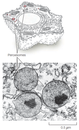 2.Nitrogen metabolism
Many nitrogenous compounds are metabolized in peroxisomes such as Urea and Purines. The enzymes like Urate oxidase participate in it and richly present in Peroxisomes.
3.Biosynthesis of Cholesterol, Bile salts and Plasmalogens.
In addition to providing a compartment for oxidation reactions, peroxisomes are involved in biosynthesis of lipids. In animal cells, Cholesterol and Dolichol are synthesized in peroxisomes as well as in the ER. In the liver, peroxisomes are also involved in the synthesis of Bile acids, which are derived from cholesterol. In addition, peroxisomes contain enzymes required for the synthesis of Plasmalogens —A family of phospholipids in which one of the hydrocarbon chains is joined to glycerol by an ether bond rather than by an ester bond. Plasmalogens are important membrane components in some tissues, particularly heart and brain, although they are absent in others.
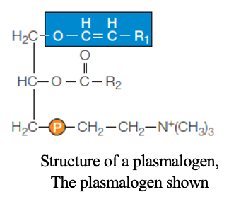 4.Enzymes for production of Hydrogen peroxide (H2O2) and its control.
Peroxisomes are so named because they usually contain one or more enzymes that use molecular oxygen to remove hydrogen atoms from specific organic substrates and produce hydrogen peroxide (H2O2). Catalase uses the H2O2 generated by other enzymes in the organelle to oxidize a variety of other substrates—including phenols, formic acid, formaldehyde, and alcohol—by the “peroxidation” reaction. This type of oxidation reaction is particularly important in liver and kidney cells, where the peroxisomes detoxify various toxic molecules that enter the blood-stream. About 25% of the ethanol we drink is oxidized to acetaldehyde in this way. Peroxisomes are also important in plants. Two types of plant peroxisomes have been studied extensively. One is present in leaves, where it participates in Photorespiration.
The other type of peroxisome is present in germinating seeds, where it converts the fatty acids stored in seed lipids into the sugars needed for the growth of the young plant.
 In 1961 Bill Breidenbach reported Glyoxylate cycle. He further reported that enzymes for Glyoxylate cycle are not present in mitochondria. In 1967 Breidenbach and R. W., Beevers H. reported that the enzymes of Glyoxylate cycle are present in specialized organelles present in seeds called Glyoxysomes. These Glyoxysomes are present in seedlings of plant speciesthat store carbon and energy reserves in the seed as fat (primarily triacylglycerols). In such species, stored triacylglycerols are mobilized and converted to sucrose during early postgerminative development by a sequence of events that includes β oxidation of fatty acids as well as a pathway known as the glyoxylate cycle.
Glyoxysomes are found only in the tissues where the fat is stored (endosperm or cotyledons, depending on the species) and are present only for the relatively short period of time required for the seedling to deplete its supply of stored fat. Once they fulfill their role in the seedling, the glyoxysomes are converted to peroxisomes.
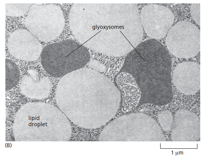
ENDOMEMBRANOUS SYSTEM/h2>
- While each of the membranous organelles is distinct in terms of its structure and function, many of these are considered together as an Endomembrane system because their functions are coordinated. The endomembrane system include Endoplasmic reticulum (ER), Golgi complex, Lysosomes and Vacuoles.
Endoplasmic Reticulum
It was discovered independently by Porter and Thompson (1945). The term endoplasmic reticulum was used by Keith Porter and Kallman in 1953 to identify a fine reticulum (network) in endoplasm of the cell. In 1953 Watson proposed that ER is continuous with Nuclear membrane.
 Endoplasmic reticulum
It is an irregular 3-D network of membranous tubules, forming a continuous sheet enclosing a single internal space. The extent of endoplasmic reticulum varies from cell to cell. Normally it forms 30-60% of membrane system of the cell which increases the internal surface 30-40 times as compared to external surface.
Structure of ER
Endoplasmic reticulum consists of membrane lined channels or spaces. The channels or spaces contain a fluid called Endoplasmic matrix, which is quite different from cytoplasmic matrix present outside the reticulum. The membranes of endoplasmic reticulum are 50-60 Å thick.
Endoplasmic reticulum
It is an irregular 3-D network of membranous tubules, forming a continuous sheet enclosing a single internal space. The extent of endoplasmic reticulum varies from cell to cell. Normally it forms 30-60% of membrane system of the cell which increases the internal surface 30-40 times as compared to external surface.
Structure of ER
Endoplasmic reticulum consists of membrane lined channels or spaces. The channels or spaces contain a fluid called Endoplasmic matrix, which is quite different from cytoplasmic matrix present outside the reticulum. The membranes of endoplasmic reticulum are 50-60 Å thick.
 Endoplasmic reticulum consists of three parts,
 1.Cisternae: They are flat interconnected sac-like parts of the endoplasmic reticulum which are 40-50 nm in diameter. The cisternae are found in bundles where they lie parallel to one another. They occur in the cells actively involved in synthetic activity.
2.Tubules: They are tube-like extensions which may be connected with cisternae or vesicles to form a reticular system. The tubules can be irregular or regular, branched or unbranched with a diameter of 50-100 nm.
3.Vesicles: They are oval or rounded sacs of 25 - 500 nm in diameter. The vesicles appear as small vacuoles. They remain isolated in the cytoplasm. The vesicles are also called Microsomes.
4.
 Endoplasmic reticulum is quite extensive in metabolically active cells (e.g., cells of pancreas, liver), simple in storage cells (in the form of tubules in adipose cells), reduced in spermatocytes (in the form of a few vesicles), and absent in eggs, mature erythrocytes, embryonic cells, resting cells, prokaryotic cells, etc.
 In the cells that are involved in active secretion, the Endoplasmic reticulum (ER) is studded on its outer surface with ribosomes and, because of its appearance such ER is referred to as Rough endoplasmic reticulum (RER). Whereas the cells, which produce large quantities of lipid, have ER that is not studded with ribosomes. Such ER is known as Smooth ER (SER). RER is greatly developed in those cells which are involved in the synthesis of Secretory Proteins such as Pancreatic cells, while SER is developed in those cells which participate in steroid biosynthesis and detoxification.
 The endoplasmic reticulum, apart from giving mechanical support to cytoplasmic matrix, also performs a number of functions:
⁕Synthesis of secretory (serum proteins), lysosomal or membrane proteins on the ER membrane and its transport through ER lumen,
⁕Synthesis of lipids,
⁕Detoxification of drugs and toxins (Xenobiotics)
However in Muscle cells the SER gets modified and store Ca2+ ions and called Sarcoplasmic Reticulum. Most proteins that are synthesised at ER, are glycoproteins which, after being transported to Golgi apparatus, are modified therein. Depending upon the target site, and the different fates, specific groups are added to the proteins here and then sent to their proper location enclosed in the vesicles that bud off from Golgi apparatus.
 Membrane vesicles
Sphaerosomes
Sphaerosomes are single membrane bound spherical structures present in cytoplasmic matrix. They are associated with synthesis and storage of lipids. They develop from endoplasmic reticulum. In certain plants, the sphaerosomes may also posses lysosomal activity.
Lomasomes
Lomasomes are also single membrane bound spherical structures present in between plasma membrane and cell wall of Fungus. They are filled by the enzymes which help in digestion of food. They are synthesized by the ER.
Golgi Apparatus:
Golgi complex is a complex cytoplasmic structure made up of smooth membrane saccules or cisternae, a network of tubules with vesicles and vacuoles, which takes part in membrane transformation, secretion and production of complex biochemicals. It is surrounded by an organelle free cytoplasm called zone of exclusion or Golgi ground substance. They are present in all eukaryotic cells except RBC, sieve tube elements, sperms of Bryophytes and Pteridophytes.
It was first seen by George (1867) but is named after Italian scientist Camillo Golgi, who in 1898 recognised the apparatus in the nerve cells of barn owl and cat by means of metallic impregnation method, he called them as Apparato Reticular Inferno (Internal branching apparatus). In 1910 Perroncito coined Dictyosomes for Golgi Bodies. In 1914 Cajal called them as Golgi bodies after great Italian cytologist Camillo Golgi. They are also known as Baker’s Bodies, Dalton’s complexes and Terrific police man of the cell.
 It is a membranous organelle composed of flattened sac-like cisternae stacked on one another. These cisternae resemble smooth ER. There are usually 3-8 cisternae in a stack, though the number and size of Golgi stacks vary with the cell type and with the metabolic activity of the cell. Some cells have one large stack, whereas others—especially active secretory cells—have hundreds or even thousands of Golgi stacks. Besides cisternae, tubules, vesicles and vacuoles also constitute Golgi Bodies.
On the outer edges of the cisternae, a complex network of tubules and vesicles exists; this structure appears to have a definite polarity.
The two ends or faces are quite different from one another. The sacs on the forming face (cis – face) are associated with ER and differ from the sacs present on maturing face (trans - face) in thickness, its contents and the degree of vesicle formation.
 It appears that materials are transported from cis to trans face by vesicles that keep budding off from the cisternal edge to the next sac and so on.
While most of the eukaryotic cells possess Golgi apparatus, many fungi and ciliated protozoans have well-formed Golgi bodies. The cisternae in these organisms may have one stack or many. These stacks are also called Dictyosomes.
 The important function of Golgi apparatus is to package the material and prepare for secretions. The material to be secreted moves from ER to the Golgi apparatus, during which vesicles are budded off from the ER. These vesicles travel to Golgi apparatus and fuse with cis – cisternae. Therefore, Golgi apparatus is closely associated with ER in structural as well.
1.Secretion. All glandular cells depend upon Golgi complex for concentrating and pouring their secretion to the outside. Secretion vesicles of the glands usually contain Zymogen Granules. They are sent out of the cells through exocytosis or reverse pinocytosis.
2.Transformation of Membranes. Golgi complex brings about membrane transformation, that is, converting one type of membrane (e.g., that of ER) into other types (e.g., selectively permeable plasma membrane, differentiated membrane of lysosome). The complex also takes part in the recycling of plasma membrane.
3.Glycoprotein. Proteins synthesised by the rough endoplasmic reticulum reach the cisternae of the Golgi apparatus. Here, they combine with carbohydrates to form glycoproteins.
4.Special Simple Carbohydrates. Sialic acid and galactose are synthesised inside Golgi complex.
5.Hormones. The production of hormones by endocrine glands is mediated through Golgi apparatus.
6.Complex Carbohydrates. Most of the complex carbohydrates, other than glycogen and starch, are synthesised inside the Golgi complex, e.g., pectic compounds, mucopolysaccharides, hyaluronic acid, chondroitin sulphate, hemicellulose, etc.
7.Matrix. Matrix of connective tissue is formed by Golgi complex of its cells.
8.Synthesis of Pigments. In Chick embryo the retinal pigment has been observed to be synthesised by Golgi complex.
9.Formation of Acrosome. Acrosome is an important constituent of the tip of animal sperms which helps in digesting away the covering sheath of the egg or ovum during fertilization
10.Vitellogenesis. In oocytes of animals, Golgi apparatus functions as the centre around which yolk is deposited. The process is called vitellogenesis.
11.Root Hair. The formation of root hair from their mother cells is believed to take place through the agency of Golgi apparatus.
12.Formation of Lysosomes. Some of the vesicles or vacuoles of the Golgi apparatus store digestive enzymes obtained through endoplasmic reticulum in the inactive state. They function as primary lysosomes.
13.Cell wall formation. In plant cells they help in the formation of cell wall by cell plate method.
Lysosomes
They were discovered accidently by a Belgian scientist, Christian de Duve, in 1955 through fractionation technique. The organelles were observed under electron microscope by Novikoff (1956). He also coined the term, Lysosomes. Lysosomes (Gk. lysis- digestive or loose, soma- body) are small vesicles which are bounded by a single membrane and contain hydrolytic enzymes in the form of minute crystalline or semi crystalline granules of 5-8 nm. They are also called Suicidal Begs of the cell. Lysosomes are known to contain more than 60 different enzymes, and have more than 50 membrane proteins. All the enzymes do not occur in the same lysosome but there are different sets of enzymes in different types of lysosomes.
The important enzymes are acid phosphatases, sulphatases, proteases, nucleases, lipases and glycosidases, They are also called acid hydrolases because these digestive enzymes usually function in acidic medium or pH less than 7. Acidic conditions are maintained inside the lysosomes by pumping of H+ or protons into them.
 These are formed by budding off vesicles from Golgi apparatus. They are bound by a single membrane measuring about 500 nm. They are involved in intracellular digestion and contain the enzymes needed to digest all types of macromolecules. The digestive enzymes are synthesised on RER and packed into the lysosomes. The portion of SER near the Golgi apparatus may also bud off lysosomes. The covering membrane of lysosomes keeps the hydrolytic enzymes out of contact from the cellular contents.
 They have been recorded in fungi, Euglena, root tip cells of Maize, Cotton and Pea seeds. In other plants the function of lysosomes is shared by sphaerosomes, aleurone grains and vacuoles. In animals, lysosomes are abundant in leucocytes, macrophages, Kupffer's cells and similar cells with phagocytic activity. The diameter varies from 0.2-0.8 μm but sometimes it may grow to a very large size (upto 5 μm in leucocytes, kidney cells, etc.).
The Lysosomes are believed to be formed by the joint activity of ER and Golgi complex. The precursors of hydrolytic enzymes are mostly synthesised at the rough endoplasmic reticulum. The latter transfers them to the forming face of Golgi complex either directly or from smooth endoplasmic reticulum through its vesicles. In Golgi complex the precursors are changed to enzymes. The enzymes are then packed in larger vesicles which are pinched off as lysosomes from the maturing face. Portions of smooth endoplasmic reticulum lying near Golgi apparatus may also bud off lysosomes directly. Lysosomes do not normally burst in the cytoplasm. All materials which are to be acted by lysosome enzymes must enter them. Rather the materials are usually enclosed inside vesicles and the vacuoles fuse with the lysosomes for digestion of materials. Lysosomes take part in intracellular digestion of various types of materials of endogenous or exogenous origin. Extracellular digestion can be performed by them under certain conditions. They help in removing various toxic substances including carcinogens.
 The covering membrane becomes fragile in the absence of the oxygen, or the presence of excess of vitamins A and E, male and female hormones, bile salts, X-rays and ultra-violet rays. These substances are called Membrane Labilisers. The membrane is protected from these agencies by Cortisone, Cortisol, Chloroquine and a type of cholesterol, which are termed as Membrane Stabilisers.
Lysosomes exhibit Polymorphism. Depending upon their morphology and function, there are two types of lysosomes- Primary and Secondary. Seconday Lysosomes further divide into Autophagosome, Hetrophagosome and Residual bodies.
 1. Primary Lysosomes: They are newly pinched off vesicles from the Golgi apparatus. The primary lysosomes are small in size. They contain hydrolytic enzymes in the form of granules.
2. Secondary Lysosomes: They are also called heterophagosomes or digestive vacuoles. A secondary lysosome is formed by the fusion of membrane vesicles with lysosome (having hydrolytic or digestive enzymes). Secondary Lysosomes are of three types,
Autophagosome
Autophagic Vacuoles (Autophagosomes, Autolysosomes). They are produced by the fusion of a number of primary lysosomes around worn out or degenerate intracellular organelles. The latter are digested. The phenomenon is also called autophagy or autodigestion. It helps in disposal of cell debris. Therefore, lysosomes are also called disposal bags or disposal units. The digested products are made available to the cell for new synthesis. Lysosomes are, therefore, also known as recycling centres. Besides removing worn out organelles, old or diseased cells, the autophagic vacuoles are also used in removing internal obstructions. Autophagic vacuoles provide nourishment during starvation (de Duve, 1967).
Hetrophagosome
They are also called heterophagosomes or digestive vacuoles. A Hetrophagosome is formed by the fusion of food containing phagosome with lysosome (having hydrolytic or digestive enzymes). Digestion occurs. The digested food passes out into the cytoplasm. Ultimately, the secondary lysosome is left with only undigested or indigestible food.
Residual Bodies
(Residual or Tertiary Lysosomes). They are those lysosomes in which only indigestible food materials have been left. The residual bodies or lysosomes pass outwardly and fuse with the plasma membrane to throw out the debris into external environment by exocytosis or ephagy. Sometimes, residual bodies remain inside the cells due to
(i)Failure of exocytosis and
(ii)Absence of some hydrolytic enzymes. This leads to pathological diseases called Storage Disorders like hepatitis and polynephritis. Ageing is also due to them.
Lysosomes storage disorders
 Microbodies
 are also single membrane bound organelles, associated with oxidation reaction other than those of respiration. These organelles often possesses a crystalloid core and granular matrix. There are two types of microbodies namely Peroxisomes and Glyoxysomes. The peroxisomes have enzymes for peroxide biosynthesis. These occur in most of the animals and plants, but are quite common in photosynthetic cells. Their number can be 70-100 per mesophyll cell wherein they interact with mitochondria and chloroplasts to take part in photorespiration. The glyoxysomes usually occur in fat-rich plant cells and are associated with triglyceride metabolism through glyoxylate cycle.
Microbodies
Microbodies are also single membrane bound organelles, associated with oxidation reaction other than those of respiration. These organelles often possesses a crystalloid core and granular matrix. There are two types of microbodies namely Peroxisomes and Glyoxysomes. The peroxisomes have enzymes for peroxide biosynthesis. These occur in most of the animals and plants, but are quite common in photosynthetic cells. Their number can be 70-100 per mesophyll cell wherein they interact with mitochondria and chloroplasts to take part in photorespiration. The glyoxysomes usually occur in fat-rich plant cells and are associated with triglyceride metabolism through glyoxylate cycle.
 Peroxisomes
A peroxisome is a membrane-bound organelle (formerly known as a microbody), found in the cytoplasm of virtually all eukaryotic cells. Peroxisomes are oxidative organelles. Peroxisomes owe their name to hydrogen peroxide generating and scavenging activities. They perform key roles in lipid metabolism and the conversion of reactive oxygen species. Peroxisomes (microbodies) were first described by a Swedish doctoral student, J. Rhodin in 1954. They were identified as organelles by the Belgian cytologist Christian de Duve in 1967. De Duve and co-workers discovered that peroxisomes contain several oxidases involved in the production of hydrogen peroxide (H2O2) as well as catalase involved in the decomposition of H2O2 to oxygen and water. Due to their role in peroxide metabolism, De Duve named them “Peroxisomes”, replacing the formerly used morphological term “microbodies”.
Structure:
Peroxisomes are small (0.1–1 µm diameter) subcellular compartments (organelles) with a fine, granular matrix and surrounded by a single biomembrane which are located in the cytoplasm of a cell. The number, size and protein composition of peroxisomes are variable and depend on cell type and environmental conditions.
The Peroxisomes are present in all eukaryotic cells and participate in following metabolic pathways,
1.Oxidation of fatty acids
2.Nitrogen metabolism
3.Biosynthesis of Cholesterol, Bile salts and Plasmalogens.
4.Enzymes for production of Hydrogen peroxide (H2O2) and its control.
1.Oxidation of fatty acids:
In peroxisomes both α and β oxidation of fats take place. Particularly oxidation of vary long and polyunsaturated fatty acids take place in peroxisomes apporx, 20 to 50% fatty acids are oxidized in Peroxisomes.
Peroxisomes
A peroxisome is a membrane-bound organelle (formerly known as a microbody), found in the cytoplasm of virtually all eukaryotic cells. Peroxisomes are oxidative organelles. Peroxisomes owe their name to hydrogen peroxide generating and scavenging activities. They perform key roles in lipid metabolism and the conversion of reactive oxygen species. Peroxisomes (microbodies) were first described by a Swedish doctoral student, J. Rhodin in 1954. They were identified as organelles by the Belgian cytologist Christian de Duve in 1967. De Duve and co-workers discovered that peroxisomes contain several oxidases involved in the production of hydrogen peroxide (H2O2) as well as catalase involved in the decomposition of H2O2 to oxygen and water. Due to their role in peroxide metabolism, De Duve named them “Peroxisomes”, replacing the formerly used morphological term “microbodies”.
Structure:
Peroxisomes are small (0.1–1 µm diameter) subcellular compartments (organelles) with a fine, granular matrix and surrounded by a single biomembrane which are located in the cytoplasm of a cell. The number, size and protein composition of peroxisomes are variable and depend on cell type and environmental conditions.
The Peroxisomes are present in all eukaryotic cells and participate in following metabolic pathways,
1.Oxidation of fatty acids
2.Nitrogen metabolism
3.Biosynthesis of Cholesterol, Bile salts and Plasmalogens.
4.Enzymes for production of Hydrogen peroxide (H2O2) and its control.
1.Oxidation of fatty acids:
In peroxisomes both α and β oxidation of fats take place. Particularly oxidation of vary long and polyunsaturated fatty acids take place in peroxisomes apporx, 20 to 50% fatty acids are oxidized in Peroxisomes.
 2.Nitrogen metabolism
Many nitrogenous compounds are metabolized in peroxisomes such as Urea and Purines. The enzymes like Urate oxidase participate in it and richly present in Peroxisomes.
3.Biosynthesis of Cholesterol, Bile salts and Plasmalogens.
In addition to providing a compartment for oxidation reactions, peroxisomes are involved in biosynthesis of lipids. In animal cells, Cholesterol and Dolichol are synthesized in peroxisomes as well as in the ER. In the liver, peroxisomes are also involved in the synthesis of Bile acids, which are derived from cholesterol. In addition, peroxisomes contain enzymes required for the synthesis of Plasmalogens —A family of phospholipids in which one of the hydrocarbon chains is joined to glycerol by an ether bond rather than by an ester bond. Plasmalogens are important membrane components in some tissues, particularly heart and brain, although they are absent in others.
 4.Enzymes for production of Hydrogen peroxide (H2O2) and its control.
Peroxisomes are so named because they usually contain one or more enzymes that use molecular oxygen to remove hydrogen atoms from specific organic substrates and produce hydrogen peroxide (H2O2). Catalase uses the H2O2 generated by other enzymes in the organelle to oxidize a variety of other substrates—including phenols, formic acid, formaldehyde, and alcohol—by the “peroxidation” reaction. This type of oxidation reaction is particularly important in liver and kidney cells, where the peroxisomes detoxify various toxic molecules that enter the blood-stream. About 25% of the ethanol we drink is oxidized to acetaldehyde in this way. Peroxisomes are also important in plants. Two types of plant peroxisomes have been studied extensively. One is present in leaves, where it participates in Photorespiration.
The other type of peroxisome is present in germinating seeds, where it converts the fatty acids stored in seed lipids into the sugars needed for the growth of the young plant.
 In 1961 Bill Breidenbach reported Glyoxylate cycle. He further reported that enzymes for Glyoxylate cycle are not present in mitochondria. In 1967 Breidenbach and R. W., Beevers H. reported that the enzymes of Glyoxylate cycle are present in specialized organelles present in seeds called Glyoxysomes. These Glyoxysomes are present in seedlings of plant speciesthat store carbon and energy reserves in the seed as fat (primarily triacylglycerols). In such species, stored triacylglycerols are mobilized and converted to sucrose during early postgerminative development by a sequence of events that includes β oxidation of fatty acids as well as a pathway known as the glyoxylate cycle.
Glyoxysomes are found only in the tissues where the fat is stored (endosperm or cotyledons, depending on the species) and are present only for the relatively short period of time required for the seedling to deplete its supply of stored fat. Once they fulfill their role in the seedling, the glyoxysomes are converted to peroxisomes.

RIBOSOMES
- In 1943 Albert Claude first observed the tiny particles or granules of ribonucleoprotein and lipid under an electron microscope and he called them as Microsomes. Later on romanian born American Cell Biologist George E. Palade discovered it as dense particles or granules of the cytoplasm in 1955. In 1958, Richard B. Roberts proposed the name “Ribosome”.
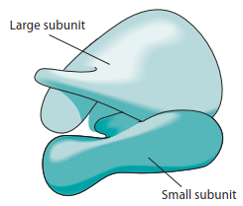 George E. Palade and other scientists discovered that ribosomes are the protein synthesizer in the cells and for this reason, in 1974; Palade was awarded the Nobel Prize for his great job. Ribosomes are remarkably plentiful in cells. A mammalian cell contains about 10 billion protein molecules and most of them are produced from the ribosomes. One active eukaryotic cell contains about 10 million ribosomes while the prokaryotic cell (Escherichia coli) contain about 15000-20000 ribosomes which are equal about 25% of the total cell mass. The average size of the ribosomes of the Escherichia coli is 200 Å in diameter.
 Types of Ribosome
Ribosomes are of two types on the basis of the size and sedimentation coefficient (S) such as the 70S and 80S. Here, ‘S’ refers to Svedberg unit. This is a sedimentation coefficient which shows how fast a cell organelle sediments in an ultracentrifuge.
70S Ribosome: This type of ribosome is comparatively smaller and has sedimentation coefficient 70S which consists of two subunits such as large 50S subunit and small 30S subunit and they are linked together. The molecular weight is 2.7×10⁶ Daltons. They are found in all prokaryotic cells.
 In eukaryotes ribosomes are of two types,
 Organellar Ribosomes are present in Mitochondria and Chloroplast, they are of 55S to 70S and of Prokaryotic type, however in human mitochondria Ribosomes are of 55S.
Cytosolic ribosomes are of 80S type and present in all eukaryotic cells. 80S Ribosome: This type of ribosome is comparatively larger and has sedimentation coefficient 80S which consists of large circular 60S subunit and small elliptical 40S subunit. The molecular weight is 40×106 Daltons.
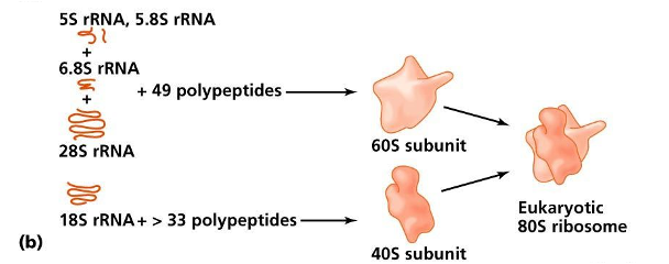 Chemical Structure of Ribosome
Chemically, ribosomes consist of ribosomal proteins and rRNA (ribosomal RNA). In the prokaryotic cells, ribosomes have 40% protein and 60% rRNA while in eukaryotic cells, ribosomes are made up of about 50% protein and 50% rRNA. Besides these, ribosomes also contain trace amounts of Magnesium (Mg), Calcium(Ca) and manganese(Mn).
 Functions: Ribosomes are factories of protein synthesis. During protein synthesis the two subunits of ribosomes joins with each other along with m-RNA. During protein synthesis many ribosomes join with the same m-RNA and form Polysomes.
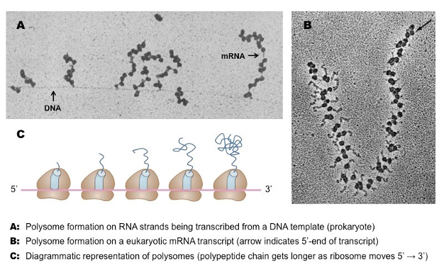 For the joining of two subunits of ribosomes Mg2+ is required.
RIBOSOMES
- In 1943 Albert Claude first observed the tiny particles or granules of ribonucleoprotein and lipid under an electron microscope and he called them as Microsomes. Later on romanian born American Cell Biologist George E. Palade discovered it as dense particles or granules of the cytoplasm in 1955. In 1958, Richard B. Roberts proposed the name “Ribosome”.
 George E. Palade and other scientists discovered that ribosomes are the protein synthesizer in the cells and for this reason, in 1974; Palade was awarded the Nobel Prize for his great job. Ribosomes are remarkably plentiful in cells. A mammalian cell contains about 10 billion protein molecules and most of them are produced from the ribosomes. One active eukaryotic cell contains about 10 million ribosomes while the prokaryotic cell (Escherichia coli) contain about 15000-20000 ribosomes which are equal about 25% of the total cell mass. The average size of the ribosomes of the Escherichia coli is 200 Å in diameter.
 Types of Ribosome
Ribosomes are of two types on the basis of the size and sedimentation coefficient (S) such as the 70S and 80S. Here, ‘S’ refers to Svedberg unit. This is a sedimentation coefficient which shows how fast a cell organelle sediments in an ultracentrifuge.
70S Ribosome: This type of ribosome is comparatively smaller and has sedimentation coefficient 70S which consists of two subunits such as large 50S subunit and small 30S subunit and they are linked together. The molecular weight is 2.7×10⁶ Daltons. They are found in all prokaryotic cells.
 In eukaryotes ribosomes are of two types,
 Organellar Ribosomes are present in Mitochondria and Chloroplast, they are of 55S to 70S and of Prokaryotic type, however in human mitochondria Ribosomes are of 55S.
Cytosolic ribosomes are of 80S type and present in all eukaryotic cells. 80S Ribosome: This type of ribosome is comparatively larger and has sedimentation coefficient 80S which consists of large circular 60S subunit and small elliptical 40S subunit. The molecular weight is 40×106 Daltons.
 Chemical Structure of Ribosome
Chemically, ribosomes consist of ribosomal proteins and rRNA (ribosomal RNA). In the prokaryotic cells, ribosomes have 40% protein and 60% rRNA while in eukaryotic cells, ribosomes are made up of about 50% protein and 50% rRNA. Besides these, ribosomes also contain trace amounts of Magnesium (Mg), Calcium(Ca) and manganese(Mn).
 Functions: Ribosomes are factories of protein synthesis. During protein synthesis the two subunits of ribosomes joins with each other along with m-RNA. During protein synthesis many ribosomes join with the same m-RNA and form Polysomes.
 For the joining of two subunits of ribosomes Mg2+ is required.
MITOCHONDRIA
- Mitochondria are called as Power House of the cell. Mitochondria were seen by Kolliker in 1850 in insect flight muscle cells and called them as Sarcosome. In 1890 Altmann studied them and called them as Bioplast. In 1898 Benda stained them with Cystal Violet and coined the word Mitochondria. In 1900 Michaelis stained them with the help of Janus Green B. In 1912 Kinsbury called them as Center of Aerobic Respiration. In 1913 Warburg discovered respiratory enzymes in them. In 1930 Hans Kreb reported Kreb Cycle enzymes in the matrix of mitochondria.
Mitochondria are present in all eukaryotic cell but they are secondarily lost in RBC. Numbers of mitochondria varies from cell to cell.
A single Mitochondria is present in Chlorella, Microasterias, Micromonas and Trypanosoma.
o20-24 are present in Sperms,
o300-400 in Kidney cells,
o500-1000 in Liver cells,
o50,000 in Chaos chaos,
o1,400,00-1,500,00 in eggs of sea urchin,
o3,00,000 in eggs of some amphibians and,
o5,00,000 in cells of insect flight muscles.
Size of Mitochondria:
They are about 1.5 to 10 μm in length and 0.25-1.0 μm in diameter though large mitochondrial upto 10 μm are present in pancreatic cell.
Structure of Mitochondria:
A mitochondrion is bounded by two highly specialized membranes—one surrounding the other. These membranes, called the outer and inner mitochondrial membranes, create two mitochondrial compartments:
A large internal space called the matrix and
A much narrower intermembrane space.
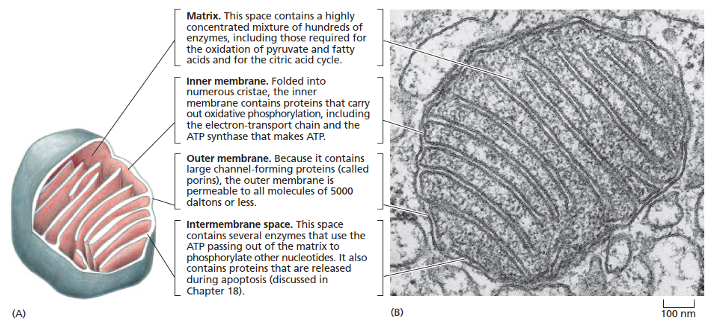 When purified mitochondria are gently fractionated into separate components and their contents analyzed, each of the membranes, and the spaces they enclose, are found to contain a unique collection of proteins.
The outer membrane contains many molecules of a transport protein called Porin, which, forms wide aqueous channels through the lipid bilayer. As a result, the outer membrane is like a sieve that is permeable to all molecules of 5000 daltons or less, including small proteins. This makes the intermembrane space chemically equivalent to the cytosol with respect to the small molecules and inorganic ions it contains.
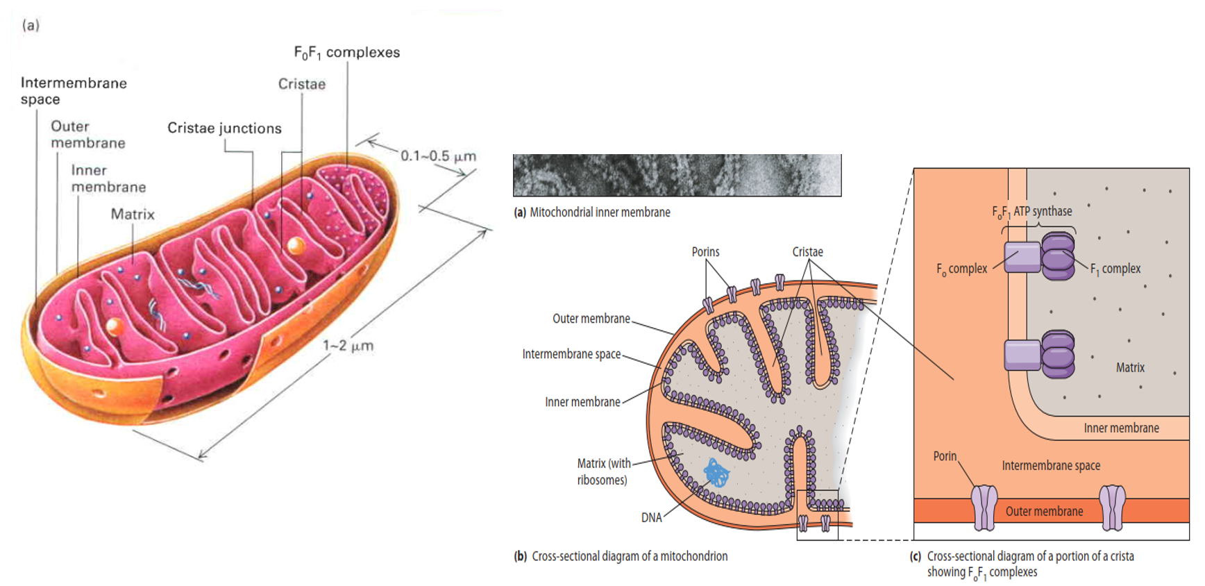 In contrast, the inner membrane, like other membranes in the cell, is impermeable to the passage of ions and most small molecules, except where a path is provided by specific membrane transport proteins. The mitochondrial matrix therefore contains only molecules that are selectively transported into the matrix across the inner membrane, and so its contents are highly specialized. The inner membrane is highly specialized. Its lipid bilayer contains a high proportion of the "double“ phospholipid Cardiolipin, which has four fatty acids rather than two and may help to make the membrane especially impermeable to ions.
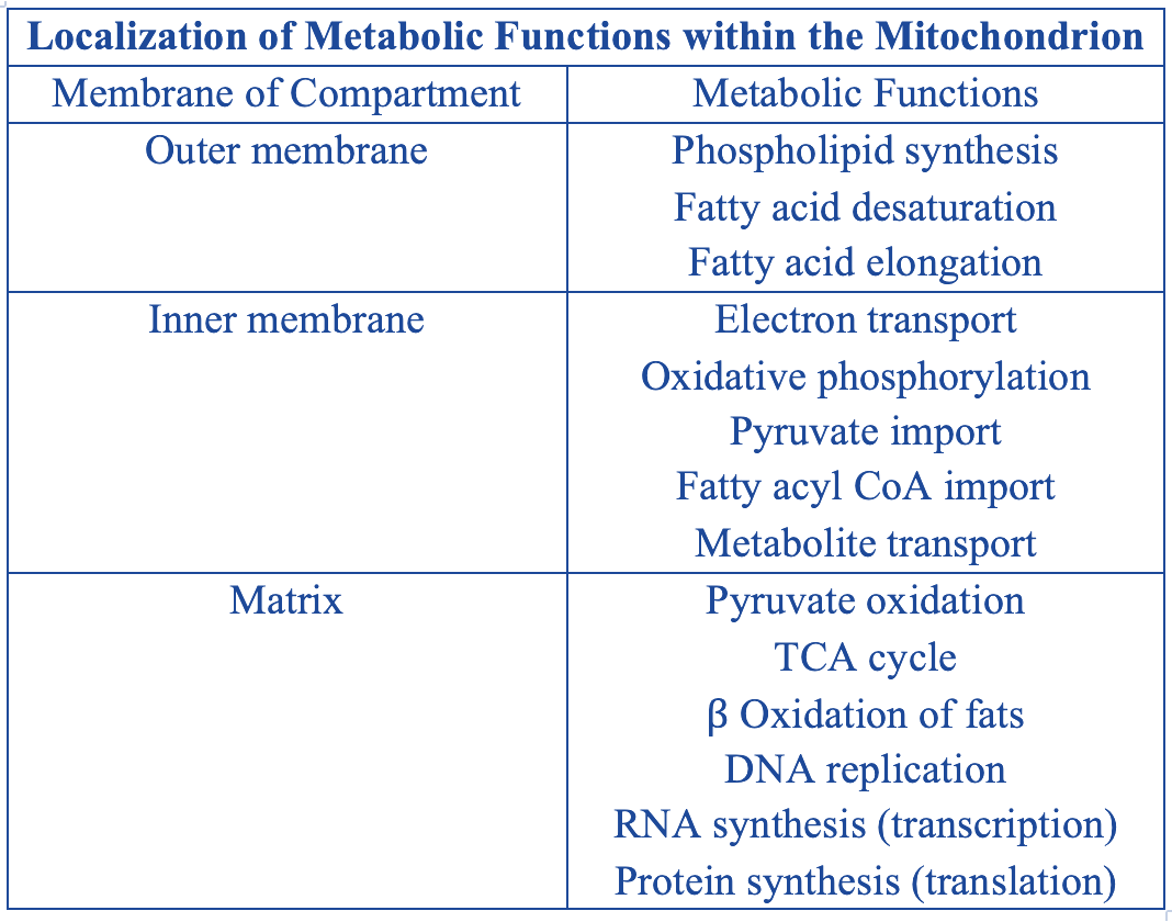 The inner mitochondrial membrane is the site of Oxidative phosphorylation, and it contains the proteins of the electron-transport chain, the proton pumps, and the ATP synthase required for ATP production. It also contains a variety of transport proteins that allow the entry of selected small molecules—such as pyruvate and fatty acids.
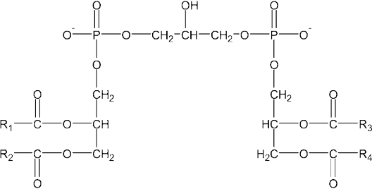 The inner membrane forms many folds called cristae, which increase its surface area. The shape of cristae is variable in mitochondria of different species, for example, fungal mitochondria often have plate-like cristae, while in Euglena cristae may be vesicle-shaped. Besides this the number of Cristae also varies from cell to cell for example the number of cristae in a cardiac muscle cell is three time more than in the mitochondrion of a liver cell. The inner membrane encloses the Mitochondrial matrix, which is quite dense and is loaded with ribosomes, DNA and often large calcium phosphate granules.
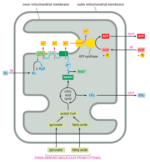 Mitochondrial ribosomes are smaller than the cytoplasmic ribosomes, and are similar to the bacterial ribosomes in number of characters, e.g., size, composition of subunits, etc. Mitochondrial DNA is a closed circular molecule much like bacterial DNA. The enzymes of Kreb’s Cycle are also present in the matrix of mitochondria.
In 1967, Lynn Margulis suggested not only the endosymbiotic origin of mitochondria and plastids, but also an endosymbiotic origin of the eukaryotic cell itself. In her model, eukaryotic cells originated in a series of endosymbioses involving distinct bacterial cells. There are various evidences in favor of this theory,
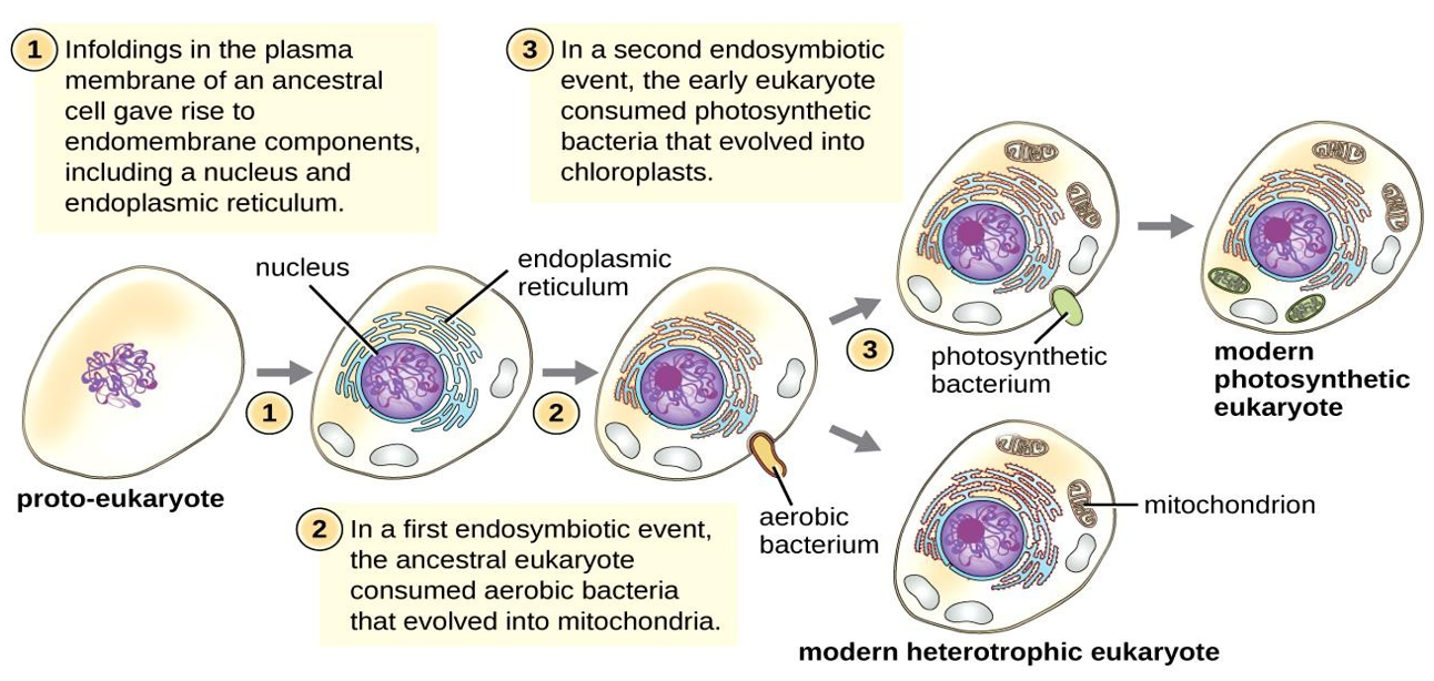 ⁕Like a cell mitochondria arise from pre-existing mitochondria.
⁕Mitochondria has its own DNA and its own protein synthesizing machinery, like ribosomes, t-RNAs and own amino acyl synthetases.
⁕Many proteins of Mitochondrial metabolism are synthesized by mitochondria themselves.
⁕Like a cell mitochondria arise from pre-existing mitochondria.
⁕Mitochondria has its own DNA and its own protein synthesizing machinery, like ribosomes, t-RNAs and own amino acyl synthetases.
⁕Many proteins of Mitochondrial metabolism are synthesized by mitochondria themselves.MITOCHONDRIA
- Mitochondria are called as Power House of the cell. Mitochondria were seen by Kolliker in 1850 in insect flight muscle cells and called them as Sarcosome. In 1890 Altmann studied them and called them as Bioplast. In 1898 Benda stained them with Cystal Violet and coined the word Mitochondria. In 1900 Michaelis stained them with the help of Janus Green B. In 1912 Kinsbury called them as Center of Aerobic Respiration. In 1913 Warburg discovered respiratory enzymes in them. In 1930 Hans Kreb reported Kreb Cycle enzymes in the matrix of mitochondria.
Mitochondria are present in all eukaryotic cell but they are secondarily lost in RBC. Numbers of mitochondria varies from cell to cell.
A single Mitochondria is present in Chlorella, Microasterias, Micromonas and Trypanosoma.
o20-24 are present in Sperms,
o300-400 in Kidney cells,
o500-1000 in Liver cells,
o50,000 in Chaos chaos,
o1,400,00-1,500,00 in eggs of sea urchin,
o3,00,000 in eggs of some amphibians and,
o5,00,000 in cells of insect flight muscles.
Size of Mitochondria:
They are about 1.5 to 10 μm in length and 0.25-1.0 μm in diameter though large mitochondrial upto 10 μm are present in pancreatic cell.
Structure of Mitochondria:
A mitochondrion is bounded by two highly specialized membranes—one surrounding the other. These membranes, called the outer and inner mitochondrial membranes, create two mitochondrial compartments:
A large internal space called the matrix and
A much narrower intermembrane space.
 When purified mitochondria are gently fractionated into separate components and their contents analyzed, each of the membranes, and the spaces they enclose, are found to contain a unique collection of proteins.
The outer membrane contains many molecules of a transport protein called Porin, which, forms wide aqueous channels through the lipid bilayer. As a result, the outer membrane is like a sieve that is permeable to all molecules of 5000 daltons or less, including small proteins. This makes the intermembrane space chemically equivalent to the cytosol with respect to the small molecules and inorganic ions it contains.
 In contrast, the inner membrane, like other membranes in the cell, is impermeable to the passage of ions and most small molecules, except where a path is provided by specific membrane transport proteins. The mitochondrial matrix therefore contains only molecules that are selectively transported into the matrix across the inner membrane, and so its contents are highly specialized. The inner membrane is highly specialized. Its lipid bilayer contains a high proportion of the "double“ phospholipid Cardiolipin, which has four fatty acids rather than two and may help to make the membrane especially impermeable to ions.
 The inner mitochondrial membrane is the site of Oxidative phosphorylation, and it contains the proteins of the electron-transport chain, the proton pumps, and the ATP synthase required for ATP production. It also contains a variety of transport proteins that allow the entry of selected small molecules—such as pyruvate and fatty acids.
 The inner membrane forms many folds called cristae, which increase its surface area. The shape of cristae is variable in mitochondria of different species, for example, fungal mitochondria often have plate-like cristae, while in Euglena cristae may be vesicle-shaped. Besides this the number of Cristae also varies from cell to cell for example the number of cristae in a cardiac muscle cell is three time more than in the mitochondrion of a liver cell. The inner membrane encloses the Mitochondrial matrix, which is quite dense and is loaded with ribosomes, DNA and often large calcium phosphate granules.
 Mitochondrial ribosomes are smaller than the cytoplasmic ribosomes, and are similar to the bacterial ribosomes in number of characters, e.g., size, composition of subunits, etc. Mitochondrial DNA is a closed circular molecule much like bacterial DNA. The enzymes of Kreb’s Cycle are also present in the matrix of mitochondria.
In 1967, Lynn Margulis suggested not only the endosymbiotic origin of mitochondria and plastids, but also an endosymbiotic origin of the eukaryotic cell itself. In her model, eukaryotic cells originated in a series of endosymbioses involving distinct bacterial cells. There are various evidences in favor of this theory,
 ⁕Like a cell mitochondria arise from pre-existing mitochondria.
⁕Mitochondria has its own DNA and its own protein synthesizing machinery, like ribosomes, t-RNAs and own amino acyl synthetases.
⁕Many proteins of Mitochondrial metabolism are synthesized by mitochondria themselves.
⁕Like a cell mitochondria arise from pre-existing mitochondria.
⁕Mitochondria has its own DNA and its own protein synthesizing machinery, like ribosomes, t-RNAs and own amino acyl synthetases.
⁕Many proteins of Mitochondrial metabolism are synthesized by mitochondria themselves.PLASTIDS/h2>
- Plastid is a double membrane-bound organelle involved in the synthesis and storage of food, commonly found within the cells of photosynthetic plants.
Plastids were discovered and named by Ernst Haeckel in 1866, but A. F. W. Schimper was the first to provide a clear definition. They are necessary for essential life processes, like photosynthesis and food storage.
A plastid containing green pigment (chlorophyll) is called Chloroplast whereas a plastid containing pigments apart from green is called a Chromoplast. A plastid that lacks pigments is called a Leucoplast and is involved mainly in food storage.
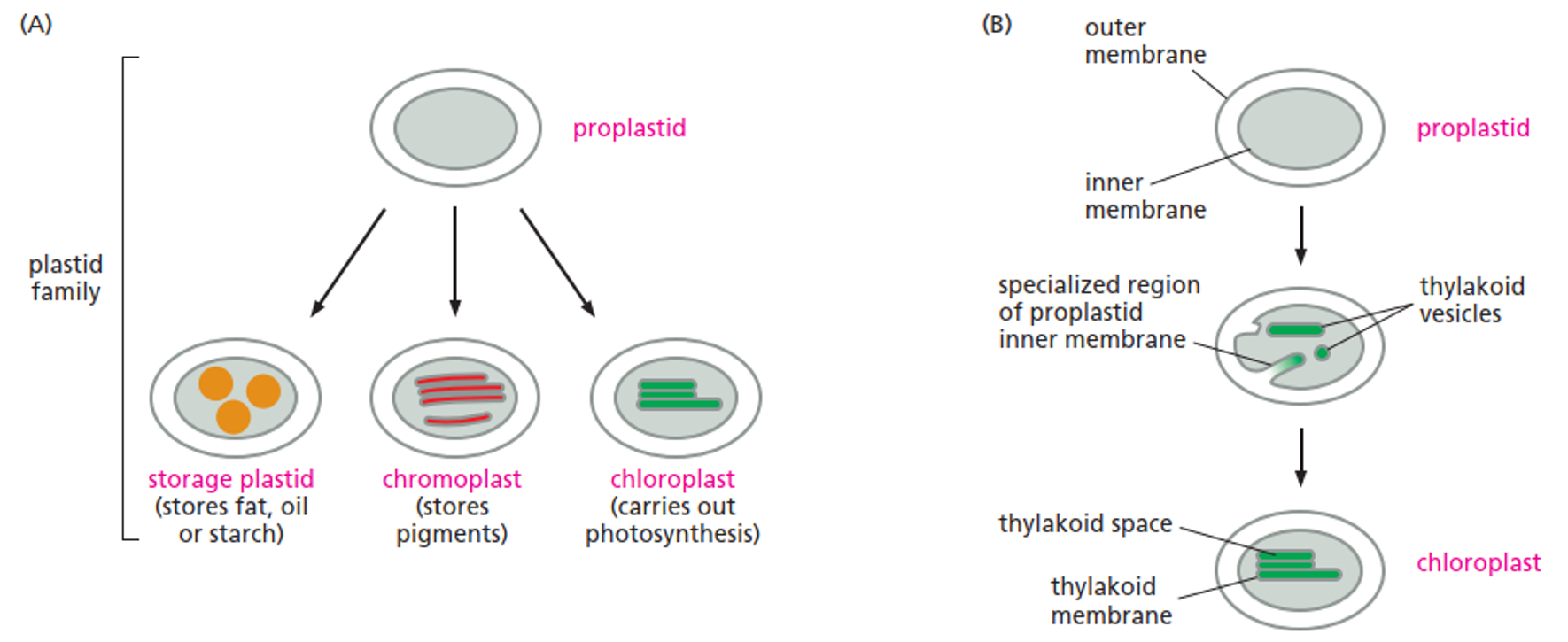 In a plant cells plastids arise from Proplastids.
Proplastids:
These organelles are colorless rounded amoeboid in nature and found in merismatic and very young plant cells. They are 0.4 to 1.0 μm in diameter. During differentiation they can give rise to other kind of plastids such as Leucoplast, Chloroplast and Chromoplasts.
 Leucoplast:
They are colorless plastids formed by the Proplastids. In Leucoplast lamellae are not organized into Grana. They are found in endosperm, tubers, roots and other non-photosynthetic tissues of plants. They serve various functions, for example, storage of starch, lipids, or proteins. Plastids of these three types are respectively known as Amyloplasts, Elaioplasts, and Proteinoplasts.
A leucoplast may also perform biosynthetic functions such as the synthesis of fatty acids, amino acids, and various other compounds.
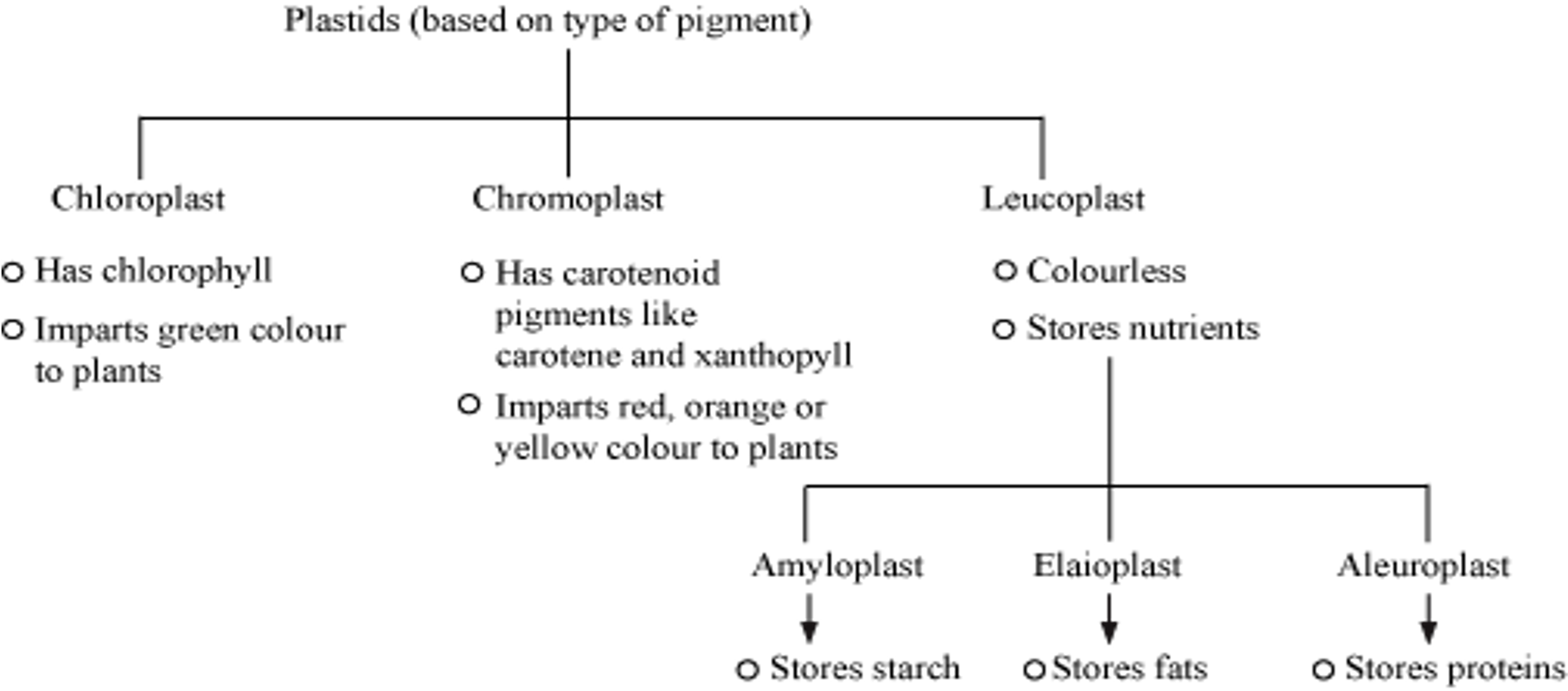 Besides storing and synthesis of biomolecules Leucoplast can also form Chloroplast and Chromoplast. During changing into Chloroplast they first change into Etioplasts, which are pre-granal, immature chloroplasts but can also be chloroplasts that have been deprived of light, lack active pigment and can be considered leucoplasts. After several minutes exposure to light, etioplasts begin to transform into functioning chloroplasts and cease being leucoplasts.
Plastids: Chromoplast
Chromoplasts are plastids, heterogeneous organelles responsible for pigment synthesis and storage in specific photosynthetic eukaryotes. Chromoplasts are found in fruits, flowers, roots, and stressed and aging leaves, and are responsible for their distinctive colors. This is always associated with a massive increase in the accumulation of carotenoid pigments.
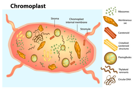 The conversion of chloroplasts to chromoplasts in ripening is a classic example. Chromoplasts synthesize and store pigments such as orange carotene, yellow xanthophylls, and various other red pigments. As such, their color varies depending on what pigment they contain. The main evolutionary purpose of chromoplasts is probably to attract pollinators or eaters of colored fruits, which help disperse seeds. However, they are also found in roots such as carrots and sweet potatoes. They allow the accumulation of large quantities of water-insoluble compounds in otherwise watery parts of plants.
Chloroplast:
They were first observed by Schimper which called them as Autoplast in 1883. Plant chloroplasts are large organelles (5–10 μm long) that, like mitochondria, are bounded by a double membrane called the chloroplast envelope.
Shape:
Gerneally they are disc shaped but their shape varies from cell to cell for example, they are
Collar like in Ulothrix,
Ribbon shaped in Spirogyra,
Cup shaped in Chlamydomonas,
Stellate in Zygnema and
Reticulate in Oedogonium
Number:
The number of chloroplast also varies from cell for example a single chloroplast is present in the cells of Chlamydomonas, Chlorella, Ulothrix, Oedogonium and Sprirogyra indica. Usually a plant cell has 20-40 chloroplasts but several hundred are found in the internodal cells of Chara.
Chloroplasts are located predominantly in the mesophyll cells of leaves. They are usually present around the central vacuole of a mesophyll cell. Chloroplasts of higher plants are generally lens-shaped, approximately 2 to 4 μm wide and 5 to 10 μm long, and typically numbering 20 to 40 per cell. Their dimensions make chloroplasts giants among organelles as large as an entire mammalian red blood cell. Like a cell chloroplasts arise by fission from preexisting chloroplasts (or their non pigmented precursors, which are called proplastids). Chloroplasts were identified as the site of photosynthesis in 1881 in an ingenious experiment by the German biologist T. Engelmann.
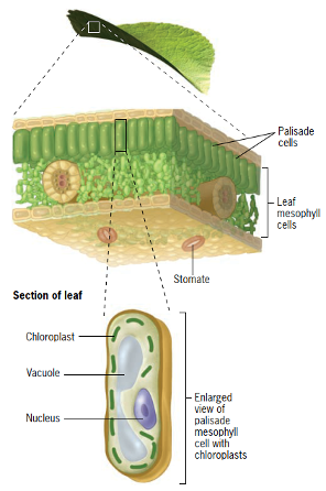 The outer covering of a chloroplast consists of an envelope composed of two membranes separated by a narrow space. Like the outer membrane of a mitochondrion, the outer membrane of a chloroplast envelope contains several different porins. Although these proteins have relatively large channels (in the range of 1 nm), they exhibit some selectivity toward various solutes. The inner membrane of the envelope is highly impermeable; substances moving through this membrane do so only with the aid of a variety of transporters. In addition to the inner and outer membranes of the envelope, chloroplasts have a third internal membrane system, called the Thylakoid membrane.
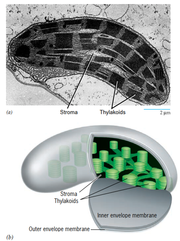 The thylakoid membrane forms a network of flattened discs called thylakoids, which are frequently arranged in stacks called Grana.
Because of this three-membrane structure, the internal organization of chloroplasts is more complex than that of mitochondria. In particular, their three membranes divide chloroplasts into three distinct internal compartments:
1.The inter-membrane space between the two membranes of the chloroplast envelope;
2.The Stroma, which lies inside the envelope but outside the thylakoid membrane; and
3.The Thylakoid lumen. At places inner membrane is connected to Thylakoids. Thylakoids were first described by Menke in 1961. They are also known as Baggy Trousers. In higher plants Thylakoids are of two types,
1.Granal Thylakoids
2.Agranal Thylakoids or Stromal
Agranal Thylakoids forms a network of tubules which join with Granal Thylakoids. Thylakoids contain 50% of the protein and all the components involved in the essential steps of Photosynthesis.
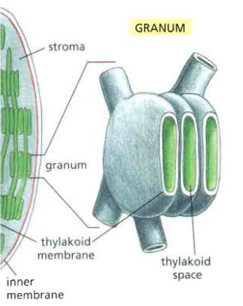 Stroma or Matrix :
The ground substance of a chloroplast is known as Matrix or Stroma. It is a crystallo-colloidal complex that is made of 50% soluble proteins. Like Mitochondria it has its own t-RNA, Ribosomes, and its own DNA called or Chloroplast or cp-DNA is naked, usually circular but in some algae it is linear. A chloroplast may have several copies of it. DNA makes the chloroplast genetically autonomous. With the help of ribosomes the chloroplast is able to synthesize most of the enzymes required by it. The
important enzymes present in chloroplast are those that take part in synthesis of photosynthetic pigments, photolysis of water, enzymes for photophosphorylation, and carbon fixation. In stroma Plasto-globuli are present. Plastoglobuli are lipid droplets 10-500 nm diameter. They may contain some enzymes, Vitamin K and Quinones. The chloroplast matrix of higher plants may store starch temporarily, as starch grains. It is as Assimilation starch. In green algae (e.g., Spirogyra, Ulothrix), the chloroplasts possesses special starch storing structures called Pyrenoids.
Origin:
Like Mitochondria Chloroplast is also semi-autonomous and has an endosymbiotic origin.
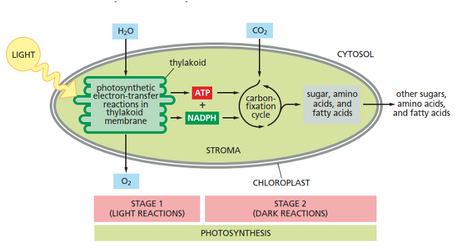 Nucleus is the largest cell organelle in an animal cell and second largest cell in the plant cell. It is also called control house of the cell. It was first observed by Anton von Leeuwenhoek in 1692 in fish RBC and by Fontana in 1781 but described by Robert Brown in 1831 in orchid root cells. Nucleus is a relatively large organelle controlling all the activities of the eukaryotic cells. Some cells have more than one nucleus, e.g., binucleate cells have 2 nuclei per cell such as Paramoecium caudatum and 3 nuclei are present in Paramoecium aurelia. Many animal cells are multinucleate such condition is called Syncytial and present in Skeletal muscle cell, Ascaris, Opalina and Polykaryocytes of Bone marrow while multinucleate condition in plant cell is called Coenocytic such as latex vessels cell and in Rhizopus.
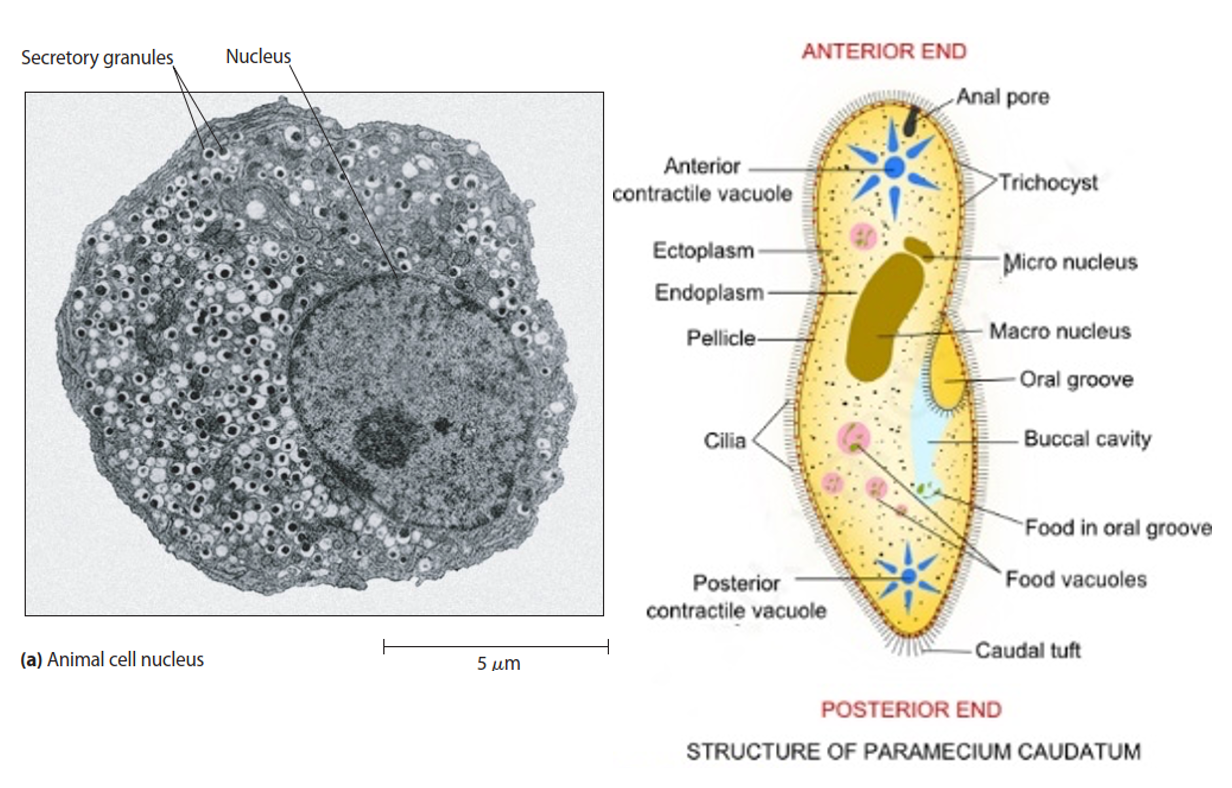 However, some cells lack nucleus (anucleate) at maturity, such as, mammalian RBCs and Sieve tube cells (food conducting elements in vascular plants). That the nucleus is a store house of hereditary information was proved by a Danish biologist Joachim Hammerling (1943) on the basis of his studies on Acetabularia. He demonstrated in 1943 that the nucleus determines the characters of the cell and ultimately the characters of the individual. He conducted certain experiments using two species of a green alga, Acetabularia.
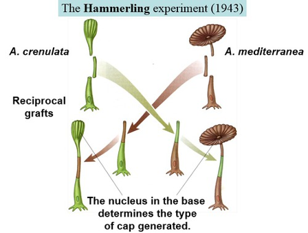 The two species, namely A. crenulata and A. mediterranea used in this experiment differ in the shape of their caps. While in A. crenulata the cap has loose rays, in A. mediterranea an umbrella-like cap is found. The nucleus in both the species is situated in rhizoid at the bottom of stalk.
If cap is cut off, it will develop again and its shape will be that of the original type. However, if after removing the caps, stalk of one species, is grafted on rhizoid (containing the nucleus) of the other species, shape of cap will be determined by nucleus and not by stalk. If the nucleus belongs to A. crenulata, shape of cap will be of the crenulata type and if the nucleus comes from A. mediterranea, cap will be of mediterranea type. When both nuclei are present, shape of cap will be intermediate. This experiment demonstrated clearly that characters of an individual are controlled by nucleus of the cell or cells.
Size: It occupies 15-25% of the cell mass. It depends upon cell size, ploidy level, cell maturity and cell function for example it occupies 60% cell size in thymus cells and 10% in parenchymatous cell.
Position: In most of the cells the nucleus is central but in plant cells and adipocytes nucleus is peripheral.
Shape: Shapes of nucleus is also varies,
It is flat in squamous cells,
Reniform in or kidney shaped in macronculeus of Paramoecium,
Horseshoe shaped in monocytes and
Multilobed in WBCs or Leucocytes

PLASTIDS/h2>
- Plastid is a double membrane-bound organelle involved in the synthesis and storage of food, commonly found within the cells of photosynthetic plants.
Plastids were discovered and named by Ernst Haeckel in 1866, but A. F. W. Schimper was the first to provide a clear definition. They are necessary for essential life processes, like photosynthesis and food storage.
A plastid containing green pigment (chlorophyll) is called Chloroplast whereas a plastid containing pigments apart from green is called a Chromoplast. A plastid that lacks pigments is called a Leucoplast and is involved mainly in food storage.
 In a plant cells plastids arise from Proplastids.
Proplastids:
These organelles are colorless rounded amoeboid in nature and found in merismatic and very young plant cells. They are 0.4 to 1.0 μm in diameter. During differentiation they can give rise to other kind of plastids such as Leucoplast, Chloroplast and Chromoplasts.
 Leucoplast:
They are colorless plastids formed by the Proplastids. In Leucoplast lamellae are not organized into Grana. They are found in endosperm, tubers, roots and other non-photosynthetic tissues of plants. They serve various functions, for example, storage of starch, lipids, or proteins. Plastids of these three types are respectively known as Amyloplasts, Elaioplasts, and Proteinoplasts.
A leucoplast may also perform biosynthetic functions such as the synthesis of fatty acids, amino acids, and various other compounds.
 Besides storing and synthesis of biomolecules Leucoplast can also form Chloroplast and Chromoplast. During changing into Chloroplast they first change into Etioplasts, which are pre-granal, immature chloroplasts but can also be chloroplasts that have been deprived of light, lack active pigment and can be considered leucoplasts. After several minutes exposure to light, etioplasts begin to transform into functioning chloroplasts and cease being leucoplasts.
Plastids: Chromoplast
Chromoplasts are plastids, heterogeneous organelles responsible for pigment synthesis and storage in specific photosynthetic eukaryotes. Chromoplasts are found in fruits, flowers, roots, and stressed and aging leaves, and are responsible for their distinctive colors. This is always associated with a massive increase in the accumulation of carotenoid pigments.
 The conversion of chloroplasts to chromoplasts in ripening is a classic example. Chromoplasts synthesize and store pigments such as orange carotene, yellow xanthophylls, and various other red pigments. As such, their color varies depending on what pigment they contain. The main evolutionary purpose of chromoplasts is probably to attract pollinators or eaters of colored fruits, which help disperse seeds. However, they are also found in roots such as carrots and sweet potatoes. They allow the accumulation of large quantities of water-insoluble compounds in otherwise watery parts of plants.
Chloroplast:
They were first observed by Schimper which called them as Autoplast in 1883. Plant chloroplasts are large organelles (5–10 μm long) that, like mitochondria, are bounded by a double membrane called the chloroplast envelope.
Shape:
Gerneally they are disc shaped but their shape varies from cell to cell for example, they are
Collar like in Ulothrix,
Ribbon shaped in Spirogyra,
Cup shaped in Chlamydomonas,
Stellate in Zygnema and
Reticulate in Oedogonium
Number:
The number of chloroplast also varies from cell for example a single chloroplast is present in the cells of Chlamydomonas, Chlorella, Ulothrix, Oedogonium and Sprirogyra indica. Usually a plant cell has 20-40 chloroplasts but several hundred are found in the internodal cells of Chara.
Chloroplasts are located predominantly in the mesophyll cells of leaves. They are usually present around the central vacuole of a mesophyll cell. Chloroplasts of higher plants are generally lens-shaped, approximately 2 to 4 μm wide and 5 to 10 μm long, and typically numbering 20 to 40 per cell. Their dimensions make chloroplasts giants among organelles as large as an entire mammalian red blood cell. Like a cell chloroplasts arise by fission from preexisting chloroplasts (or their non pigmented precursors, which are called proplastids). Chloroplasts were identified as the site of photosynthesis in 1881 in an ingenious experiment by the German biologist T. Engelmann.
 The outer covering of a chloroplast consists of an envelope composed of two membranes separated by a narrow space. Like the outer membrane of a mitochondrion, the outer membrane of a chloroplast envelope contains several different porins. Although these proteins have relatively large channels (in the range of 1 nm), they exhibit some selectivity toward various solutes. The inner membrane of the envelope is highly impermeable; substances moving through this membrane do so only with the aid of a variety of transporters. In addition to the inner and outer membranes of the envelope, chloroplasts have a third internal membrane system, called the Thylakoid membrane.
 The thylakoid membrane forms a network of flattened discs called thylakoids, which are frequently arranged in stacks called Grana.
Because of this three-membrane structure, the internal organization of chloroplasts is more complex than that of mitochondria. In particular, their three membranes divide chloroplasts into three distinct internal compartments:
1.The inter-membrane space between the two membranes of the chloroplast envelope;
2.The Stroma, which lies inside the envelope but outside the thylakoid membrane; and
3.The Thylakoid lumen. At places inner membrane is connected to Thylakoids. Thylakoids were first described by Menke in 1961. They are also known as Baggy Trousers. In higher plants Thylakoids are of two types,
1.Granal Thylakoids
2.Agranal Thylakoids or Stromal
Agranal Thylakoids forms a network of tubules which join with Granal Thylakoids. Thylakoids contain 50% of the protein and all the components involved in the essential steps of Photosynthesis.
 Stroma or Matrix :
The ground substance of a chloroplast is known as Matrix or Stroma. It is a crystallo-colloidal complex that is made of 50% soluble proteins. Like Mitochondria it has its own t-RNA, Ribosomes, and its own DNA called or Chloroplast or cp-DNA is naked, usually circular but in some algae it is linear. A chloroplast may have several copies of it. DNA makes the chloroplast genetically autonomous. With the help of ribosomes the chloroplast is able to synthesize most of the enzymes required by it. The
important enzymes present in chloroplast are those that take part in synthesis of photosynthetic pigments, photolysis of water, enzymes for photophosphorylation, and carbon fixation. In stroma Plasto-globuli are present. Plastoglobuli are lipid droplets 10-500 nm diameter. They may contain some enzymes, Vitamin K and Quinones. The chloroplast matrix of higher plants may store starch temporarily, as starch grains. It is as Assimilation starch. In green algae (e.g., Spirogyra, Ulothrix), the chloroplasts possesses special starch storing structures called Pyrenoids.
Origin:
Like Mitochondria Chloroplast is also semi-autonomous and has an endosymbiotic origin.
 Nucleus is the largest cell organelle in an animal cell and second largest cell in the plant cell. It is also called control house of the cell. It was first observed by Anton von Leeuwenhoek in 1692 in fish RBC and by Fontana in 1781 but described by Robert Brown in 1831 in orchid root cells. Nucleus is a relatively large organelle controlling all the activities of the eukaryotic cells. Some cells have more than one nucleus, e.g., binucleate cells have 2 nuclei per cell such as Paramoecium caudatum and 3 nuclei are present in Paramoecium aurelia. Many animal cells are multinucleate such condition is called Syncytial and present in Skeletal muscle cell, Ascaris, Opalina and Polykaryocytes of Bone marrow while multinucleate condition in plant cell is called Coenocytic such as latex vessels cell and in Rhizopus.
 However, some cells lack nucleus (anucleate) at maturity, such as, mammalian RBCs and Sieve tube cells (food conducting elements in vascular plants). That the nucleus is a store house of hereditary information was proved by a Danish biologist Joachim Hammerling (1943) on the basis of his studies on Acetabularia. He demonstrated in 1943 that the nucleus determines the characters of the cell and ultimately the characters of the individual. He conducted certain experiments using two species of a green alga, Acetabularia.
 The two species, namely A. crenulata and A. mediterranea used in this experiment differ in the shape of their caps. While in A. crenulata the cap has loose rays, in A. mediterranea an umbrella-like cap is found. The nucleus in both the species is situated in rhizoid at the bottom of stalk.
If cap is cut off, it will develop again and its shape will be that of the original type. However, if after removing the caps, stalk of one species, is grafted on rhizoid (containing the nucleus) of the other species, shape of cap will be determined by nucleus and not by stalk. If the nucleus belongs to A. crenulata, shape of cap will be of the crenulata type and if the nucleus comes from A. mediterranea, cap will be of mediterranea type. When both nuclei are present, shape of cap will be intermediate. This experiment demonstrated clearly that characters of an individual are controlled by nucleus of the cell or cells.
Size: It occupies 15-25% of the cell mass. It depends upon cell size, ploidy level, cell maturity and cell function for example it occupies 60% cell size in thymus cells and 10% in parenchymatous cell.
Position: In most of the cells the nucleus is central but in plant cells and adipocytes nucleus is peripheral.
Shape: Shapes of nucleus is also varies,
It is flat in squamous cells,
Reniform in or kidney shaped in macronculeus of Paramoecium,
Horseshoe shaped in monocytes and
Multilobed in WBCs or Leucocytes

NUCLEUS
- EM studies have shown that nucleus consist of,
1.Nuclear Envelope
2.Nuclear Matrix
3.Nucleoplasm
4.Nucleolus
5.Chromatin
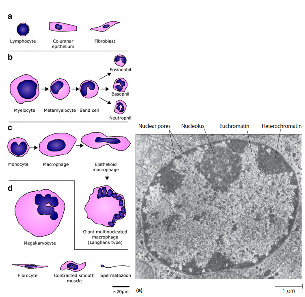 1.Nuclear Envelope:
Transmission electron microscopy revealed that the nucleus is bounded by a nuclear envelope composed of two membranes the inner and outer nuclear membranes separated by a perinuclear space measuring about 20–40 nm across. The outer nuclear membrane is continuous with the endoplasmic reticulum, making the perinuclear space continuous with the lumen of the ER. Like membranes of the rough ER, the outer membrane is often studded on its outer surface with ribosomes engaged in protein synthesis. Several proteins in the outer membrane bind to actin and intermediate filaments of the cell’s cytoskeleton, thereby anchoring the nucleus within the cell and providing a mechanism for nuclear movements.
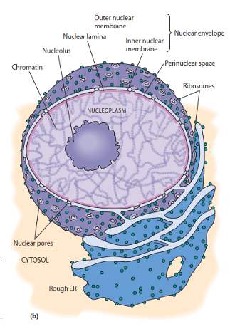 Tubular invaginations of the nuclear envelope, collectively known as the nucleoplasmic reticulum, may project into the internal nuclear space and increase the area of the nucleus that makes direct contact with the inner nuclear membrane. One of the most distinctive features of the nuclear envelope is the presence of specialized channels called Nuclear pores, which are especially easy to see when the nuclear envelope is examined by freeze-fracture microscopy. Each pore is a small cylindrical channel extending through both membranes of the nuclear envelope, thereby providing direct continuity between the cytosol and the nucleoplasm. The number of pores varies greatly with cell type and activity. A typical mammalian nucleus has about 3000–4000 pores, or about 10–20 pores per square micrometer of membrane surface area.
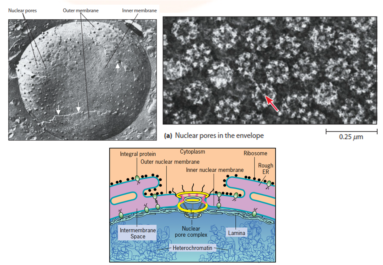 At each pore, the inner and outer membranes of the nuclear envelope are fused together, forming a channel that is lined with an intricate protein structure called the nuclear pore complex (NPC). The NPC has an outer diameter of 120 nm and is built from about 30 different proteins called nucleoporins. In electron micrographs, the most striking feature of the pore complex is its octagonal symmetry. It is made up of rings of eight subunits arranged in an octagonal pattern. In other views, the eight subunits are seen to protrude on both the cytoplasmic and nucleoplasmic sides of the envelope. Nuclear pore complex regulate the transport of solutes in between cytoplasm and nucleoplasm, it also participate in maturation of Ribosomes.
 2. Nuclear Matrix
In the early 1970s researchers discovered that an insoluble fibrous network retaining the overall shape of the nucleus remains behind after more than 95% of the chromatin has been removed by a combination of nuclease and detergent treatments. This network, called the nuclear matrix or nucleoskeleton, is thought to help maintain the shape of the nucleus and provide an organizing skeleton for the chromatin fibers. Underlying the inner nuclear membrane is the nuclear lamina, a filamentous meshwork that provides structural support to the nucleus. The nuclear lamina is composed of 60- to 80-kilodalton (kd) fibrous proteins called lamins, along with associated proteins. Nucleolamins help in stability of nuclear membrane and chromatin.
3. Nucleoplasm: (Strasburger, 1882). It is a transparent, semi-fluid and colloidal substance which fills the nucleus. It contains nucleosides and a number of enzymes (e.g., DNA polymerase, RNA polymerase, nucleoside phosphorylase) which are required for the synthesis and functioning of DNA, RNA, nucleoproteins, etc. Some of the proteins present in nucleoplasm are essential for spindle formation.
4. Nucleolus: It was first discovered by Fontana in 1781, described by Wagner in 1840 and provided with its present name by Bowman in 1840. Nucleolus is a naked, round or slightly irregular structure which is attached to the chromatin at a specific region called Nuclear Organiser Region (NOR). Commonly 1-4 nucleoli are found in a nucleus. Up to 1600 nucleoli are reported in the oocytes of Xenopus. A covering membrane is absent around nucleolus. Calcium seems to be essential for maintaining its configuration. Nucleolus has four components-
⁕Amorphous matrix,
⁕Granular part,
⁕Fibrillate portion and
⁕Chromatin part
(a) Amorphous Matrix. It is the homogeneous ground substance of the nucleolus. Matrix is formed of protein.
(b) Granular Portion. It consists of granules of the size of 150-200 Å which lie scattered in the amorphous matrix. The granules are formed of protein and RNA in the ratio of 2 : 1. They are believed to be precursors of ribosomes.
(c) Fibrillar Portion (Nucleolonema). It is formed of a large number of small fibrils that are 50-80 Å long. The fibrils are made up of both protein and RNA and are believed to be precursors of granules.
(d) Chromatin Portion. It is that part of chromatin which is associated with nucleolus. Depending upon its position nucleolar chromatin is of two types- perinucleolar and intranucleolar. The perinucleolar chromatin lies around the periphery of the nucleolus. It gives rise to in growths or trabeculae which produce the intranucleolar chromatin.
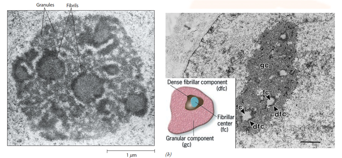 The nucleolus is the site for ribosomal RNA synthesis. The nucleoli are larger and more numerous in cells that are actively involved in protein synthesis. Isolated nucleoli contain the nucleolus organizer region (NOR) -a stretch of DNA carrying multiple copies of rRNA genes. These multiple rRNA genes occur in all genomes and are thus an important example of repeated DNA that carries genetic information. The number of copies of the rRNA genes varies greatly from species to species, but animal cells generally contain hundreds of copies and plant cells often contain thousands. The multiple copies are grouped into one or more NORs, which may reside on more than one chromosome; in each NOR, the multiple gene copies are tandemly arranged. They are also called secondary constriction.
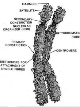 In humans secondary constriction is present in chromosome no. 13,14,15, 21 & 22.
Chromatin: It is hereditary DNA-protein fibrillate complex which is named so because of its ability to get stained with certain basic dyes (Gk. chroma- colour, Flemming, 1879). Chromatin occurs in the form of fine overlapping and coiled fibers which appear to produce a network called chromatin reticulum. Chromatin fibers are distributed throughout the nucleoplasm. They are differentiated into two regions- Euchromatin and Heterochromatin. Euchromatin is narrow (30-80 Å thick) lightly stained and diffused fibrous part which forms the bulk of chromatin. Heterochromatin is wider (250 Å thick), darkly stained and condensed granular part which is attached here and there on the euchromatin. Depending upon the size of granules formed by heterochromatin they are called chromocentres, karyosomes or false nucleoli.
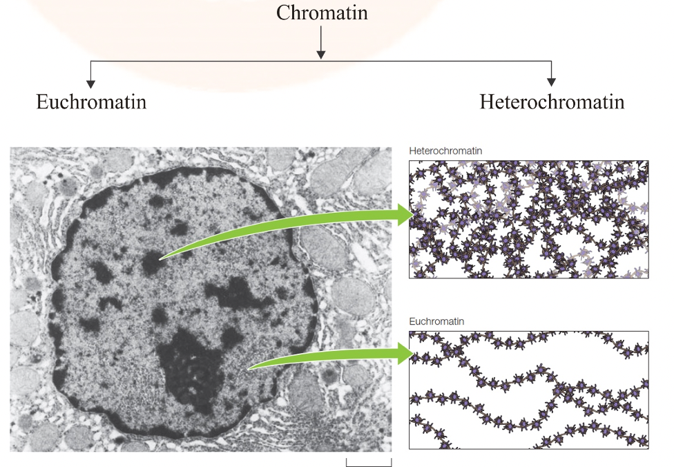 Euchromatin
 is a lightly packed form of chromatin (DNA, RNA, and protein) that is enriched in genes, and is often (but not always) under active transcription. Euchromatin comprises the most active portion of the genome within the cell nucleus, 92% of the human genome is euchromatic.
It enters into replication first and lightly stained. The basic structure of euchromatin is an elongated, open, 10 nm microfibril. It has a beads on a string structure or Nucleosome structure. Nucleosomes were first observed as particles in the electron microscope by Don and Ada Olins in 1974, and their existence and structure (as histone octamers surrounded by approximately 200 base pairs of DNA) were proposed by Roger Kornberg.
Euchromatin
Euchromatin is a lightly packed form of chromatin (DNA, RNA, and protein) that is enriched in genes, and is often (but not always) under active transcription. Euchromatin comprises the most active portion of the genome within the cell nucleus, 92% of the human genome is euchromatic.
It enters into replication first and lightly stained. The basic structure of euchromatin is an elongated, open, 10 nm microfibril. It has a beads on a string structure or Nucleosome structure. Nucleosomes were first observed as particles in the electron microscope by Don and Ada Olins in 1974, and their existence and structure (as histone octamers surrounded by approximately 200 base pairs of DNA) were proposed by Roger Kornberg.
 In 1974, using the data from nuclease digestion and other types of information, Roger Kornberg, proposed that DNA and histones are organized into repeating subunits, called nucleosomes. Nucleosome is formed by the octamer of histone proteins, around this octamer 200 bps of DNA is wrapped. The formation of nucleosomes converts a DNA molecule into a chromatin thread that is approximately one-third the length of the initial piece of DNA, and it provides the first level of DNA packing.
All four of the histones that make up the octamer are relatively small proteins, with a high proportion of positively charged amino acids (lysine and arginine). The positive charges help the histones bind tightly to the negatively charged sugar phosphate backbone of DNA. These numerous electrostatic interactions explain in part why DNA of virtually any sequence can bind to a histone octamer. Each individual nucleosome core particle consists of a complex of eight histone proteins-two molecules each of histones H2A, H2B, H3, and H4-and double-stranded DNA that is 147 nucleotide pairs long. The histone octamer forms a protein core around which the double-stranded DNA is wound. Each nucleosome core particle is separated from the next by a region of linker DNA, which can vary in length from a few nucleotide pairs up to about 80. On average, therefore, nucleosomes repeat at intervals of about 200 nucleotide pairs. For example, a diploid human cell with 6.4 x 10⁹ nucleotide pairs contains approximately 30 million nucleosomes.
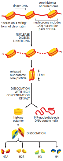 In interphase nucleus generally chromatin exists in two forms Euchromatin and Heterochromatin. Usually Euchromatin exists in Beads on a string form while Heterochromatin are at higher condensation level i.e. 30 nm chromatin fiber which is the Solenoid form which is formed with the help of H1 histone protein.
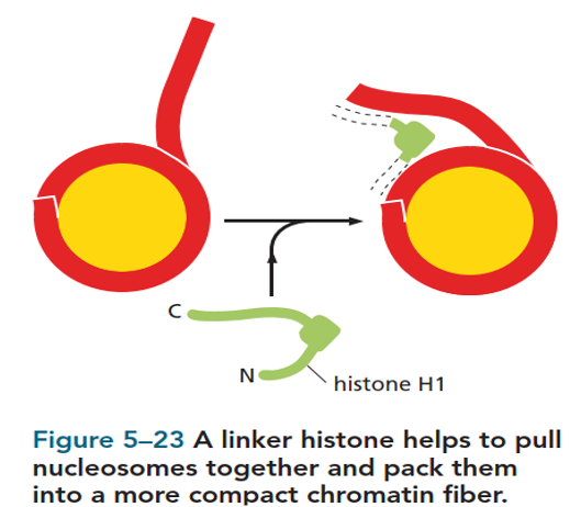
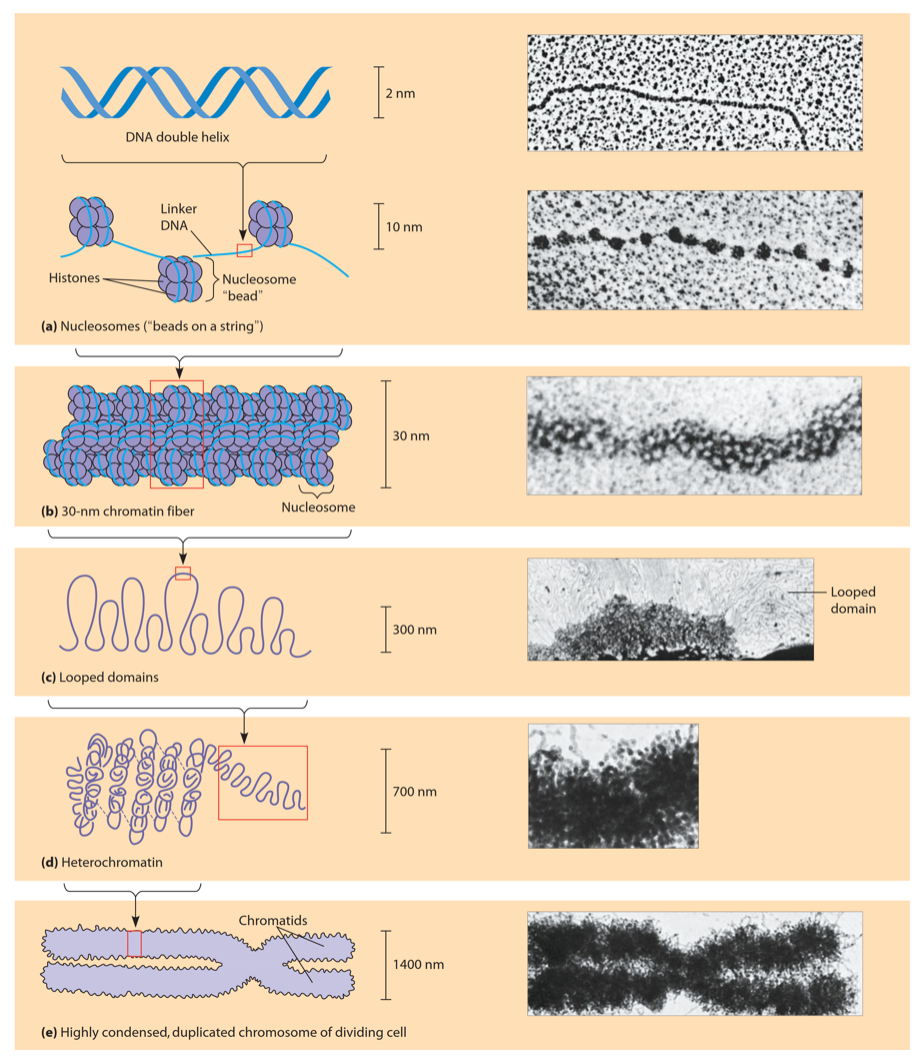
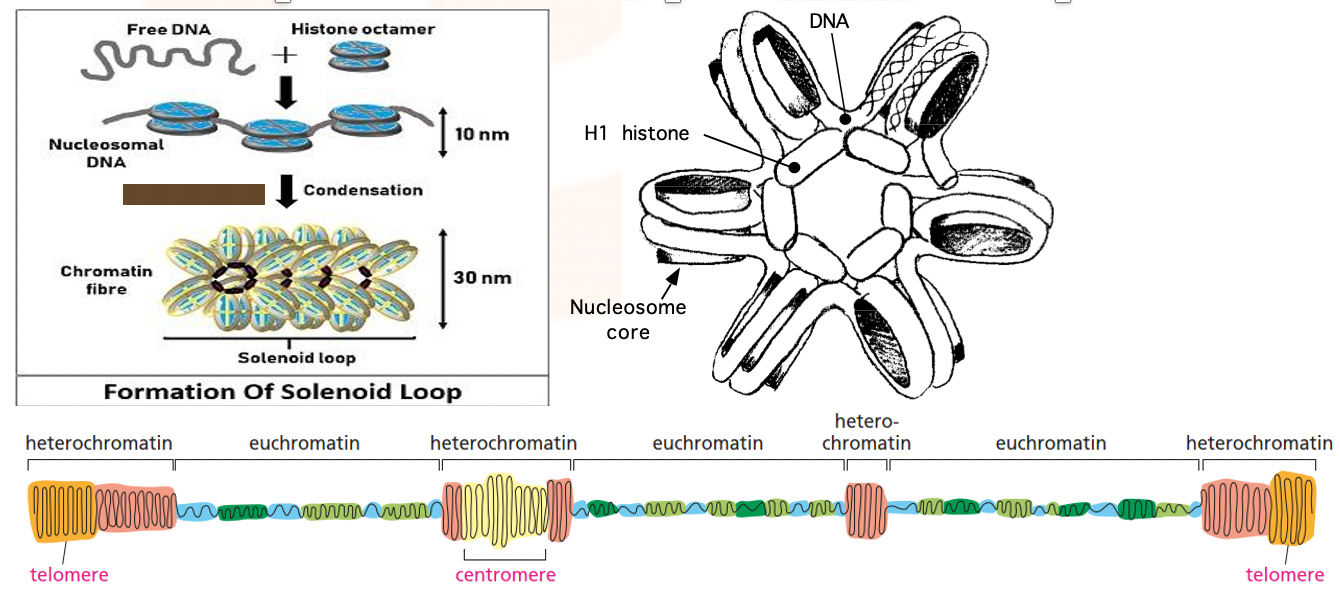 Chromosomes is the condensed form of chromatin visible during dividing or M-Phasee. The chromosomes are capable of self-reproduction and maintaining morphological and physiological properties through successive generations. They are capable of transmitting the contained hereditary material to the next generation. Hence these are known as ‘Hereditary vehicles.
•Chromosomes were first observed by Karl Negali in 1842 in plant cells as rod-shaped bodies in plant cells.
•In 1848 Hofmeister Observed chromosomes as darkly stained bodies in pollen mother cells of Tradecscantia.
•In 1888 Waldayer coined the term chromosome.
•In 1902 Walter S. Sutton proposed ‘Theory of Chromosomal basis of Heredity.
The salient features of chromosome theory of heredity are: -
•The somatic cells of an organism which are derived by the repeated divisions of the zygote are all diploid, i.e. these consist of two identical sets of chromosomes. One set of chromosomes is received from mother (maternal set) through ovum and the other set is inherited from father through sperm (paternal set). These two chromosomes of one type constitute the homologous pair.
•The chromosomes retain their structural uniqueness, individuality and their continuity throughout the life-cycle of an organism.
•Each chromosome carries specific determiners or Mendelian factors and plays a significant role in the development of an organism from the zygote.
•The behavior of chromosomes during meiosis, which occurs at the time of gamete formation provides an evidence that genes or determiners are located in the chromosomes. This also explains the mechanism of segregation of characteristics at the time of gamete formation.
Number of Chromosomes:
The number of chromosomes in the somatic cells of higher animals and plants is known as diploid or somatic or zygotic number, while in the gametes (sperms and eggs) it is haploid, gametic or reduced. The number of chromosomes is constant in all the somatic cells of all the individuals of a species. Chromosome number is used in the identification of species and in tracing the relationship within the species.
The lowest no of chromosomes are present in,
Parascaris = No of chromosomes are 2,
Mucor hiemalis = No of chromosomes are 2,
Happlopappus gracilis = No of chromosomes 2n = 4
While highest no of chromosomes are present in,
In plants it is Adder’s Tongue fern
Ophioglossum retiulatum = 2n = 1262 while in animals,
Aulocantha = 2n = 1632
Chromosomes is the condensed form of chromatin visible during dividing or M-Phasee. The chromosomes are capable of self-reproduction and maintaining morphological and physiological properties through successive generations. They are capable of transmitting the contained hereditary material to the next generation. Hence these are known as ‘Hereditary vehicles.
•Chromosomes were first observed by Karl Negali in 1842 in plant cells as rod-shaped bodies in plant cells.
•In 1848 Hofmeister Observed chromosomes as darkly stained bodies in pollen mother cells of Tradecscantia.
•In 1888 Waldayer coined the term chromosome.
•In 1902 Walter S. Sutton proposed ‘Theory of Chromosomal basis of Heredity.
The salient features of chromosome theory of heredity are: -
•The somatic cells of an organism which are derived by the repeated divisions of the zygote are all diploid, i.e. these consist of two identical sets of chromosomes. One set of chromosomes is received from mother (maternal set) through ovum and the other set is inherited from father through sperm (paternal set). These two chromosomes of one type constitute the homologous pair.
•The chromosomes retain their structural uniqueness, individuality and their continuity throughout the life-cycle of an organism.
•Each chromosome carries specific determiners or Mendelian factors and plays a significant role in the development of an organism from the zygote.
•The behavior of chromosomes during meiosis, which occurs at the time of gamete formation provides an evidence that genes or determiners are located in the chromosomes. This also explains the mechanism of segregation of characteristics at the time of gamete formation.
Number of Chromosomes:
The number of chromosomes in the somatic cells of higher animals and plants is known as diploid or somatic or zygotic number, while in the gametes (sperms and eggs) it is haploid, gametic or reduced. The number of chromosomes is constant in all the somatic cells of all the individuals of a species. Chromosome number is used in the identification of species and in tracing the relationship within the species.
The lowest no of chromosomes are present in,
Parascaris = No of chromosomes are 2,
Mucor hiemalis = No of chromosomes are 2,
Happlopappus gracilis = No of chromosomes 2n = 4
While highest no of chromosomes are present in,
In plants it is Adder’s Tongue fern
Ophioglossum retiulatum = 2n = 1262 while in animals,
Aulocantha = 2n = 1632- Chromosomes exhibit cyclic change in shape and size during cell cycle. In the non dividing interphase nucleus, the chromosomes form an interwoven network of line twisted but uncoiled threads of chromatin, and are invisible. During cell division the chromatin threads condense into compact structures by helical coiling. In prophase of cell division the chromosomes appear as distinct threads and by metaphase and also in anaphase these become short, compact bodies having definite shapes and sizes. In anaphase these appear as rod-shaped, V-shaped, L-shaped or J-shaped. The chromosomes are studied and described at this stage or at anaphase stage. In telophase these again uncoil to form the chromatin net.
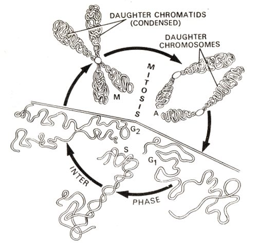 Chromosomes differ greatly in size in different organisms. Even the chromosomes of different pairs in the nucleus of the same cell have different sizes. The anaphase chromosomes range from 1 μm to 30 μm in length and 0.22 μ to 2 μ in width.
A chromosome consists of following parts,
⁕Centromere
⁕Telomere
⁕Secondary Constriction,
Centromere:
Centromere is the part of chromosome where two chromatids join with each other. At centromere specific proteins bind and form a disc like structure called Kinetochore, at kinetochore spindle fibers interact and help in the separation of sister chromatids (Chromatids which join with same centromere in a chromosome are called Sister Chromatids).
The centromeric region is also called Primary constriction. The microtubules of the chromosomal spindle fibers are attached to the centromere. Therefore, centromere is associated with the chromosomal movement during cell division. A chromosome without centromere is lost during anaphase.
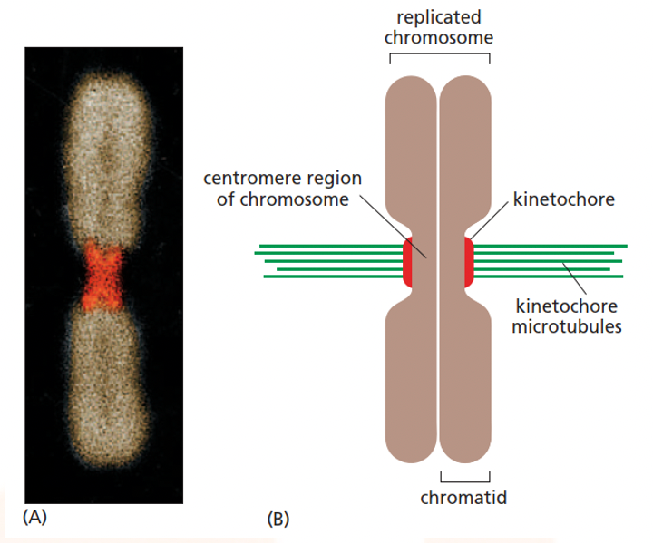 Chromosomes are classified by two ways on the basis of centromeres,
1.On the basis of number of centromeres
2.On the basis of position of centromeres
On the basis of number of centromeres:
Usually, there are two centromeres one on each chromatid but these may be more or none. Depending upon the number of centromeres, the chromosomes may be:
1.Monocentric with one centromere.
2.Dicentric with two centromeres, one in each chromatid.
3.Polycentric with more than two centromeres.
4.Acentric without centromere. Such chromosomes represent freshly broken segments of chromosomes which do not survive for long.
5.Diffused or non-located with indistinct centromere diffused throughout the length of chromosome. The microtubules of spindle fibers are attached to chromosome arms at many points. The diffused centromeres are found in insects, some algae and some groups of plants (e.g. Luzula).
2.On the basis of position of centromeres:
Based on the location of centromere the chromosomes are of four types,
1.Acrocentric are rod-shaped chromosomes with centromere occupy terminal position. These chromosomes appear ‘i’ shaped during anaphase.
2.Telocentric are rod-shaped chromosomes having subterminal centromere. These chromosomes appear ‘j’ shaped during anaphase.
3.Submetacentric are S-shaped chromosomes with centromere slightly away from the mid-point so that the two arms are unequal. These chromosomes appear ‘L’ shaped during anaphase.
4.Metacentric are large chromosomes in which centromere lies in the middle of chromosome so that the two arms are almost equal. These chromosomes appear ‘V’ shaped during anaphase.
5.
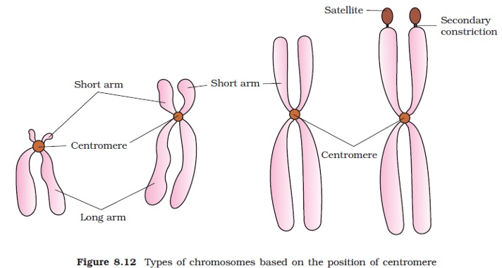 Telomere:
Telomere constitute the end of chromosomes. The number of telomere varies in between 2 to 4. Hermann J. Muller, who introduced the term telomere in 1938, demonstrated that Drosophila chromosomes without natural ends—produced by breaking chromosomes with X rays—were not transmitted to progeny. In a classical study of maize chromosomes, Barbara McClintock demonstrated that new ends of broken chromosomes are sticky and tend to fuse with each other. In contrast, the natural ends of normal (unbroken) chromosomes are stable and show no tendency to fuse with other broken or native ends. McClintock’s results indicated that telomeres must have special structures different from the ends produced by breakage of chromosomes. The telomeres of eukaryotic chromosomes have unique structures that include short nucleotide sequences present as tandem repeats. Although the sequences vary somewhat in different species, the basic repeat unit has the pattern 5’ T1–4A0–1G1–8-3’ in all but a few species. For example, the repeat sequence in humans and other vertebrates is TTAGGG, that of the protozoan Tetrahymena thermophila is TTGGGG, and that of the plant Arabidopsis thaliana is TTTAGGG. In most species, additional repetitive DNA sequences are present adjacent to telomeres; these are referred to as telomere-associated sequences.
These Telomere associated sequences can roll back on similar sequences and close the free end of the chromatids. This structure is also stabilized with the help of certain proteins. The telomeres of humans and a few other species have been shown to form structures called t-loops, in which the single strand at the 3’ terminus invades an upstream telomeric repeat (TTAGGG in mammals) and pairs with the complementary strand, displacing the equivalent strand. The DNA in these t-loops is protected from degradation and/or modification by DNA repair processes by a telomere-specific protein complex called Shelterin.
The telomeric sequences are under constant attack by exonucleases and maintained by continuous synthesis with the help of a unique enzyme Telomerase.
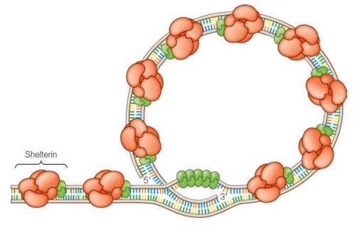 Secondary Constriction or Nucleolar Organizer
Sometimes one or both the arms of a chromosome are marked by a constriction other than the primary constriction. During interpose this area is associated with the :nucleolus and is found to participate in the formation of necleolus. It is, therefore, known as Nucleolar Organizer Region or the Secondary constriction. its location on the chromosome is marked with a lightly stained constricted region.
Nucleolar Organizer Region (NOR):
In certain chromosomes, the secondary constriction is intimately associated with the nucleolus during interphase. It contains genes coding for 18S and 28S ribosomal RNA and is responsible for the formation of necleolus. It is, therefore, known as nucleolar organizer region (NOR). Its location in the chromosome is marked by a lightly stained constricted area and is constant for a given chromosome. The terminal part of a chromosome beyond secondary constriction is called Satellite.
 A karyotype is an individual's collection of chromosomes. The term also refers to a laboratory technique that produces an image of an individual's chromosomes. The technique to generate karyotype is called Karyotyping.
Karyotypes describe the chromosome count of an organism and what these chromosomes look like under a light microscope. Attention is paid to their length, the position of the centromeres, banding pattern, any differences between the sex chromosomes, and any other physical characteristics.
The preparation and study of karyotypes is part of cytogenetics. In 1955 Joe Hin Tjio working in Albert Levan's developed Human Karyotype. In human karyotype there are 23 pairs of chromosomes in which 22 pairs are called Autosomes while one pair determine the sex and called Allosome.
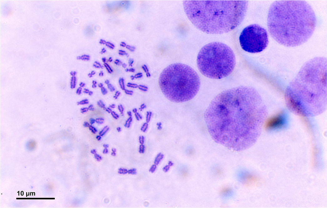 A karyotype may include the number of chromosomes, relative size, position of centromere, length of the arm, secondary constriction and satellites, features by which a particular set of chromosome can be arranged. A diagrammatic representation of karyotype (morphological characteristic of the chromosomes) of a species is called Idiogram, where the chromosomes of an organism are ordered in a series of decreasing size.
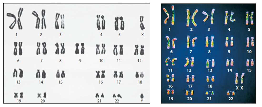 Depending upon complexity Karyotype is of two types,
 Identification of chromosomes and karyotyping can be done by following techniques:
Chromosome Banding:
Chromosome banding is the “Lengthwise variation in staining properties along a chromosome”. Chromosome banding refers to alternating light and dark regions along the length of a chromosome, produced after staining with a dye. A band is defined as the part of a chromosome that is clearly distinguishable from its adjacent segments by appearing darker or lighter with the use of one or more banding techniques. Torbjorn Caspersson when working with plants and using the highly fluorescent quinacrine mustard produced the first chromosome banding patterns, which were reported in 1968. The alternating bands of bright and dull fluorescence were called Q-bands. Quinacrine-bright bands were composed primarily of DNA rich in the bases adenine and thymine. Quinacrine mustard staining was applied to human chromosomes to produce the first-banded human karyogram in 1970. Analyses of Q-banded chromosomes led to the unequivocal identification of each human chromosome, and within each chromosome, specific regions and bands, since then variety of banding techniques are developed.
Classification of Banding Techniques
Based on
•GC and AT rich regions.
•Constitutive Heterochromatin Region.
Banding is always carried out in metaphase chromosomes whose size has condensed and whose diameter is increased are used for chromosome banding studies after fixing the stage.
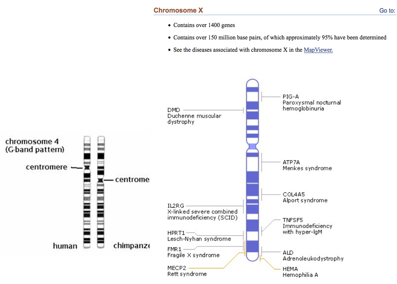 Banding Techniques
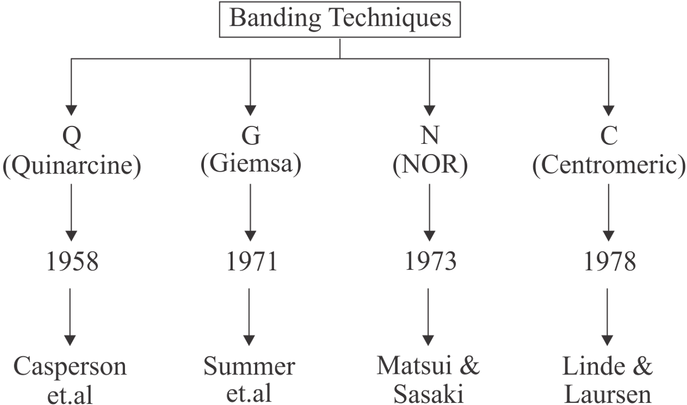 Besides Banding many new and powerful techniques are developed such as Fluorescence in situ hybridization (FISH) and multicolour fluorescence in situ hybridization (McFISH) which have been extensively used in animals and plants. Here using DNA probes, labelled with radioactive or nonradioactive molecules are used to locate the positions of specific DNA sequences on chromosomes. In McFISH, DNA may also be labelled with fluorochromes to allow the use of one or more colors to locate the position of one or more DNA sequences simultaneously on the same chromosomes.
Both FISH and McFISH are highly specific and are widely used in Medical Diagnosis, Evolutionary and population studies.
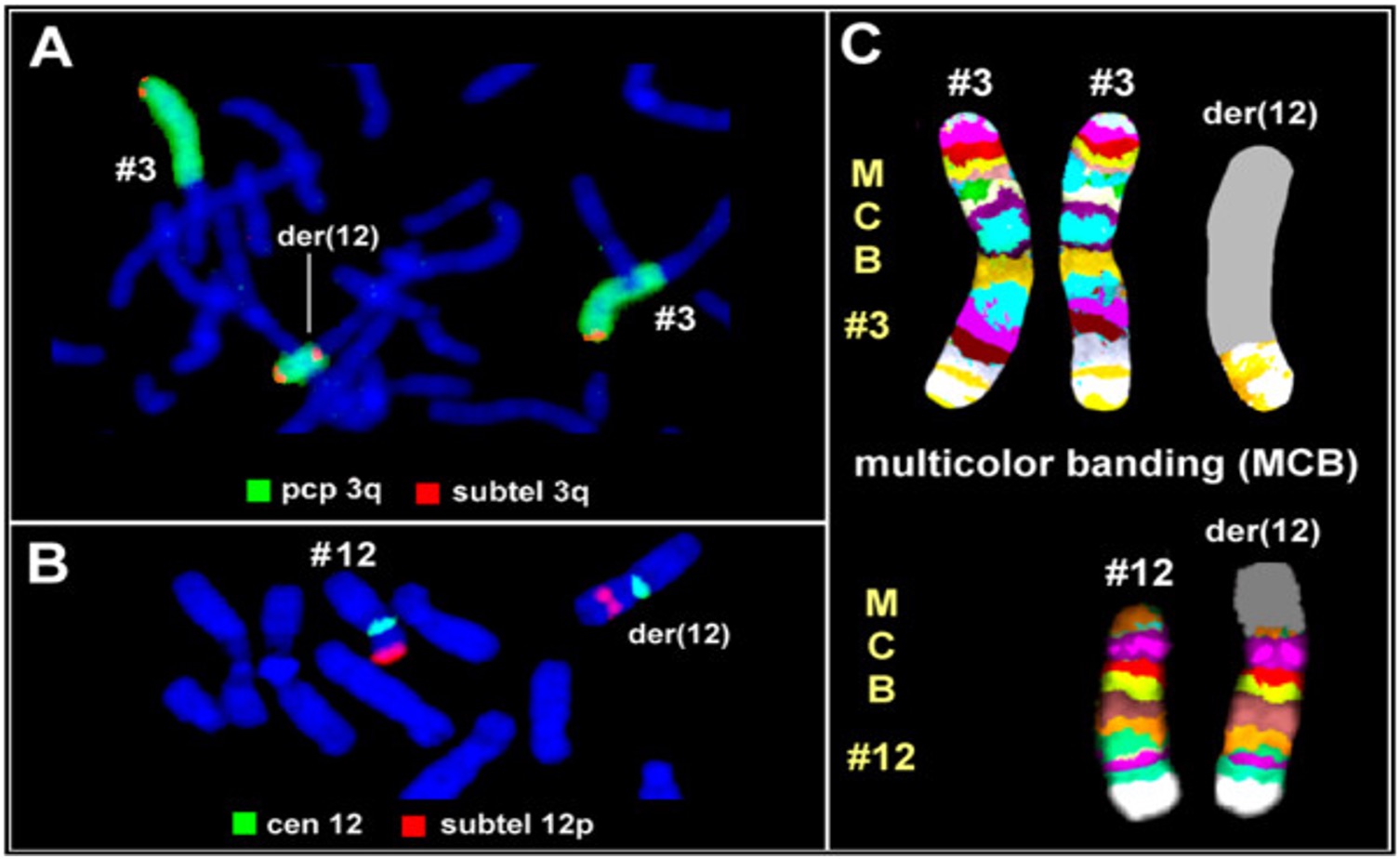
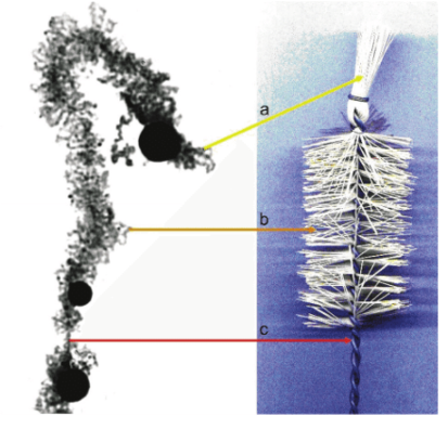 Large chromosomes
Chromosomes transform into the lampbrush form during the diplotene stage of meiotic prophase I due to an active transcription of many genes. They are highly extended meiotic half-bivalents, each consisting of 2 sister chromatids. Lampbrush chromosomes are clearly visible even in the light microscope, where they are seen to be organized into a series of chromomeres with large chromatin loops extended laterally. Amphibian and avian lampbrush chromosomes can be micro-surgically isolated from oocyte nucleus (germinal vesicle) with either forceps or needles In the homologous sections of the bivalent, chromatin is condensed (spirally twisted) or decondensed in the form of side loops – two per each chromosome and four at the level of the bivalent.
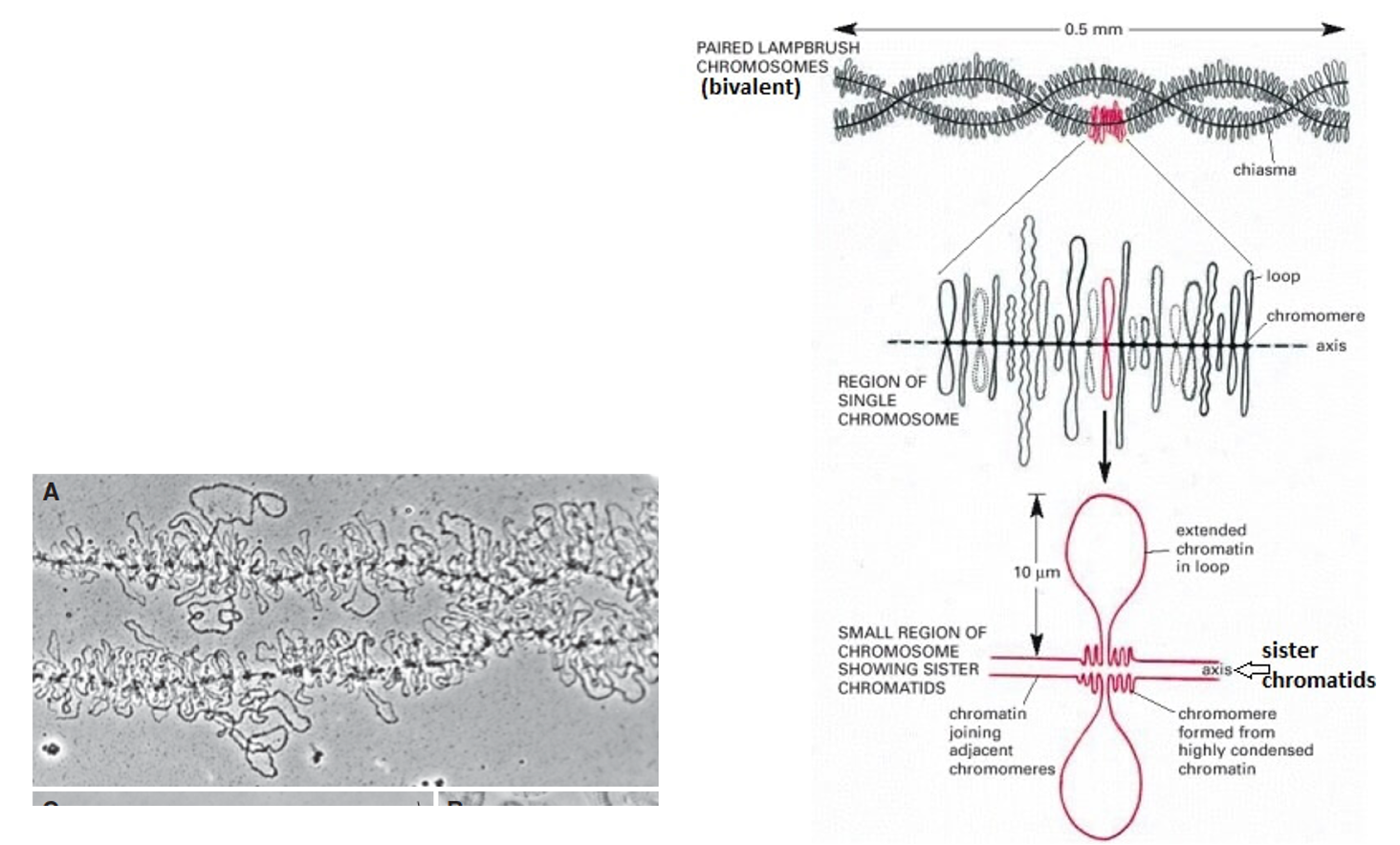 Lampbrush chromosome loops are considered an example of open chromatin. Their analogues are thought to be the “puffs” of polytenic chromosomes. They differ between each other. Polytenic chromosomes are made up of parallel chromatids, whereas lampbrush chromosome chromatin constitutes of a single DNA helix. The average length of a typical lampbrush chromosome loop is 10-15 μm, though some can be as long as50 or even 100 μm. Lampbrush chromosomes of various species have a very similar structure and perform the same function.
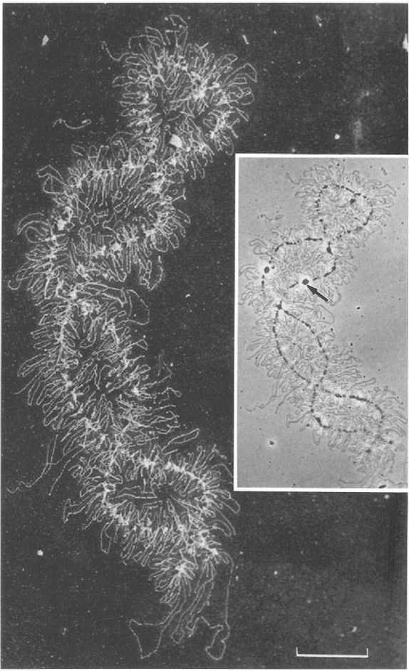 Polytene chromosomes
 are specific interphase chromosomes consisting of thousands of deoxyribonucleic acid (DNA) strands. For this reason they are very large and display a characteristic band–interband morphology. Polyteny arises in tissues, organs and at developmental stages when there is need for the rapid development of an organ at an unaltered high level of function. Organs containing cells with polytene chromosomesn are, as a rule, involved in intense secretory functions accomplished during a short time against a background of rapid growth.
Polytene chromosomes were discovered by Balbiani (1881) in larval salivary glands, Malpighian tubules, intestine, hypoderm and muscles of Chironomus plumosus as a cylindrical cord that repeatedly unrivalled and filled the nucleus. He called this structure a ‘Permanent Spireme’. Nine years later, in 1890, he found a permanent spireme in the macronucleus anlage of the infusorian Loxophyllum meleagris.
Polytene chromosomes
Polytene chromosomes are specific interphase chromosomes consisting of thousands of deoxyribonucleic acid (DNA) strands. For this reason they are very large and display a characteristic band–interband morphology. Polyteny arises in tissues, organs and at developmental stages when there is need for the rapid development of an organ at an unaltered high level of function. Organs containing cells with polytene chromosomesn are, as a rule, involved in intense secretory functions accomplished during a short time against a background of rapid growth.
Polytene chromosomes were discovered by Balbiani (1881) in larval salivary glands, Malpighian tubules, intestine, hypoderm and muscles of Chironomus plumosus as a cylindrical cord that repeatedly unrivalled and filled the nucleus. He called this structure a ‘Permanent Spireme’. Nine years later, in 1890, he found a permanent spireme in the macronucleus anlage of the infusorian Loxophyllum meleagris.
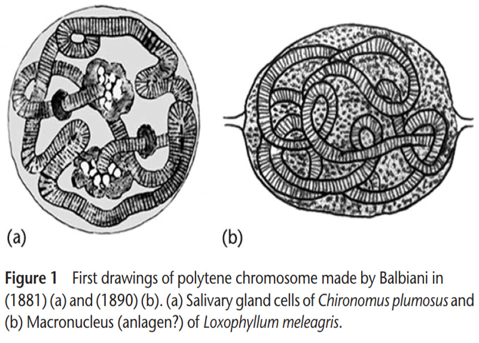 The relationship between polytene chromosomes (known as ‘giant chromosomes’ at that time) and regular mitotic chromosomes was first proposed by Rambousek (1912) and then by Kostoff (1930), Koltzoff (1934) and Bauer (1935). The giant size of the salivary gland chromosomes was explained by Koltzoff (1934) as being the consequence of multi strandedness. The term ‘Polytene’ was proposed by Koller (1935) and adopted by Darlington (1937).
In 1933–1934 scientists using squashed preparations, showed that the ‘Spireme’ is not continuous but consists of separate elements whose number is close to the haploid number of the mitotic chromosomes. Each element is formed as a result of the tight synapsis (joining together) of homologous chromosomes. Each chromosome has a definite and constant morphology and is composed of segments, displaying a distinctive transverse-banding pattern.
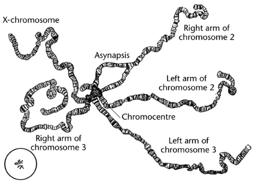
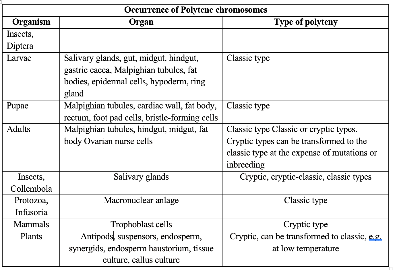
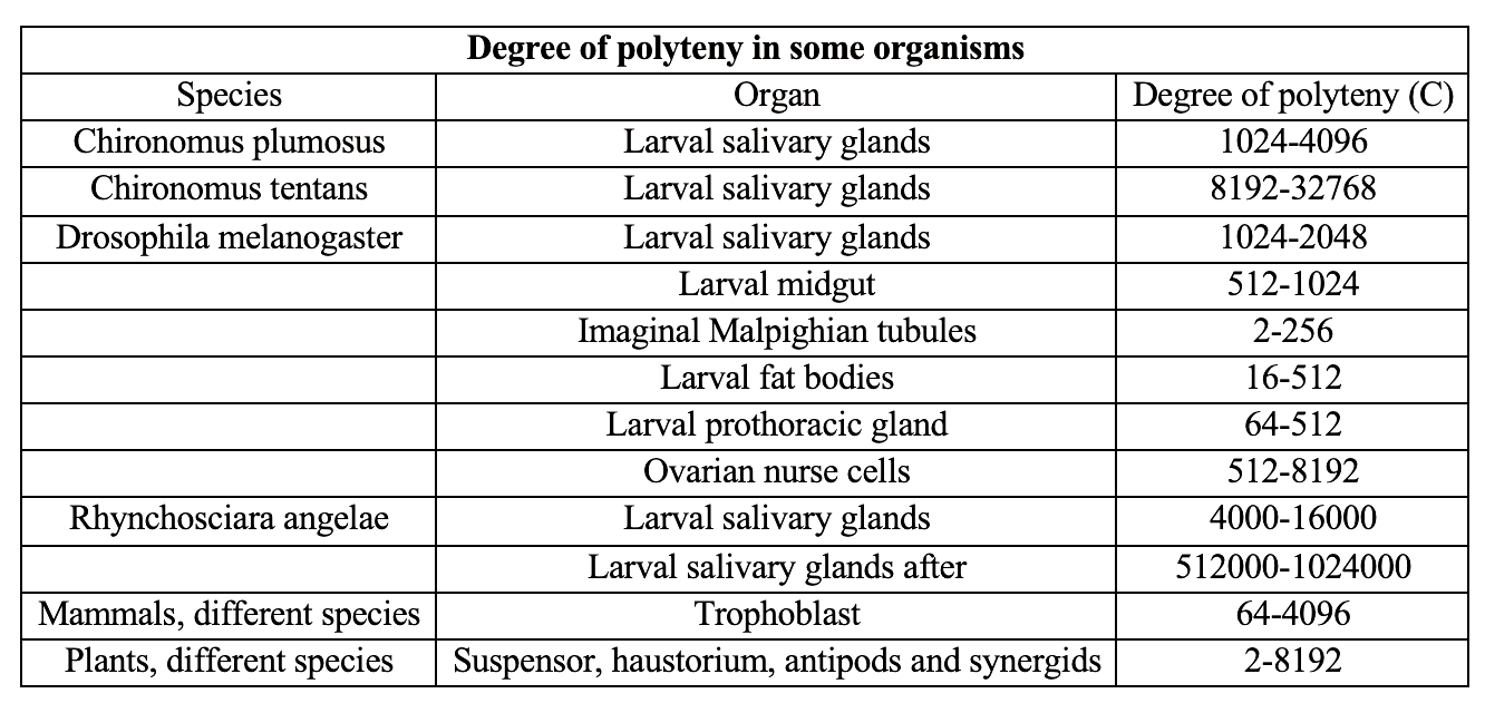 Cells with polytene chromosomes differ in the following ways from mitotically dividing cells and those undergoing endomitosis. First, the formation of polytene chromosomes is associated with the elimination of the entire mechanism of mitosis after each deoxyribonucleic acid (DNA) doubling, as a result of which the cell cycle consists of just two periods, synthetic (S) and intersynthetic (G). The polytenization cell cycle is set during midembryogenesis in Drosophila melanogaster. Second, at the end of each replication period, DNA strands do not segregate; rather, they remain paired to each other to different degrees rapid growth. The features of polyteny provide the conditions necessary to accomplish these functions. In many polytene nuclei, non homologous chromosomes are joined by their centromeric regions to form a common chromocentre. Chromocentres in polytene chromosomes correspond to heterochromatin in mitotic chromosomes. Along the linear axis of each chromatid, variation in the extent of coiling of the DNA and its associated proteins leads to variation in the concentration of the chromatin. Regions of high concentrations are known as chromomeres. For each chromatid the pattern of chromomeres is highly specific so that in the polytene chromosome homologous chromomeres align alongside each other exactly
and usually appear to fuse as a band across the polytene element.
The banding of polytene chromosome is generally such a stable and specific feature of their organization that the individual bands can be recognized, mapped and assigned reference numbers. In the interchromomeric or inter band regions, the DNA and protein concentration is lower than in the bands.
Interbands
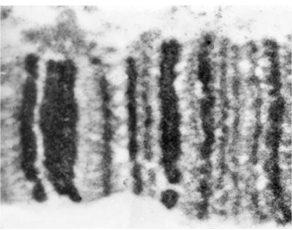
NUCLEUS
- EM studies have shown that nucleus consist of,
1.Nuclear Envelope
2.Nuclear Matrix
3.Nucleoplasm
4.Nucleolus
5.Chromatin
 1.Nuclear Envelope:
Transmission electron microscopy revealed that the nucleus is bounded by a nuclear envelope composed of two membranes the inner and outer nuclear membranes separated by a perinuclear space measuring about 20–40 nm across. The outer nuclear membrane is continuous with the endoplasmic reticulum, making the perinuclear space continuous with the lumen of the ER. Like membranes of the rough ER, the outer membrane is often studded on its outer surface with ribosomes engaged in protein synthesis. Several proteins in the outer membrane bind to actin and intermediate filaments of the cell’s cytoskeleton, thereby anchoring the nucleus within the cell and providing a mechanism for nuclear movements.
 Tubular invaginations of the nuclear envelope, collectively known as the nucleoplasmic reticulum, may project into the internal nuclear space and increase the area of the nucleus that makes direct contact with the inner nuclear membrane. One of the most distinctive features of the nuclear envelope is the presence of specialized channels called Nuclear pores, which are especially easy to see when the nuclear envelope is examined by freeze-fracture microscopy. Each pore is a small cylindrical channel extending through both membranes of the nuclear envelope, thereby providing direct continuity between the cytosol and the nucleoplasm. The number of pores varies greatly with cell type and activity. A typical mammalian nucleus has about 3000–4000 pores, or about 10–20 pores per square micrometer of membrane surface area.
 At each pore, the inner and outer membranes of the nuclear envelope are fused together, forming a channel that is lined with an intricate protein structure called the nuclear pore complex (NPC). The NPC has an outer diameter of 120 nm and is built from about 30 different proteins called nucleoporins. In electron micrographs, the most striking feature of the pore complex is its octagonal symmetry. It is made up of rings of eight subunits arranged in an octagonal pattern. In other views, the eight subunits are seen to protrude on both the cytoplasmic and nucleoplasmic sides of the envelope. Nuclear pore complex regulate the transport of solutes in between cytoplasm and nucleoplasm, it also participate in maturation of Ribosomes.
 2. Nuclear Matrix
In the early 1970s researchers discovered that an insoluble fibrous network retaining the overall shape of the nucleus remains behind after more than 95% of the chromatin has been removed by a combination of nuclease and detergent treatments. This network, called the nuclear matrix or nucleoskeleton, is thought to help maintain the shape of the nucleus and provide an organizing skeleton for the chromatin fibers. Underlying the inner nuclear membrane is the nuclear lamina, a filamentous meshwork that provides structural support to the nucleus. The nuclear lamina is composed of 60- to 80-kilodalton (kd) fibrous proteins called lamins, along with associated proteins. Nucleolamins help in stability of nuclear membrane and chromatin.
3. Nucleoplasm: (Strasburger, 1882). It is a transparent, semi-fluid and colloidal substance which fills the nucleus. It contains nucleosides and a number of enzymes (e.g., DNA polymerase, RNA polymerase, nucleoside phosphorylase) which are required for the synthesis and functioning of DNA, RNA, nucleoproteins, etc. Some of the proteins present in nucleoplasm are essential for spindle formation.
4. Nucleolus: It was first discovered by Fontana in 1781, described by Wagner in 1840 and provided with its present name by Bowman in 1840. Nucleolus is a naked, round or slightly irregular structure which is attached to the chromatin at a specific region called Nuclear Organiser Region (NOR). Commonly 1-4 nucleoli are found in a nucleus. Up to 1600 nucleoli are reported in the oocytes of Xenopus. A covering membrane is absent around nucleolus. Calcium seems to be essential for maintaining its configuration. Nucleolus has four components-
⁕Amorphous matrix,
⁕Granular part,
⁕Fibrillate portion and
⁕Chromatin part
(a) Amorphous Matrix. It is the homogeneous ground substance of the nucleolus. Matrix is formed of protein.
(b) Granular Portion. It consists of granules of the size of 150-200 Å which lie scattered in the amorphous matrix. The granules are formed of protein and RNA in the ratio of 2 : 1. They are believed to be precursors of ribosomes.
(c) Fibrillar Portion (Nucleolonema). It is formed of a large number of small fibrils that are 50-80 Å long. The fibrils are made up of both protein and RNA and are believed to be precursors of granules.
(d) Chromatin Portion. It is that part of chromatin which is associated with nucleolus. Depending upon its position nucleolar chromatin is of two types- perinucleolar and intranucleolar. The perinucleolar chromatin lies around the periphery of the nucleolus. It gives rise to in growths or trabeculae which produce the intranucleolar chromatin.
 The nucleolus is the site for ribosomal RNA synthesis. The nucleoli are larger and more numerous in cells that are actively involved in protein synthesis. Isolated nucleoli contain the nucleolus organizer region (NOR) -a stretch of DNA carrying multiple copies of rRNA genes. These multiple rRNA genes occur in all genomes and are thus an important example of repeated DNA that carries genetic information. The number of copies of the rRNA genes varies greatly from species to species, but animal cells generally contain hundreds of copies and plant cells often contain thousands. The multiple copies are grouped into one or more NORs, which may reside on more than one chromosome; in each NOR, the multiple gene copies are tandemly arranged. They are also called secondary constriction.
 In humans secondary constriction is present in chromosome no. 13,14,15, 21 & 22.
Chromatin: It is hereditary DNA-protein fibrillate complex which is named so because of its ability to get stained with certain basic dyes (Gk. chroma- colour, Flemming, 1879). Chromatin occurs in the form of fine overlapping and coiled fibers which appear to produce a network called chromatin reticulum. Chromatin fibers are distributed throughout the nucleoplasm. They are differentiated into two regions- Euchromatin and Heterochromatin. Euchromatin is narrow (30-80 Å thick) lightly stained and diffused fibrous part which forms the bulk of chromatin. Heterochromatin is wider (250 Å thick), darkly stained and condensed granular part which is attached here and there on the euchromatin. Depending upon the size of granules formed by heterochromatin they are called chromocentres, karyosomes or false nucleoli.
 Euchromatin
 is a lightly packed form of chromatin (DNA, RNA, and protein) that is enriched in genes, and is often (but not always) under active transcription. Euchromatin comprises the most active portion of the genome within the cell nucleus, 92% of the human genome is euchromatic.
It enters into replication first and lightly stained. The basic structure of euchromatin is an elongated, open, 10 nm microfibril. It has a beads on a string structure or Nucleosome structure. Nucleosomes were first observed as particles in the electron microscope by Don and Ada Olins in 1974, and their existence and structure (as histone octamers surrounded by approximately 200 base pairs of DNA) were proposed by Roger Kornberg.
Euchromatin
Euchromatin is a lightly packed form of chromatin (DNA, RNA, and protein) that is enriched in genes, and is often (but not always) under active transcription. Euchromatin comprises the most active portion of the genome within the cell nucleus, 92% of the human genome is euchromatic.
It enters into replication first and lightly stained. The basic structure of euchromatin is an elongated, open, 10 nm microfibril. It has a beads on a string structure or Nucleosome structure. Nucleosomes were first observed as particles in the electron microscope by Don and Ada Olins in 1974, and their existence and structure (as histone octamers surrounded by approximately 200 base pairs of DNA) were proposed by Roger Kornberg.
 In 1974, using the data from nuclease digestion and other types of information, Roger Kornberg, proposed that DNA and histones are organized into repeating subunits, called nucleosomes. Nucleosome is formed by the octamer of histone proteins, around this octamer 200 bps of DNA is wrapped. The formation of nucleosomes converts a DNA molecule into a chromatin thread that is approximately one-third the length of the initial piece of DNA, and it provides the first level of DNA packing.
All four of the histones that make up the octamer are relatively small proteins, with a high proportion of positively charged amino acids (lysine and arginine). The positive charges help the histones bind tightly to the negatively charged sugar phosphate backbone of DNA. These numerous electrostatic interactions explain in part why DNA of virtually any sequence can bind to a histone octamer. Each individual nucleosome core particle consists of a complex of eight histone proteins-two molecules each of histones H2A, H2B, H3, and H4-and double-stranded DNA that is 147 nucleotide pairs long. The histone octamer forms a protein core around which the double-stranded DNA is wound. Each nucleosome core particle is separated from the next by a region of linker DNA, which can vary in length from a few nucleotide pairs up to about 80. On average, therefore, nucleosomes repeat at intervals of about 200 nucleotide pairs. For example, a diploid human cell with 6.4 x 10⁹ nucleotide pairs contains approximately 30 million nucleosomes.
 In interphase nucleus generally chromatin exists in two forms Euchromatin and Heterochromatin. Usually Euchromatin exists in Beads on a string form while Heterochromatin are at higher condensation level i.e. 30 nm chromatin fiber which is the Solenoid form which is formed with the help of H1 histone protein.


 Chromosomes is the condensed form of chromatin visible during dividing or M-Phasee. The chromosomes are capable of self-reproduction and maintaining morphological and physiological properties through successive generations. They are capable of transmitting the contained hereditary material to the next generation. Hence these are known as ‘Hereditary vehicles.
•Chromosomes were first observed by Karl Negali in 1842 in plant cells as rod-shaped bodies in plant cells.
•In 1848 Hofmeister Observed chromosomes as darkly stained bodies in pollen mother cells of Tradecscantia.
•In 1888 Waldayer coined the term chromosome.
•In 1902 Walter S. Sutton proposed ‘Theory of Chromosomal basis of Heredity.
The salient features of chromosome theory of heredity are: -
•The somatic cells of an organism which are derived by the repeated divisions of the zygote are all diploid, i.e. these consist of two identical sets of chromosomes. One set of chromosomes is received from mother (maternal set) through ovum and the other set is inherited from father through sperm (paternal set). These two chromosomes of one type constitute the homologous pair.
•The chromosomes retain their structural uniqueness, individuality and their continuity throughout the life-cycle of an organism.
•Each chromosome carries specific determiners or Mendelian factors and plays a significant role in the development of an organism from the zygote.
•The behavior of chromosomes during meiosis, which occurs at the time of gamete formation provides an evidence that genes or determiners are located in the chromosomes. This also explains the mechanism of segregation of characteristics at the time of gamete formation.
Number of Chromosomes:
The number of chromosomes in the somatic cells of higher animals and plants is known as diploid or somatic or zygotic number, while in the gametes (sperms and eggs) it is haploid, gametic or reduced. The number of chromosomes is constant in all the somatic cells of all the individuals of a species. Chromosome number is used in the identification of species and in tracing the relationship within the species.
The lowest no of chromosomes are present in,
Parascaris = No of chromosomes are 2,
Mucor hiemalis = No of chromosomes are 2,
Happlopappus gracilis = No of chromosomes 2n = 4
While highest no of chromosomes are present in,
In plants it is Adder’s Tongue fern
Ophioglossum retiulatum = 2n = 1262 while in animals,
Aulocantha = 2n = 1632
Chromosomes is the condensed form of chromatin visible during dividing or M-Phasee. The chromosomes are capable of self-reproduction and maintaining morphological and physiological properties through successive generations. They are capable of transmitting the contained hereditary material to the next generation. Hence these are known as ‘Hereditary vehicles.
•Chromosomes were first observed by Karl Negali in 1842 in plant cells as rod-shaped bodies in plant cells.
•In 1848 Hofmeister Observed chromosomes as darkly stained bodies in pollen mother cells of Tradecscantia.
•In 1888 Waldayer coined the term chromosome.
•In 1902 Walter S. Sutton proposed ‘Theory of Chromosomal basis of Heredity.
The salient features of chromosome theory of heredity are: -
•The somatic cells of an organism which are derived by the repeated divisions of the zygote are all diploid, i.e. these consist of two identical sets of chromosomes. One set of chromosomes is received from mother (maternal set) through ovum and the other set is inherited from father through sperm (paternal set). These two chromosomes of one type constitute the homologous pair.
•The chromosomes retain their structural uniqueness, individuality and their continuity throughout the life-cycle of an organism.
•Each chromosome carries specific determiners or Mendelian factors and plays a significant role in the development of an organism from the zygote.
•The behavior of chromosomes during meiosis, which occurs at the time of gamete formation provides an evidence that genes or determiners are located in the chromosomes. This also explains the mechanism of segregation of characteristics at the time of gamete formation.
Number of Chromosomes:
The number of chromosomes in the somatic cells of higher animals and plants is known as diploid or somatic or zygotic number, while in the gametes (sperms and eggs) it is haploid, gametic or reduced. The number of chromosomes is constant in all the somatic cells of all the individuals of a species. Chromosome number is used in the identification of species and in tracing the relationship within the species.
The lowest no of chromosomes are present in,
Parascaris = No of chromosomes are 2,
Mucor hiemalis = No of chromosomes are 2,
Happlopappus gracilis = No of chromosomes 2n = 4
While highest no of chromosomes are present in,
In plants it is Adder’s Tongue fern
Ophioglossum retiulatum = 2n = 1262 while in animals,
Aulocantha = 2n = 1632- Chromosomes exhibit cyclic change in shape and size during cell cycle. In the non dividing interphase nucleus, the chromosomes form an interwoven network of line twisted but uncoiled threads of chromatin, and are invisible. During cell division the chromatin threads condense into compact structures by helical coiling. In prophase of cell division the chromosomes appear as distinct threads and by metaphase and also in anaphase these become short, compact bodies having definite shapes and sizes. In anaphase these appear as rod-shaped, V-shaped, L-shaped or J-shaped. The chromosomes are studied and described at this stage or at anaphase stage. In telophase these again uncoil to form the chromatin net.
 Chromosomes differ greatly in size in different organisms. Even the chromosomes of different pairs in the nucleus of the same cell have different sizes. The anaphase chromosomes range from 1 μm to 30 μm in length and 0.22 μ to 2 μ in width.
A chromosome consists of following parts,
⁕Centromere
⁕Telomere
⁕Secondary Constriction,
Centromere:
Centromere is the part of chromosome where two chromatids join with each other. At centromere specific proteins bind and form a disc like structure called Kinetochore, at kinetochore spindle fibers interact and help in the separation of sister chromatids (Chromatids which join with same centromere in a chromosome are called Sister Chromatids).
The centromeric region is also called Primary constriction. The microtubules of the chromosomal spindle fibers are attached to the centromere. Therefore, centromere is associated with the chromosomal movement during cell division. A chromosome without centromere is lost during anaphase.
 Chromosomes are classified by two ways on the basis of centromeres,
1.On the basis of number of centromeres
2.On the basis of position of centromeres
On the basis of number of centromeres:
Usually, there are two centromeres one on each chromatid but these may be more or none. Depending upon the number of centromeres, the chromosomes may be:
1.Monocentric with one centromere.
2.Dicentric with two centromeres, one in each chromatid.
3.Polycentric with more than two centromeres.
4.Acentric without centromere. Such chromosomes represent freshly broken segments of chromosomes which do not survive for long.
5.Diffused or non-located with indistinct centromere diffused throughout the length of chromosome. The microtubules of spindle fibers are attached to chromosome arms at many points. The diffused centromeres are found in insects, some algae and some groups of plants (e.g. Luzula).
2.On the basis of position of centromeres:
Based on the location of centromere the chromosomes are of four types,
1.Acrocentric are rod-shaped chromosomes with centromere occupy terminal position. These chromosomes appear ‘i’ shaped during anaphase.
2.Telocentric are rod-shaped chromosomes having subterminal centromere. These chromosomes appear ‘j’ shaped during anaphase.
3.Submetacentric are S-shaped chromosomes with centromere slightly away from the mid-point so that the two arms are unequal. These chromosomes appear ‘L’ shaped during anaphase.
4.Metacentric are large chromosomes in which centromere lies in the middle of chromosome so that the two arms are almost equal. These chromosomes appear ‘V’ shaped during anaphase.
5.
 Telomere:
Telomere constitute the end of chromosomes. The number of telomere varies in between 2 to 4. Hermann J. Muller, who introduced the term telomere in 1938, demonstrated that Drosophila chromosomes without natural ends—produced by breaking chromosomes with X rays—were not transmitted to progeny. In a classical study of maize chromosomes, Barbara McClintock demonstrated that new ends of broken chromosomes are sticky and tend to fuse with each other. In contrast, the natural ends of normal (unbroken) chromosomes are stable and show no tendency to fuse with other broken or native ends. McClintock’s results indicated that telomeres must have special structures different from the ends produced by breakage of chromosomes. The telomeres of eukaryotic chromosomes have unique structures that include short nucleotide sequences present as tandem repeats. Although the sequences vary somewhat in different species, the basic repeat unit has the pattern 5’ T1–4A0–1G1–8-3’ in all but a few species. For example, the repeat sequence in humans and other vertebrates is TTAGGG, that of the protozoan Tetrahymena thermophila is TTGGGG, and that of the plant Arabidopsis thaliana is TTTAGGG. In most species, additional repetitive DNA sequences are present adjacent to telomeres; these are referred to as telomere-associated sequences.
These Telomere associated sequences can roll back on similar sequences and close the free end of the chromatids. This structure is also stabilized with the help of certain proteins. The telomeres of humans and a few other species have been shown to form structures called t-loops, in which the single strand at the 3’ terminus invades an upstream telomeric repeat (TTAGGG in mammals) and pairs with the complementary strand, displacing the equivalent strand. The DNA in these t-loops is protected from degradation and/or modification by DNA repair processes by a telomere-specific protein complex called Shelterin.
The telomeric sequences are under constant attack by exonucleases and maintained by continuous synthesis with the help of a unique enzyme Telomerase.
 Secondary Constriction or Nucleolar Organizer
Sometimes one or both the arms of a chromosome are marked by a constriction other than the primary constriction. During interpose this area is associated with the :nucleolus and is found to participate in the formation of necleolus. It is, therefore, known as Nucleolar Organizer Region or the Secondary constriction. its location on the chromosome is marked with a lightly stained constricted region.
Nucleolar Organizer Region (NOR):
In certain chromosomes, the secondary constriction is intimately associated with the nucleolus during interphase. It contains genes coding for 18S and 28S ribosomal RNA and is responsible for the formation of necleolus. It is, therefore, known as nucleolar organizer region (NOR). Its location in the chromosome is marked by a lightly stained constricted area and is constant for a given chromosome. The terminal part of a chromosome beyond secondary constriction is called Satellite.
 A karyotype is an individual's collection of chromosomes. The term also refers to a laboratory technique that produces an image of an individual's chromosomes. The technique to generate karyotype is called Karyotyping.
Karyotypes describe the chromosome count of an organism and what these chromosomes look like under a light microscope. Attention is paid to their length, the position of the centromeres, banding pattern, any differences between the sex chromosomes, and any other physical characteristics.
The preparation and study of karyotypes is part of cytogenetics. In 1955 Joe Hin Tjio working in Albert Levan's developed Human Karyotype. In human karyotype there are 23 pairs of chromosomes in which 22 pairs are called Autosomes while one pair determine the sex and called Allosome.
 A karyotype may include the number of chromosomes, relative size, position of centromere, length of the arm, secondary constriction and satellites, features by which a particular set of chromosome can be arranged. A diagrammatic representation of karyotype (morphological characteristic of the chromosomes) of a species is called Idiogram, where the chromosomes of an organism are ordered in a series of decreasing size.
 Depending upon complexity Karyotype is of two types,
 Identification of chromosomes and karyotyping can be done by following techniques:
Chromosome Banding:
Chromosome banding is the “Lengthwise variation in staining properties along a chromosome”. Chromosome banding refers to alternating light and dark regions along the length of a chromosome, produced after staining with a dye. A band is defined as the part of a chromosome that is clearly distinguishable from its adjacent segments by appearing darker or lighter with the use of one or more banding techniques. Torbjorn Caspersson when working with plants and using the highly fluorescent quinacrine mustard produced the first chromosome banding patterns, which were reported in 1968. The alternating bands of bright and dull fluorescence were called Q-bands. Quinacrine-bright bands were composed primarily of DNA rich in the bases adenine and thymine. Quinacrine mustard staining was applied to human chromosomes to produce the first-banded human karyogram in 1970. Analyses of Q-banded chromosomes led to the unequivocal identification of each human chromosome, and within each chromosome, specific regions and bands, since then variety of banding techniques are developed.
Classification of Banding Techniques
Based on
•GC and AT rich regions.
•Constitutive Heterochromatin Region.
Banding is always carried out in metaphase chromosomes whose size has condensed and whose diameter is increased are used for chromosome banding studies after fixing the stage.
 Banding Techniques
 Besides Banding many new and powerful techniques are developed such as Fluorescence in situ hybridization (FISH) and multicolour fluorescence in situ hybridization (McFISH) which have been extensively used in animals and plants. Here using DNA probes, labelled with radioactive or nonradioactive molecules are used to locate the positions of specific DNA sequences on chromosomes. In McFISH, DNA may also be labelled with fluorochromes to allow the use of one or more colors to locate the position of one or more DNA sequences simultaneously on the same chromosomes.
Both FISH and McFISH are highly specific and are widely used in Medical Diagnosis, Evolutionary and population studies.

 Large chromosomes
Chromosomes transform into the lampbrush form during the diplotene stage of meiotic prophase I due to an active transcription of many genes. They are highly extended meiotic half-bivalents, each consisting of 2 sister chromatids. Lampbrush chromosomes are clearly visible even in the light microscope, where they are seen to be organized into a series of chromomeres with large chromatin loops extended laterally. Amphibian and avian lampbrush chromosomes can be micro-surgically isolated from oocyte nucleus (germinal vesicle) with either forceps or needles In the homologous sections of the bivalent, chromatin is condensed (spirally twisted) or decondensed in the form of side loops – two per each chromosome and four at the level of the bivalent.
 Lampbrush chromosome loops are considered an example of open chromatin. Their analogues are thought to be the “puffs” of polytenic chromosomes. They differ between each other. Polytenic chromosomes are made up of parallel chromatids, whereas lampbrush chromosome chromatin constitutes of a single DNA helix. The average length of a typical lampbrush chromosome loop is 10-15 μm, though some can be as long as50 or even 100 μm. Lampbrush chromosomes of various species have a very similar structure and perform the same function.
 Polytene chromosomes
 are specific interphase chromosomes consisting of thousands of deoxyribonucleic acid (DNA) strands. For this reason they are very large and display a characteristic band–interband morphology. Polyteny arises in tissues, organs and at developmental stages when there is need for the rapid development of an organ at an unaltered high level of function. Organs containing cells with polytene chromosomesn are, as a rule, involved in intense secretory functions accomplished during a short time against a background of rapid growth.
Polytene chromosomes were discovered by Balbiani (1881) in larval salivary glands, Malpighian tubules, intestine, hypoderm and muscles of Chironomus plumosus as a cylindrical cord that repeatedly unrivalled and filled the nucleus. He called this structure a ‘Permanent Spireme’. Nine years later, in 1890, he found a permanent spireme in the macronucleus anlage of the infusorian Loxophyllum meleagris.
Polytene chromosomes
Polytene chromosomes are specific interphase chromosomes consisting of thousands of deoxyribonucleic acid (DNA) strands. For this reason they are very large and display a characteristic band–interband morphology. Polyteny arises in tissues, organs and at developmental stages when there is need for the rapid development of an organ at an unaltered high level of function. Organs containing cells with polytene chromosomesn are, as a rule, involved in intense secretory functions accomplished during a short time against a background of rapid growth.
Polytene chromosomes were discovered by Balbiani (1881) in larval salivary glands, Malpighian tubules, intestine, hypoderm and muscles of Chironomus plumosus as a cylindrical cord that repeatedly unrivalled and filled the nucleus. He called this structure a ‘Permanent Spireme’. Nine years later, in 1890, he found a permanent spireme in the macronucleus anlage of the infusorian Loxophyllum meleagris.
 The relationship between polytene chromosomes (known as ‘giant chromosomes’ at that time) and regular mitotic chromosomes was first proposed by Rambousek (1912) and then by Kostoff (1930), Koltzoff (1934) and Bauer (1935). The giant size of the salivary gland chromosomes was explained by Koltzoff (1934) as being the consequence of multi strandedness. The term ‘Polytene’ was proposed by Koller (1935) and adopted by Darlington (1937).
In 1933–1934 scientists using squashed preparations, showed that the ‘Spireme’ is not continuous but consists of separate elements whose number is close to the haploid number of the mitotic chromosomes. Each element is formed as a result of the tight synapsis (joining together) of homologous chromosomes. Each chromosome has a definite and constant morphology and is composed of segments, displaying a distinctive transverse-banding pattern.


 Cells with polytene chromosomes differ in the following ways from mitotically dividing cells and those undergoing endomitosis. First, the formation of polytene chromosomes is associated with the elimination of the entire mechanism of mitosis after each deoxyribonucleic acid (DNA) doubling, as a result of which the cell cycle consists of just two periods, synthetic (S) and intersynthetic (G). The polytenization cell cycle is set during midembryogenesis in Drosophila melanogaster. Second, at the end of each replication period, DNA strands do not segregate; rather, they remain paired to each other to different degrees rapid growth. The features of polyteny provide the conditions necessary to accomplish these functions. In many polytene nuclei, non homologous chromosomes are joined by their centromeric regions to form a common chromocentre. Chromocentres in polytene chromosomes correspond to heterochromatin in mitotic chromosomes. Along the linear axis of each chromatid, variation in the extent of coiling of the DNA and its associated proteins leads to variation in the concentration of the chromatin. Regions of high concentrations are known as chromomeres. For each chromatid the pattern of chromomeres is highly specific so that in the polytene chromosome homologous chromomeres align alongside each other exactly
and usually appear to fuse as a band across the polytene element.
The banding of polytene chromosome is generally such a stable and specific feature of their organization that the individual bands can be recognized, mapped and assigned reference numbers. In the interchromomeric or inter band regions, the DNA and protein concentration is lower than in the bands.
Interbands
