LOCOMOTION AND MOVEMENT for Class 11 BIOLOGY
INTRODUCTION
- Movement is one of the significant features of living beings. Animals and plants exhibit a wide range of movements. Streaming of protoplasm in the unicellular organisms like Amoeba is a simple form of movement. Movement of cilia, flagella and tentacles are shown by many organisms.
Human beings can move limbs, jaws, eyelids, tongue, etc. Some of the movements result in a change of place or location. Such voluntary movements are called locomotion. Walking, running, climbing, flying, swimming are all some forms of locomotory movements. Locomotory structures need not be different from those affecting other types of movements.
For example, in Paramecium, cilia helps in the movement of food through cytopharynx and in locomotion as well.
Hydra can use its tentacles for capturing its prey and also use them for locomotion. We use limbs for changes in body postures and locomotion as well. The above observations suggest that movements and locomotion cannot be studied separately. The two may be linked by stating that all locomotions are movements but all movements are not locomotions.
Methods of locomotion performed by animals vary with their habitats and the demand of the situation. However, locomotion is generally for search of food, shelter, mate, suitable breeding grounds, favourable climatic conditions or to escape from enemies/predators.
Locomotion requires a perfect coordinated activity of the muscular, skeletal and neural system. Study of movement is called kinesiology.
CHAPTER WEIGHTAGE : The important areas of the chapter usually are , JOINTS (1-2 marks or a short note in 3 marks), DISORDERS , SKELETAL SYSTEM (sliding filament theory & ultrstructure of skeletal muscle are very important topics) . Number of questions which can be expected are 2-3
INTRODUCTION
- Movement is one of the significant features of living beings. Animals and plants exhibit a wide range of movements. Streaming of protoplasm in the unicellular organisms like Amoeba is a simple form of movement. Movement of cilia, flagella and tentacles are shown by many organisms.
Human beings can move limbs, jaws, eyelids, tongue, etc. Some of the movements result in a change of place or location. Such voluntary movements are called locomotion. Walking, running, climbing, flying, swimming are all some forms of locomotory movements. Locomotory structures need not be different from those affecting other types of movements.
For example, in Paramecium, cilia helps in the movement of food through cytopharynx and in locomotion as well.
Hydra can use its tentacles for capturing its prey and also use them for locomotion. We use limbs for changes in body postures and locomotion as well. The above observations suggest that movements and locomotion cannot be studied separately. The two may be linked by stating that all locomotions are movements but all movements are not locomotions.
Methods of locomotion performed by animals vary with their habitats and the demand of the situation. However, locomotion is generally for search of food, shelter, mate, suitable breeding grounds, favourable climatic conditions or to escape from enemies/predators.
Locomotion requires a perfect coordinated activity of the muscular, skeletal and neural system. Study of movement is called kinesiology.
CHAPTER WEIGHTAGE
The important areas of the chapter usually are , JOINTS , DISORDERS , SKELETAL SYSTEM . Number of questions which can be expected are 1-2.
MOVEMENT IN ANIMALS
- Cells of the human body exhibit three main types of movements, namely, amoeboid, ciliary and muscular.
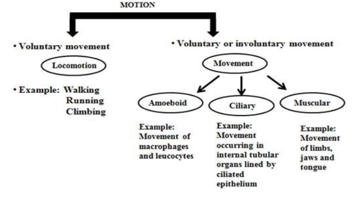
MOVEMENT IN ANIMALS
- Cells of the human body exhibit three main types of movements, namely, amoeboid, ciliary and muscular.

SKELETAL TISSUE
- Skeletal system consists of a framework of bones and cartilages.
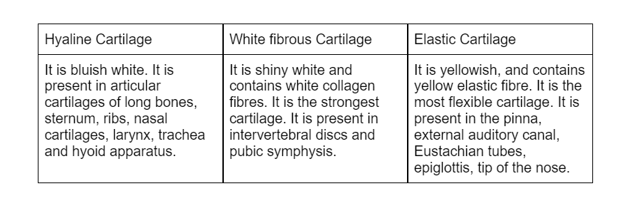
- CARTILAGE(Gristle) -
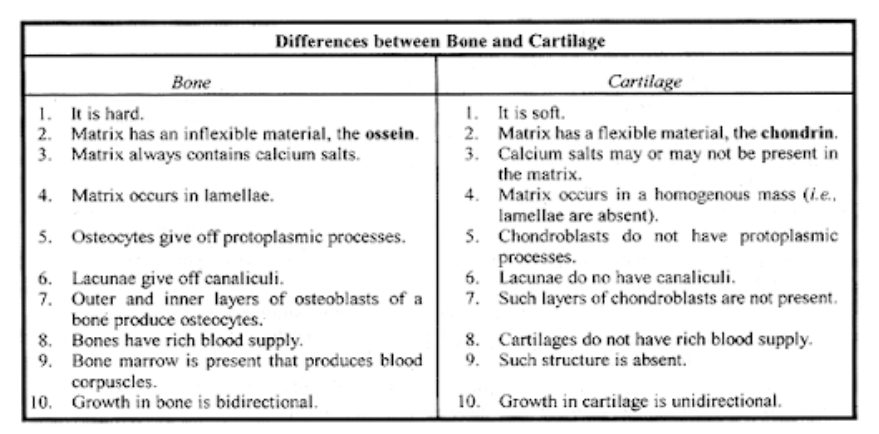
- The intercellular material of cartilage is solid,pliable and resists compression. The slightly pliable matrix is due to chondroitin salts. It is largely avascular. It contains chondroblast and chondrocyte cells.
- Chondroblasts are young cells, small and with small projections(filopodia). A cartilage cell is present in a fluid filled space, the cartilage lacuna. Chondrocytes are mature cells, large in size. Cells of this tissue are enclosed in small cavities within the matrix secreted by them. The chondroblasts are metabolically more active cells than chondrocytes.
- Most of the cartilages in vertebrate embryos are replaced by bones in adults. Cartilage is present in the tip of nose, outer ear, joints, between adjacent bones of vertebral column, limbs and hands in adults.
- Growth of cartilage is always from the periphery i.e. unidirectional because perichondrium surrounds the cartilage only on one side.
- BONE:
- It is the hardest tissue that provides structural frame to the body. Bones have a hard and non-pliable ground substance rich in calcium salts and collagen fibres which give bone its strength.


Skeletal System
- Skeletal system ka wo part jo body ki long axis ke along hai wo axial skeleton kehlata hai and isme 80 bones hoti hai. Skull, vertebral column, sternum and ribs issi ka parta hai. Iss central part jo bones attached hoti hai wo appendicular skeleton kehlati hai jisme 126 bones hoti hai. Girdles and limbs appendicular skeleton ka part hai.
- Skull me 8 bones cranium yani ki brain box form karti hai, 14 facial bones hoti hai, 1 hyoid bone jiske tongue bone bhi kehte hai and dono kaano me 3-3 ear ossicles hote hai.
- 1 frontal bone jo ki forehead and eye orbit ki roof bnati hai.
- 2 parietal bone cranium ki roof me situated hai.
- 1 occipital bone cranium ki back side me hoti hai jisme ke hole hota hai jiske hum foramen magnum kehte hai and issi hole ke through brain se spinal cord emergy hoti hai.
- Issi bone me 2 condyles hote hai jo ki vertebral colum ki first bone yani ki atlas ke sath articulate karte hai yani ki join karte hai and yahi joint aapke cranium ko aapki spinal bones se jodta hai. Human ki occipital bone me 2 condyles hone ke karan humans ke skull ko dicondylic skull kaha jata hai.
- 2 temporal bones hoti hai right and left side ek ek. Yeah ear ke paas wali bones hai basically jiske roughly kanpatti bhi bol dete hai.
- 1 Ethmoid bone nasal cavity ki roof bnati hai and it gives rise to 2 nasal conchae jo ki nasal cavity me project karte hai.
- 1 Sphenoid bone hoti hai jo ki butterfly shaped bone hai and yeah bone basically baki sabhi cranial bones ke sath articulate karti hai. Issi me choti si cavity hoti hai jiske hum sells turcica bolte hai and issi cavity me pituitary gland situated hota hai.
- 2 Nasal bones nasal bridge form karti hai.
- 2 Infra nasal inferior nasal conchae bnati hai.
- 2 lacrymal bones eyes ki sides me hoti hai.
- 2 Palatine bones buccal cavity ki roof banti hai.
- 1 Vomer bone bhi buccal cavity ki roof me hi milti hai.
- 2 Zygomatic bones basically cheek bones kehlati hai.
- Right and left side, dono taraf 1-1 upper jaw bone yani ki maxilla hoti hai.
- 1 Mandible bone lower jaw form karti hai. Mandibe is the only movable bone of the skull.
- Hyoid bone tongue bone kehlati hai kyunki issi bone se tongue ka base attached hota hai. Yeah bone lower jaw and larynx ke beech hoti hai.
- Craniostylic jaw suspension basically aisa jaw suspension hai jisme upper jaw immovable hota hai yani ki fixed hota hai kyunki it articulates with temporal bones.
Vertebral Column - Adults me 26 vertebrae hoti hai and children me 33.
- Yeah straight nhi hota rather S shape ke curves hote hai. Total 4 curves hote hai which corresponds to 4 different regions of the spine. Inme se 2 curves primary hote hai jo ki jab baby deliver hota hai toh formed hote hai lekin baki ke 2 curves jinko hum secondary curves bolte hai wo birth ke baad development ke sath bante hai and inhi curves ke birth par na hona hi karan hai ki chota baby shuru ke kuch mahine tak apna head stable nhi kar pata toh we have to hold him carefully supporting his head and lower spine.
- Spine ke 4 regions hai : cervical (neck area), thoracic (thoracic cavity wala), lumbar (lower spine) and pelvic region.
- Thoracic and pelvic curves primary hote hai jabki cervical and lumbar curves secondary hote hai.
- Children me 7 cervical vertebrae hoti hai, 12 thoracic vertebrae, 5 lumbar, 5 sacrum bones and 4 coccygeal bones. Toh inko hum represent karte hai by :C7T12L5S5Co4
- Adult me development ke time sacrum ki paancho ek single bone me fuse hojati hai and charo coccygeal bone bhi fuse karke ek single coccyx bna leti hai toh adult ka vertebral formula hua : C7T12L5S1Co1
- The vertebral column protects the spinal cord, supports the head and serves as the point of attachment for the ribs and musculature of the back.
- Har vertebrae me ek centrum yani ki body part hota hai and kuch spinal processes neural canal ko enclose karti hai. Issi neural canal ke through spinal cord (nervous tissue) travel karta hai. Centrum basically notochord se hi develop hota hai. Jo processes nikalti hai wo basically kis direction me project karti hai unke basis pe unko naam dedete hai jaise ki transverse processes, spinous processes etc. yeah apne kaam ka nahi hai.
- Har vertebrae ki upper side pe superior zygapophyses hote hai dono taraf and lower side par 2 (right and left) zygapophyses hote hai. Ek vertebrae ka inferior zygapophyses next/neeche wali vertebrae ke superior zygapophyses ke sath articulate karta hai.
- Humans me centrum ka upper and lower part flat hota hai toh aisi vertebrae ko hum amphiplatyan vertebrae bolte hai.
- Transverse processes me vertebral arteries ke passage ke liye foramen transversarium hote hai lekin yeah 7th vertebrae me absent hota hai.
- Cervical Vertebrae
→ Cervical vertebrae ki jo spinous process hoti hai wo bifid hoti hai and 3rd-6th are typical vertebrae jabki 1st, 2nd and 7th are atypical. Atypical ka matlab hai ki general ya usual structure se kuch alag hota hai inme.
→ Pehli cervical vertebrae atlas ke naam se jani jati hai. Yeah ek ring shaped bone hai jisme centrum reduced hota hai and spinous process nahi hoti. Iss bone me occipital bone se 2 condyles se articulate karne ke liye 2 facets hote hai. Another unique feature is the presence of odontoid fossa jo ki 2nd vertebral bone se articulate karega. Yeah head ki YES movement ke liye responsible hai.
→ 2nd cervical vertebrae axis kehlati hai. Head ki NO movement me issi ka role hota hai jo ki basically rotatory movement hai atlas and axis joint ki. Iss vertebrae ka unique feature yeah hai ki iske centrum se odontoid process nikalti hai jo ki atlas bone me odontoid fossa ke sath articulate karti hai. Isme transverse processes hoti hai.
→ 7th vertebrae ka unique feature yeah hai ki yeah sabse largest cervical bone hai and isliye isko vertebrae prominens naam dia gya hai. Iski spinous process bifid nahi hoti rather ek tubercle me end hoti hai. Another feature is ki isme transversarium absent hote hai. - Sternum breast bone kehlati hai and yeah ek dagger shaped bone hai. Main 2 parts hai : upper manubrium/prosternum jo ki short part hai and lower elongated part is mesosternum. Issi mesosternum me ribs ke articulation ke liye facets hote hai and it ends into a xiphoid process.
- Ribs ke 12 pairs hote hai humans me and yeah thoracic cavity ko protect karte hai. Ribs are bicephalic yani ki yeah dono ends se articulate karti hai. Coastal cartilage ribs to sternum se jodta hai.
→Ribs ko further 3 types me divide kia ja sakta hai : true ribs, false ribs and floating ribs.
→ true ribs ko hi vertebro-sternal ribs kehte hai kyunki yeah ek end se thoracic vertebrae se articulate karti hai and dusre end se sternum se. Pehle 7 pair ko true ribs mana gya hai. First rib front me manubrium se sath articulate karti hai jabki 2-6th ribs mesosternum ke sath articulate karti hai. 7th rib mesosternum and xiphoid process se articulate karti hai.
→ False ribs ko vertebr0-chondrial ribs kaha jata hai and is category me 8th-10th ribs aati hai kyunki yeah front side me 7th rib ke costal cartilage se articulate karti hai.
→ 11th and 12th rib ko floating ribs kaha jata hai kyunki inka sternal end free hota hai and yeah keval backside se vertebral column se hi judi hui hoti hai. - Amphicoelous aisi vertebrae hai jinka centrum dono ends se concave hota hai. Ex : vertebrae of cartilaginous fishes and 8th vertebrae of frog.
- Procoelous aisi vertebrae hai jinke centrum ka anterior end concave hota hai and posterior end convex. Ex : typical vertebrae of frog and vertebrae of lizard.
- Opisthocoelous aisi vertebrae hoti hai jinka centrum anterior end se convex hota hai jabki concavity posterior end me aati hai. Ex : vertebrae of urodela.
- Acoelous aisi vertebrae hoti hai jinme dono ends pe hi concavity nahi hoti kinda flat. Ex : 9th vertebrae of frog, free caudal vertebrae of pigeon.
- Amphiplatayan vertebrae me centrum dono taraf se flat hota hai. Jaise ki human ki vertebrae jo ki intervertebral cartilaginous discs se separated hoti hai.
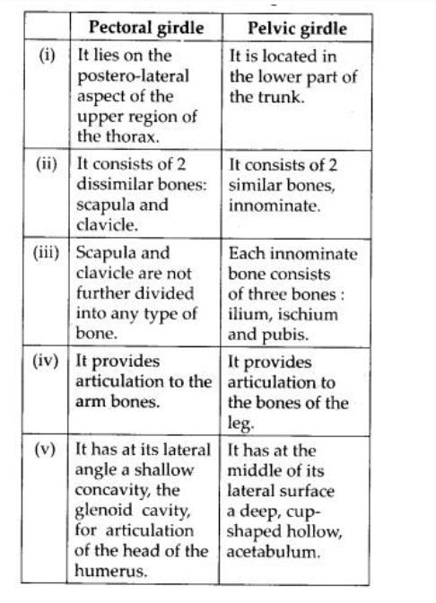
- Pectoral girdle 2 hai : ek rhs pe and dusra Lhs pe. Ek girdle me ek scapula yani ki shoulder bone hoti hai and ek clavicle yani ki collar bone hoti hai. Scapula ek triangular flat bone hai jo kamar ke upper part me located and it is superficial to the ribs. Yani ki ribs ke uper hai. Toh iski location dorsally hui between 2nd and 7th rib. Coracoid process ek bony hook shaped structure hota hai scapula pe jo project karta hai anterolaterally from the superior aspect of the scapular neck. Yeah process kafi sare ligaments and tendons ka anchor point hai. Scapula me glenoid cavity hoti hai jhaan upper arm bone yani ki humerus bone ka head articulate karta hai and this is known as glenohumeral joint or shoulder joint.
- Clavicle : Clavicle anterior side present hoti hai jo sternum ke manubrium se articulate karti hai at the sternoclavicular joint and scapula se articulate karti hai at the acromioclavicular joint. Iski shape S letter jaisi hai.
- Humans ke 2 forelimbs hai dono me 30-30 bones hoti hai which are 1 humerus upper arm me + 1 radius.1 ulna forearm me + 8 carpals/wrist bones wrist me + 5 metacarpals (palm bones) + 14 phalanges (pancho ungliyon ki haddiyan) hath me.
- Humerus ko hi funny bone bhi bol dete hai. Iska head golf ball jaisa hota hai jo ki scapula ki socket like glenoid cavity se articulate karke ball and socket joint bnata hai which we eill be discussing separately.
- Forearm ki radius choti wwali bone hai jo ki towards thumb located hai. Ulna bone longer hai and yeah little finger ki taraf located hai. Ulna bone ke (kohni ke taraf) olecranon process me end hoti hai. Yahi olecranon process elbow ki prominence form karti hai jiske hum touch bhi karke mehsoos bhi kar sakte hain. In contrast to the ulna, the radius is narrow at its proximal end and widens at its distal end.
- Wrist me 8 carpals hote hai jo ki 2 rows me fitted hai. Proximal row me 4 carpals and 4 carpals distal row me. Har carpal ka different name hai but we don’t have to remember their names.
- Thumb yani ki pollex ko chor kar har digit me 3-3 bones hoti hai jinko phalanges kehte hai. Thumb me keval 2 bones hoti hai. Phalangeal formula is : 2,3,3,3,3
- Pelvic girdle coxa naam ki bone se bna hua hai toh we have 2 coxa bones : right side wali and left side wali. Issi coxa bone ko innominate bone bhi keh dete hai and yeah 3 bones : Ilium, Ischium & Pubis ke fusion se bnti hai.
- Side view dekhenge toh upperpart Ilium bone (largest of all three bones) se banta hai, anterior part me pubis bone hoti hai and posterior part me ischium bone hai jo ki most strongest and thickest bone hai. Coxa ya fir hip bones me pubic bone ka part, anteriorly join karta hai and form karega pubic symphysis naam ke joint ko.
- Acetabulum ek depression hai (socket like) jisme thigh bone yani ki femur bone ka head fir hokr ball and socket joint bnata hai.
- Lateral view me hi dekhenge toh acetabulum ke neeche ek hole dikhta hai jiske obturator foramen kehte hai and it is surrounded by ischium and pubis bone.
- Dono Ilium bones peeche ki taraf sacrum se join karti hai and form karengi sacroiliac joint.
- Femur longest, heaviest nad strongest bone in the body. Proximal end se yeah acetabulum me articulate karegi and distal end ki taraf se yeah tibia and patella se articulate karti hai. Yeah ek replacing bone hai jo ki cartilage ko bone se replace karne ke baad banti hai. Other such bones in Human body are : humerus, radius, ulna, tibia, fibula and carpels.
- Patella bone ko kneecap bhi bola jata hai. Yeah ek sesamoid bone hai jo tendon ke ossification se banti hai. It develops from tendon of quadriceps femoris muscle. Patella bone ka ligament patellar ligament kehlata hai and wo patella ko tibial tuberosity se join karwata hai.
- Tibia shin bone kehlati hai jo ki leg ki medial, larger weight bearing bone hai.
- Fibula is parallel and lateral to the tibia, but it is smaller and it does not articulate with femur bone. Ankle joint ko stablize karne me help karti hai.
- Ankle me 7 bones hoti hai jinko tarsals bola jata hai. Inn 7 tarsals me se talus, the most superior tarsal bone, is the only bone of the foot that articulates with the fibula and tibia.
- Calcaneum (yeah bhi tarsal bone hi hai) is the heel bone.
- Foot ki sole me 5 metatarsals hoti hai.
- Phalanges basically great toe/hallux me 2 bones hoti hai jabki baki toes me 3-3 bones hoti hai.
Axial Skeleton
Cranium :
Facial Bones :
Sternum & Ribs :
Types of Vertebrae
Appendicular Skeleton :
→ Appendicular skeleton me 2 girdles and limbs aate hai.Pectoral Girdle :
Upper Extremities
Pelvic Girdle
Lower Extremities
SKELETAL SYSTEM
- Skeletal system consists of a framework of bones and a few cartilages. This system has a significant role in movement shown by the body. Imagine chewing food without jaw bones and walking around without the limb bones.
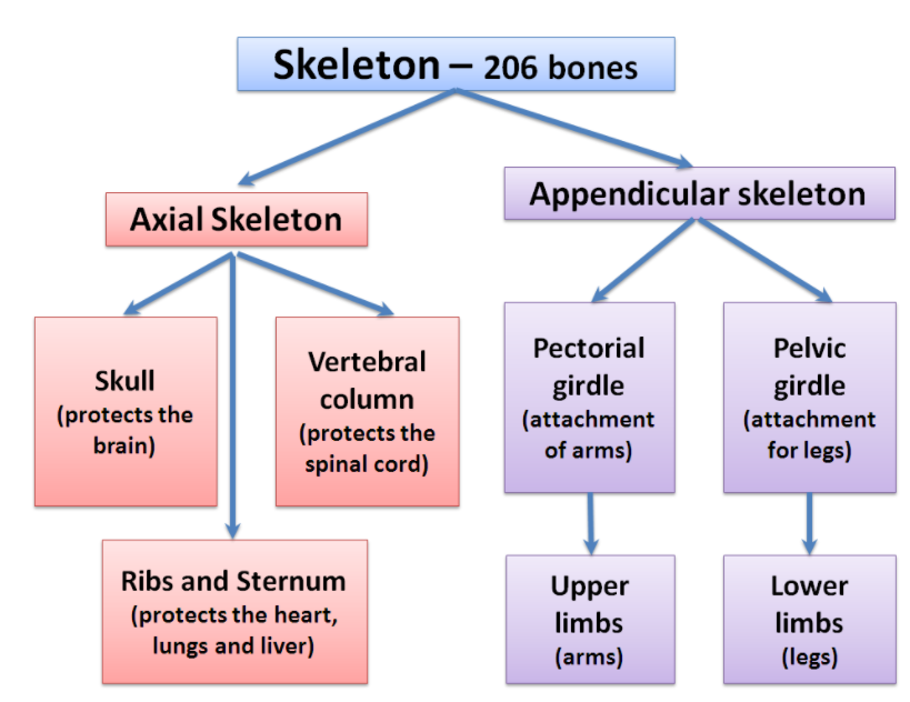
- In human beings, this system is made up of 206 bones and a few cartilages. It is grouped into two principal divisions – the axial and the appendicular skeleton.

AXIAL SKELETON
- Axial skeleton comprises 80 bones distributed along the main axis of the body. The skull, vertebral column, sternum and ribs constitute the axial skeleton.
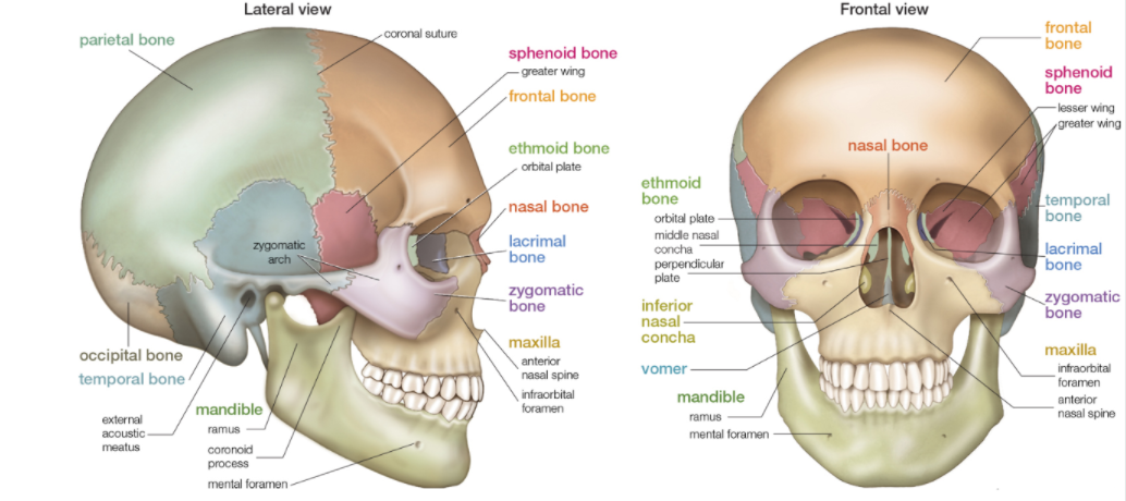
- Skull :The skull is composed of two sets of bones - cranial and facial, that totals to 22 bones.
- Cranium: Cranial bones are 8 in number. They form the hard protective outer covering, cranium for the brain.
- Frontal (1) : It forms the forehead.
- Parietals(2): These are situated just behind the frontal. It forms the main bulging top of the skull.
- Occipital(1) : It is situated just behind parietals. It forms the posterior and lower part of the cranium. The skull region articulates with the superior region of the vertebral column with the help of two occipital condyles(dicondylic skull). Foramen magnum is a large perforation in this bone.
- Temporal(2) : These form the right and left sides of the cranium.
- Sphenoid(1) : It is a butterfly shaped bone that forms the middle and anterior part of the base of the cranium. It articulates with all skull bones. It has depression called sella turcica for pituitary gland.
- Ethmoid(1) : A small, irregular sieve shaped bone in front of sphenoid and behind nasal bones.
- Facial bones:The facial region is made up of 14 skeletal elements which form the front part of the skull.
- Nasals(2) : They form the proximal part of the bridge of the nose.
- Inferior nasal conchae or turbinals(2) : Two highly coiled, processes project in nasal chamber.
- Vomer(1) :It forms a part of the septum between two nasal chambers.
- Lacrimals(2) : It is located in eye orbit and forms a part of the passages of tear duct.
- Zygmonastic(2) : These form the prominence of cheeks.
- Palatines(2) : They form the posterior part of the hard palate.
- Maxillae(2) : These bear the teeth of the upper jaw.
- Mandible(1) : It forms the entire lower jaw. It is the only movable bone of the skull.
- Hyoid proper: A single U-shaped bone called hyoid is present at the base of the buccal cavity and it is also included in the skull. It does not articulate with any bone but is simply suspended from temporal bones by means of ligament.It supports our tongue and provides insertion to some tongue muscles.
- Ear Ossicles: Each middle ear contains three tiny bones – Malleus, Incus and Stapes, collectively called Ear Ossicles.

VERTEBRAL COLUMN
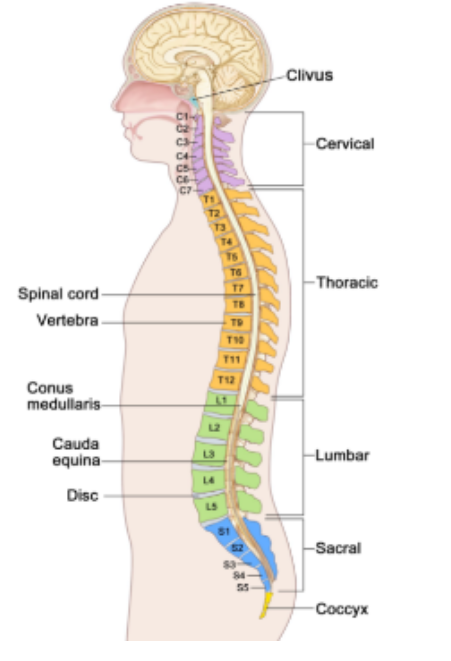
- Our vertebral column is formed by 26 serially arranged units called vertebrae and is dorsally placed. It extends from the base of the skull and constitutes the main framework of the trunk.
- The vertebral column is differentiated into cervical (7), thoracic (12), lumbar (5), sacral (1-fused) and coccygeal (1-fused) regions starting from the skull.
- Each vertebra has a central hollow portion (neural canal) through which the spinal cord passes. First vertebra is the atlas and it articulates with the occipital condyles.
- Functions
-The vertebral column protects the spinal cord, supports the head and serves as the point of attachment for the ribs and musculature of the back.
-It supports and rotates the head, protects vital organs and houses the spinal cord. - STERNUM
Sternum is a flat bone on the ventral midline of thorax.It is made up of manubrium, body and xiphoid process. - Function-
-It takes part in formation of protective thoracic baskets.
Thoracic basket = Ribs + Sternum + thoracic vertebrae
-It plays a role in respiratory mechanisms. - RIBS
There are 12 pairs of ribs. Each rib is a thin flat bone connected dorsally to the vertebral column and ventrally to the sternum. It has two articulation surfaces on its dorsal end and is hence called bicephalic. There are three types of ribs.- True ribs/vertebrosternal ribs : First seven pairs of ribs are called true ribs. Dorsally, they are attached to the thoracic vertebrae and ventrally connected to the sternum with the help of hyaline cartilage.
- False ribs/vertebrochondral ribs: The 8th, 9th and 10th pairs of ribs do not articulate directly with the sternum but join the seventh rib with the help of hyaline cartilage.
- Floating ribs: Last 2 pairs (11th and 12th) of ribs are not connected ventrally and are only connected dorsally to thoracic vertebrae so called as floating ribs.
- Functions:
-Protect the heart, large blood vessels and lungs.
-They bear respiratory muscle(external & internal intercostal muscle).

APPENDICULAR SKELETON
- The bones of the limbs along with their girdles constitute the appendicular skeleton. It includes 126 bones.
- Girdles
Pectoral and Pelvic girdle bones help in the articulation of the upper and the lower limbs respectively with the axial skeleton. Each girdle is formed of two halves.- Pectoral girdle(shoulder girdle) - Each half of the pectoral girdle consists of a clavicle and a scapula.
Clavicle/collar bone - Clavicle is a long slender bone with two curvatures.
Scapula/Shoulder bone - It is a large triangular flat bone situated in the dorsal part of the thorax between the second and the seventh ribs. The dorsal, flat, triangular body of the scapula has a slightly elevated ridge called the spine which projects as a flat, expanded process called the acromion. The clavicle articulates with this. Below the acromion is a depression called the glenoid cavity which articulates with the head of the humerus to form the shoulder joint. - Pelvic girdle(Hip girdle) - Pelvic girdle consists of two coxal bones. Each coxal bone is formed by the fusion of three bones – ilium, ischium and pubis. At the point of fusion of the above bones is a cavity called acetabulum to which the thigh bone articulates. The two halves of the pelvic girdle meet ventrally to form the pubic symphysis containing fibrous cartilage.
- Pectoral girdle(shoulder girdle) - Each half of the pectoral girdle consists of a clavicle and a scapula.
LIMB BONES
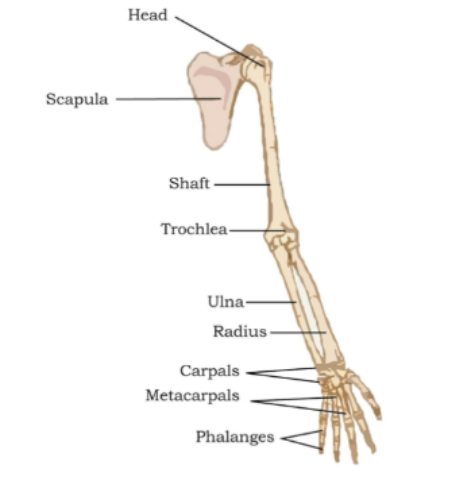
- Bone of forelimbs: Each limb is made of 30 bones.
- The bones of the hand (fore limb) are humerus(1), radius(1) and ulna(1), carpals(wrist bones-8 in number), metacarpals(palm bones-5 in number) and phalanges(digits-14 in number).
Phalangeal formula= 2,3,3,3,3 - Humerus is the longest and largest bone of the upper limb. Humerus is characterised by presence of deltoid ridge for the attachment of muscle. Radius is smaller and the ulna is longer. Radius is towards the thumb side and ulna is towards the little finger side.
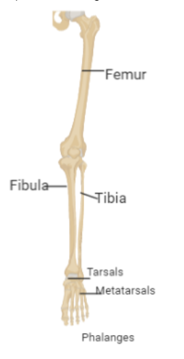
- Bones of hind limbs: Each hindlimb consists of 30 bones. These are Femur (thigh bone – the longest bone), tibia and fibula, tarsals (ankle bones – 7 in number), metatarsals (5 in number) and phalanges (digits – 14 in number).
Phalangeal formula = 2,3,3,3,3 - A cup shaped bone called patella covers the knee ventrally (knee cap). Patellar groove is found in distal end of femur. Fibula is thinner. Tibia is thicker bone and is also called shin bone. It bears body weight.


JOINTS
- The study of joints is called arthrology.
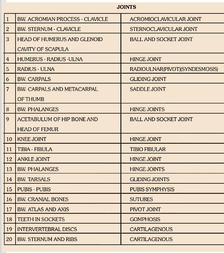
- Joints are essential for all types of movements involving the bony parts of the body.Joints are points of contact between bones, or between bones and cartilages. Force generated by the muscles is used to carry out movement through joints, where the joint acts as a fulcrum.
- Joints have been classified into three major structural forms, namely, fibrous, cartilaginous and synovial.
- Immovable/Fibrous joints: Fibrous joints do not allow any movement. This type of joint is shown by the flat skull bones which fuse end-to-end with the help of dense fibrous connective tissues in the form of sutures, to form the cranium.
- Imperfect/Cartilaginous/ Slightly movable: The bones involved are joined together with the help of cartilages. The joint between the adjacent vertebrae in the vertebral column is of this pattern and it permits limited movements.
- Synovial/Perfect/freely movable joints: Synovial joints are characterised by the presence of a fluid filled synovial cavity between the articulating surfaces of the two bones. Such an arrangement allows considerable movement. These joints help in locomotion and many other movements.
- → Ball and socket joint (between humerus and pectoral girdle)
→ Hinge joint (knee joint)
→ Pivot joint (between atlas and axis)
→ Gliding joint (between the carpals)
→ Saddle joint (between carpal and metacarpal of thumb). - It forms an internal framework and gives definite body shape and form.
- It protects delicate organs like heart, lungs etc.
- It permits growth of the body since it is living and growing.
- It provides a surface for attachment of muscles.
- It serves as levers on which muscles can act.
- It manufactures blood corpuscles in bone marrow.
- It helps in breathing and hearing.
- Acts as a mineral reserve for calcium and phosphorus.
- Functions of endoskeleton
Joints
- Different bone ke beech me jo joint hota hai ussi ko articulation ya arthrosis kehte hai. Joints ki study ko arthrology kaha jata hai. 3 types ke joints hote hai :
1.) Fixed/Immovable Joints - Inhijoint ko synarthroses bhi bola jata hai.
- Yeah bane hue hote hai dense irregular connective tissue which is rich in white collagen fibers. Ex : Sutures between skull bones, joint between teeth & teeth sockets, Epiphyseal plate between diaphysis and epiphysis of long bone.
- Yeah slightly movable joints hai and inko amphiarthroses bhi bola jata hai. In joints me bahut hi kam movement hoti hai and articulating bones ya toh hyaline cartilage ya fibrocartilage se tightly connected hoti hai. Ex : joint between the first rib and the manubrium of the sternum, at the junction of the manubrium and body of the sternum, at the intervertebral joints between the bodies of vertebrae, Distal tibiofibular joint., between tibia and fibula., pubic symphysis.
- Yeah aise joints hai jinme 2 bones ke beech me ek fluid fill cavity hoti hai which is known as synovial cavity and the fluid in synovial fluid. Dono bones articular cartilage ki layer se covered hoti hai to further reduce the friction and allow free movements of joints. Synovial joint ke bahar ek articular capsule hota hai jo synovial joint ko encapsulate karta hai. Isme 2 membranes hoti hai: outer protective fibrous layer and inner synovial membrane jo ki synovial fluid secrete karti hai.
- Gliding Joint : a simple movement in which nearly flat bone surfaces move back-and-forth and from side-to-side with respect to one another. Ex : between tarsals, between carpels, sternoclavicular joints (between the manubrium of the sternum and the clavicle), acromioclavicular joints (between the acromion of the scapula and the clavicle), sternocostal joints (between the sternum and ends of the costal cartilages at the tips of the second through seventh pairs of ribs), and vertebrocostal joints (between the heads and tubercles of ribs and bodies and transverse processes of thoracic vertebrae).
- Hinge Joint : iss joint me ek bone ka convex face dusri bone ke concave face me fit karta hai. Mostly ek bone fixed rehti hai jabki dusri move karti hai. Yeah joint uniaxial/monoaxial hai yani ki keval ek hi axis ke around movement allow karte hai jaise ki they allow flexion and extension type movement only. Examples of hinge joints are the knee (actually a modified hinge joint), elbow joint , ankle joint, and interphalangeal joints (between the phalanges of the fingers and toes).
- Pivot Joints : Ek bone ka rounded ya pointed surface dusri bone ki ring ke sath articulate karta hai. Yeah ring partly ligament and partly bone se bani hui hai. Yeah bhi uniaxial movements hi allow karte hai. Examples of pivot joints are the atlanto-axial joint, in which the atlas rotates around the axis and permits the head to turn from side-to side as when you shake your head “no”, and the radioulnar joints that enable the palms to turn anteriorly and posteriorly as the head of the radius pivots around its long axis in the radial notch of the ulna.
- Condyloid/Ellipsoid Joints : yeah ek biaxial joint hai jo 2 axis ke around movement allow karta hai - flexion-extension and adduction-abduction type of movements. Isme bhi ek bone ka oval shaped convex projection dusri bone ke oval shaped depression me fit hota hai. Examples of condyloid joints are the radiocarpal (wrist) and metacarpophalangeal joints (between the metacarpals and proximal phalanges) of the second through fifth digits.
- Saddle Joints : ek bone ka articulating surface saddle shaped (घोड़े की काठी के आकार का) hota hai and dusri bone rider ki tarah iss bone pe sit karti hai. Yeah joint bhi biaxial (flexion–extension and abduction–adduction) hai. An example of a saddle joint is the carpometacarpal joint between the trapezium of the carpus and metacarpal of the thumb.
- Ball & Socket Joint : ek bone ka ball like surface dusri bone ke cuplike depression se articulate karta hai. Yeah multiaxial joints hai jo flexion–extension, abduction– adduction, and rotation jaise movements ko allow karte hai. Examples of ball-and-socket joints are the shoulder and hip joints. At the shoulder joint, the head of the humerus fits into the glenoid cavity of the scapula. At the hip joint, the head of the femur fits into the acetabulum of the hip (coxal) bone.
2.) Cartilaginous Joints
3.) Synovial Joints
DISORDERS OF SKELETAL SYSTEM
- Arthritis: Inflammation of joints.
- Osteoporosis: Age-related disorder characterised by decreased bone mass and increased chances of fractures. Decreased levels of estrogen is a common cause.
- Gout: Inflammation of joints due to accumulation of uric acid crystals.
- Rheumatoid arthritis: It is due to inflammation of synovial membrane leading to over secretion of synovial fluid. It puts pressure on the joint and becomes painful. It is an autoimmune disorder. It mainly affects small joints of the hand and feet.
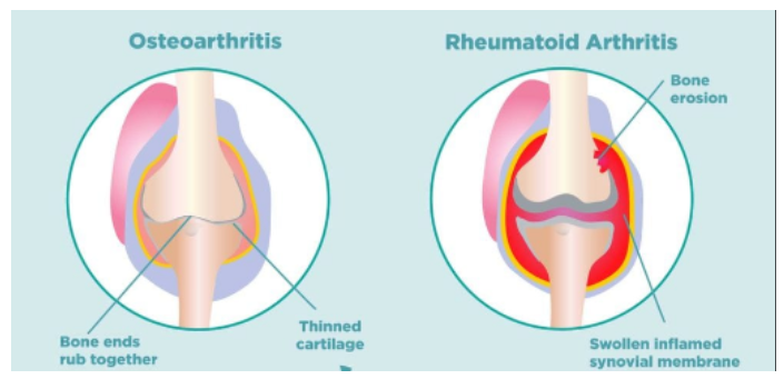
JOINTS
- The study of joints is called arthrology.
- Joints are essential for all types of movements involving the bony parts of the body.Joints are points of contact between bones, or between bones and cartilages. Force generated by the muscles is used to carry out movement through joints, where the joint acts as a fulcrum.
- Joints have been classified into three major structural forms, namely, fibrous, cartilaginous and synovial.
- Immovable/Fibrous joints: Fibrous joints do not allow any movement. This type of joint is shown by the flat skull bones which fuse end-to-end with the help of dense fibrous connective tissues in the form of sutures, to form the cranium.
- Imperfect/Cartilaginous/ Slightly movable: The bones involved are joined together with the help of cartilages. The joint between the adjacent vertebrae in the vertebral column is of this pattern and it permits limited movements.
- Synovial/Perfect/freely movable joints: Synovial joints are characterised by the presence of a fluid filled synovial cavity between the articulating surfaces of the two bones. Such an arrangement allows considerable movement. These joints help in locomotion and many other movements.
- → Ball and socket joint (between humerus and pectoral girdle)
→ Hinge joint (knee joint)
→ Pivot joint (between atlas and axis)
→ Gliding joint (between the carpals)
→ Saddle joint (between carpal and metacarpal of thumb). - It forms an internal framework and gives definite body shape and form.
- It protects delicate organs like heart, lungs etc.
- It permits growth of the body since it is living and growing.
- It provides a surface for attachment of muscles.
- It serves as levers on which muscles can act.
- It manufactures blood corpuscles in bone marrow.
- It helps in breathing and hearing.
- Acts as a mineral reserve for calcium and phosphorus.
- Functions of endoskeleton
THE MUSCULAR SYSTEM
- The study of muscles is known as myology.
- Muscle is a specialised tissue of mesodermal origin. About 40-50 per cent of the body weight of a human adult is contributed by muscles.
- They have special properties like excitability, contractility, extensibility and elasticity. Muscles have been classified using different criteria, namely location, appearance and nature of regulation of their activities.
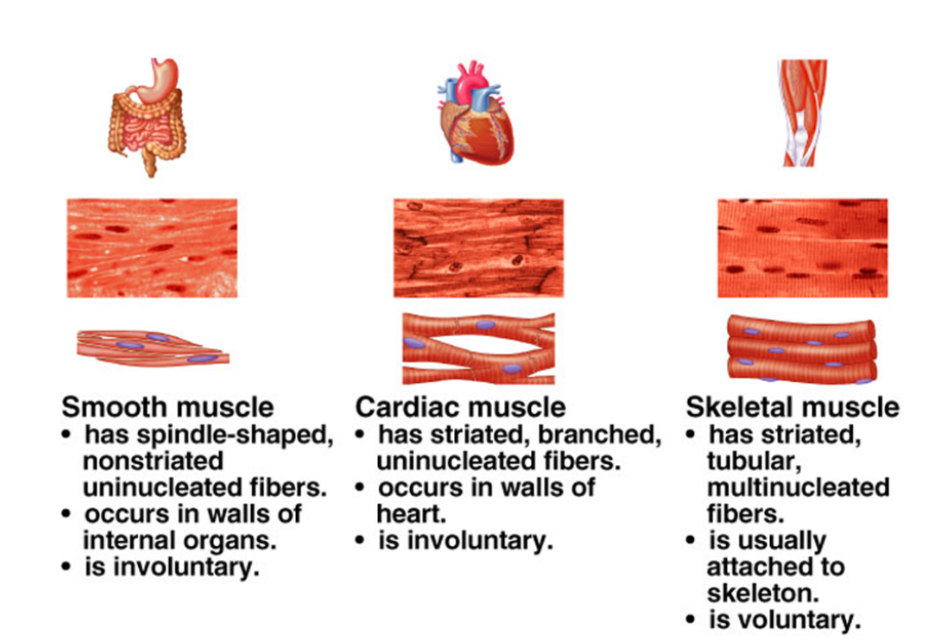
- Based on their location, three types of muscles are identified : (i) Skeletal (ii) Visceral and (iii) Cardiac.
- Skeletal muscles are closely associated with the skeletal components of the body. They have a striped appearance under the microscope and hence are called striated muscles. As their activities are under the voluntary control of the nervous system, they are known as voluntary muscles too. They are primarily involved in locomotory actions and changes of body postures.
- Visceral muscles are located in the inner walls of hollow visceral organs of the body like the alimentary canal, reproductive tract, etc. They do not exhibit any striation and are smooth in appearance. Hence, they are called smooth muscles (nonstriated muscle). Their activities are not under the voluntary control of the nervous system and are therefore known as involuntary muscles. They assist, for example, in the transportation of food through the digestive tract and gametes through the genital tract.
- Cardiac muscles are the muscles of the heart. Many cardiac muscle cells assemble in a branching pattern to form a cardiac muscle. Based on appearance, cardiac muscles are striated. They are involuntary in nature as the nervous system does not control their activities directly.
DISORDERS OF SKELETAL SYSTEM
- Arthritis: Inflammation of joints.
- Osteoporosis: Age-related disorder characterised by decreased bone mass and increased chances of fractures. Decreased levels of estrogen is a common cause.
- Gout: Inflammation of joints due to accumulation of uric acid crystals.
- Rheumatoid arthritis: It is due to inflammation of synovial membrane leading to over secretion of synovial fluid. It puts pressure on the joint and becomes painful. It is an autoimmune disorder. It mainly affects small joints of the hand and feet.

ULTRASTRUCTURE OF SKELETAL MUSCLE
- Let us examine a skeletal muscle in detail to understand the structure and mechanism of contraction.
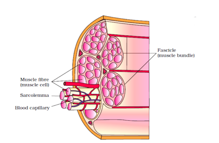
- Each organised skeletal muscle in our body is made of a number of muscle bundles or fascicles held together by a common collagenous connective tissue layer called fascia.Each muscle bundle contains a number of muscle fibres. Each muscle fibre is lined by the plasma membrane called sarcolemma enclosing the sarcoplasm containing many nuclei and numerous mitochondria called sarcomeres and numerous endoplasmic reticulum called sarcoplasmic reticulum.
- The endoplasmic reticulum, i.e., sarcoplasmic reticulum of the muscle fibres is the store house of calcium ions. A characteristic feature of the muscle fibre is the presence of a large number of parallelly arranged filaments in the sarcoplasm called myofilaments of myofibrils.
- The sarcolemma is invaginated to form T-tubules. A triad consists of a t-tubule and the segments of sarcoplasmic reticulum on either side.
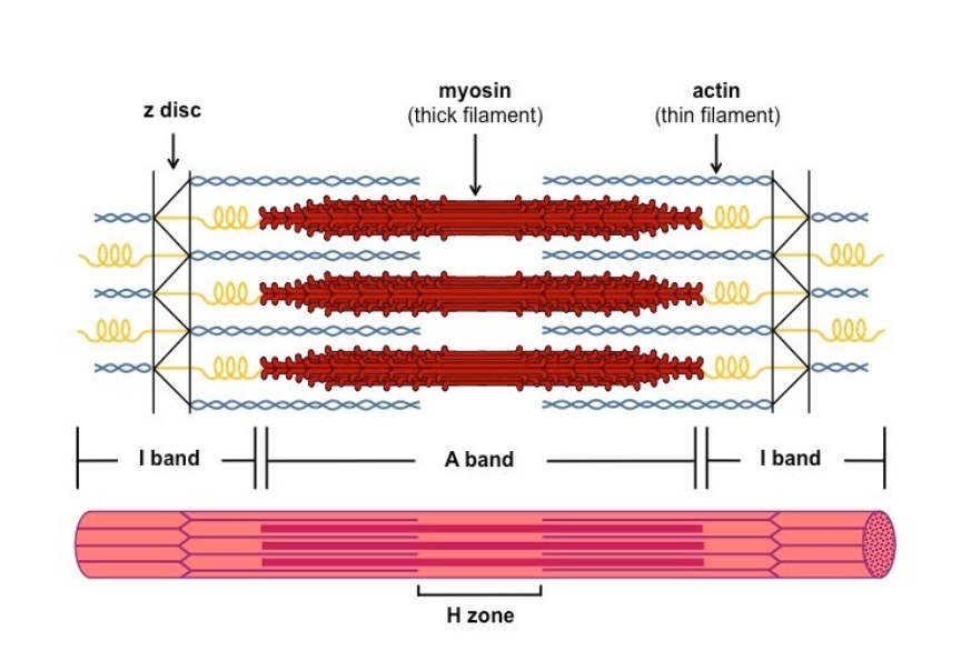
- Each myofibril has alternate dark and light bands on it. The striated appearance is due to the distribution pattern of two important proteins – Actin and Myosin.
- The light bands contain actin and are called I-band or Isotropic band, whereas the dark band called ‘A’ or Anisotropic band contains myosin. Both the proteins are arranged as rod-like structures, parallel to each other and also to the longitudinal axis of the myofibrils. Actin filaments are thinner as compared to the myosin filaments, hence are commonly called thin and thick filaments respectively.
- In the centre of each ‘I’ band is an elastic fibre called ‘Z’ line which bisects it. The thin filaments are firmly attached to the ‘Z’ line. The thick filaments in the ‘A’ band are also held together in the middle of this band by a thin fibrous membrane called ‘M’ line. The ‘A’ and ‘I’ bands are arranged alternately throughout the length of the myofibrils.
- The portion of the myofibril between two successive ‘Z’ lines is considered as the functional unit of contraction and is called a sarcomere. In a resting state, the edges of thin filaments on either side of the thick filaments partially overlap the free ends of the thick filaments leaving the central part of the thick filaments. This central part of thick filament, not overlapped by thin filaments, is called the ‘H’ zone.
Muscular Tissue
- Muscular tissue 4 basic tissues me se ek tissue hai jo ki mesodermally derived hai. Muscle tissue ki study ko hum myology kehte hai. 3 basic types ke muscle tissue hu already padh chuke hai jo ki hai : skeletal muscle tissue, cardiac muscle tissue and smooth muscles. In teeno types ki muscle tissues ke baare me and inme differences hum initial chapters me cover kar chuke hai. Iss section me hum skeletal muscle ke ultrastructure ko padhenge and iske contraction and relaxation ke bare me.
- Skin and muscle ke beech me hypodermis ya fir subcutaneous tissue layer hoti hai jo ki areolar connective tissue and adipose connective tissue se bani hui hai.
- Fascia irregular connective tissue ki ek dense sheet hai jo similar functions wali muscles ko ek sath hold karke rakhti hai. Toh basically fascia is kind of wrap around muscle and yeah muscles ki free movement ko allow karta hai and protective function toh hai hi.
- Issi fascia se connective tissue ki 3 layers originate hoti hai.
- Epimysium puri muscle ke bahar ki outer layer hai jo dense irregular connective tissue se bani hui hai.
- Perimysium muscle fibres ke around ki layer hai. Basically ek muscle ke ander bahut sare muscle fibres hote hai toh un muscle fibres ke groups (10-100 muscle fibres) ke around jo dense irregular connective tissue ki layer hoti hai usko hum perimysium kehte hai and in muscle fibres ke group ko fascicles bola jata hai toh fascia ki outer covering hi perimysium hui.
- Endomysium : ek fascicle me bahut sare muscle fibres hote hai and har ek muscle fibre ke around jo layer hoti hai usko hum endomysium kehte hai jo ki mostly reticular fibres ki bani hui hai.
- Tendon aisa connective tissue hai jo bone & muscle ko join karta hai.
- Muscle cell ko hum muscle fiber se refer karte hai jiske ander bahut sarey myofibrils arranged hote hai theek ussi tarah jaise ek moti electrical wire ne ander bahut sare lambi tarein hoti hai jo ki ek dusre se insulated bhi hoti hai mostly cases me. Khair issi muscle cell ki plama membrane ko sarcolemma kaha jata hai lekin yeah sarcolemma keval surface tak simit nahi hai. Iski extension muscle ke ander tak jati hai and yeah extension basically tube like structures hoti hai jo ki myofibrils ke nearly perpendicularly arranged hoti hai toh in tubules ko hum T tubules yani ki transverse tubules kehte hai.
- T tubules are bahar ki taraf hoti hai (they are invaginations of sarcolemma) toh inme naturally interstitial fluid hi hoga.
- Inn T tubules ka function ka action potentially ko quickly and throughout the muscle fiber uniformly propagate karwana.
- Cytoplasm jo sarcolemma ke ander enclosed hai usko hum sarcoplasm naam dete hai and isme glycogen ki amount kafi jyada hoti hai.
- Muscle me ek red colour ka protein bhi hota hai jiske hum myoglobin kehte hai wo basically oxygen ko store karke rakhta hai and jab muscle ko jarurat hogi tab oxygen ko release kardega.
- Ek muscle fiber ke ander bahut sare patle patle thread like structures hai jo ki myofibril kehlate hai and yhi muscle ki contractile unit hai. Diameter inka 2 micrometer hota hai jabki length purey muscle fiber ki length jitni hi hoti hai.
- Jaise normal baki cells me endoplasmic reticulum hota hai aise hi muscle fiber me bhi har myofibril ke around specialised smooth endoplasmic reticulum hai jo ki sarcoplasmic reticulum kehlata hai and yeah calcium ions ka storehouse hai.
- Myofibril ke ander chote protein filaments hote hai jinko hum myofilaments kehte hai and unki morphology ke basis par wo 2 types ke hote hai : thick filament jiske myosin kehte hai thin filament jiske actin kehte hai. Thin filaments ka diameter 8nm ka hota hai jabki thick filaments ka diameter 16nm hota hai. Yeahi thick and thin filaments basically contractile proteins hai jo ki contract and relax karte hai. Har ek thick filament ke liye, 2 thin filaments hote hai. Yeah Sarcomeres ki form me arranged hote hai jo ki skeletal muscle ki contractile unit hai and myofibrils ki functional unit hai.
- Toh ek myofibrils me bahut sare sarcomeres hote hai jo ki lineraly arranged hai and ek Z discs ek sarcomere ko agle se alag karti hai ya issi ko dusri bhasha me aise bol sakte hai ki 2 Z discs ke beech me ek sarcomere hota hai.
- Jab sarcomere ko microscope me dekhte hai toh hum kuch dark and kuch light bands dikhte hai. Sarcomere ka middle part ek straight dark band ki tarah dikhta hai and yeah sarcomere ka wo hissa hai jisme thin & thick filaments hote hai. A band ke center me kuch hissa aisa hota hai jhan keval myosin filaments hote hai toh usko hum H zone bolte hai. H zone ke middle me M line hoti hai jhan par supporting proteins hote hai jo ki thick filaments ko hold karke rakhte hai. I band ek light band hai jo sarcomere ke dono ends ko taraf hota hai and yeah wo part hai jisme keval think filaments hote hai toh naturally iski appearance light coloured hai. Z disc basically issi I band ke center se pass karti hai.
- Yeah alternating dark and light band hai hi muscle fibre ko microscope me striations dete hai jinke karan iss muscle ko straighted muscle bola jata hai.
- Ek muscle me overall 3 types ke proteins hote hai : ek jo contract and relax kare jaise ki myosin and actin, dusra regulatory proteins jo ki contraction and relaxation ko control karte hai and teesre hote hai structural proteins jo ki myosin and actin ko proper alignment me rakhte hai and myofibrils ko sarcolemma & extracellular matrix se link karte hai. Hum keval contractile proteins ka structure brief me dekhenge and baki sab or higher graduation me cover kia jata hai.
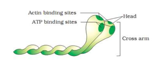
- Myosin : yeah ek motor protein hai. Motor proteins aise proteins hote hai jo ATP me chemical energy ko mechanical energy me convert karte hai. Mechanical ka yahan pe matlab hai basically pull se toh yeah myosin basically different cellular structures jo ki iss case me baki associated proteins hai unko kheechne ka kaam karega jo ki ultimately jaise ki thodi der dekhnge, wo muscle ke contraction ka karan hota hai. Ek thick filament me qareeban 300 myosin molecules hote hai and ek myosin ki shape aisi hoti hai jaise 2 golf clubs ko ek sath twist krdia ho toh two heads and two tails jaisi appearance aati hai. Har ek head pe actin binding site hoti hai and ek ATP binding site hoti hai and issi ATP binding site pe ATPase enzyme hai jo ATP → ADP + Pi reaction ko catalyse karta hai. Har ek thick filament ke charo taraf 6 thin filaments hote hai.
- Thin filament ka main component actin hai. Alag alag actin unit se ek actin filament tyaar hota hai jo ki twisted shape me hota hai. Har ek thin filament ke around 3 thick filaments hote hai. Har actin molecule pe myosin binding site hoti hai jhan par muscle contraction ke dauran myosin ka head judta hai. Relaxed state me ek tropomyosin naam ka regulatory molecule har actin molecule ki myosin binding site ko cover karke rakhta hai. Thin filament me ek or regulatory protein hota hai jiske hum troponin kehte hai. Iss troponin ki khud ki 3 units hoti hai : TpT, TpC, and TpI. TpC unit muscle contraction ke dauran jab calcium ions release hoti hai toh unke sath bind karta hai jiske karan isme conformational changes aate hai jo ki tropomyosin ko myosin binding site hai remove karta hai jiske karan ab actin filament pe myosin heads bind kar sakte hai.
Ultrastructure of Skeletal Muscle :
STRUCTURE OF CONTRACTILE PROTEINS
- Thick myofilaments
- Each myosin (thick) filament is also a polymerised protein. Many monomeric proteins called Meromyosin ,constitute one thick filament.
- Each meromyosin has two important parts, a globular head with a short arm and a tail, the former being called the heavy meromyosin (HMM) and the latter, the light meromyosin (LMM).
- The HMM component, i.e.; the head and short arm projects outwards at regular distance and angle from each other from the surface of a polymerised myosin filament and is known as cross arm.
- The globular head is an active ATPase enzyme and has binding sites for ATP and active sites for actin.
LMM lacks ATPase activity and does not combine with actin. - The thick filament consists mainly of myosin protein. Myosin contributes 55% of muscle protein by weight.
- Thin myofilaments:
- Each actin (thin) filament is made of two ‘F’ (filamentous) actins helically wound to each other. Each ‘F’ actin is a polymer of monomeric ‘G’ (Globular) actins.
- Tropomyosin also runs close to the ‘F’ actins throughout its length. In the resting state, the tropomyosin molecule lies on top of the active sites of actin strands so that attraction cannot occur between the actin and myosin to cause contraction.
- A complex protein Troponin is distributed at regular intervals on the tropomyosin.
- → TpT binds to tropomyosin as well as to the other two troponin components.
→ TpI inhibits the f-actin myosin interaction and also bonds to other components of troponin.
→ TpC is a calcium binding polypeptide. The strong affinity of the troponin for calcium ions is believed to initiate the contraction process. - In the resting state a subunit of troponin masks the active binding sites for myosin on the actin filaments.
THE MUSCULAR SYSTEM
- The study of muscles is known as myology.
- Muscle is a specialised tissue of mesodermal origin. About 40-50 per cent of the body weight of a human adult is contributed by muscles.
- They have special properties like excitability, contractility, extensibility and elasticity. Muscles have been classified using different criteria, namely location, appearance and nature of regulation of their activities.
- Based on their location, three types of muscles are identified : (i) Skeletal (ii) Visceral and (iii) Cardiac.
- Skeletal muscles are closely associated with the skeletal components of the body. They have a striped appearance under the microscope and hence are called striated muscles. As their activities are under the voluntary control of the nervous system, they are known as voluntary muscles too. They are primarily involved in locomotory actions and changes of body postures.
- Visceral muscles are located in the inner walls of hollow visceral organs of the body like the alimentary canal, reproductive tract, etc. They do not exhibit any striation and are smooth in appearance. Hence, they are called smooth muscles (nonstriated muscle). Their activities are not under the voluntary control of the nervous system and are therefore known as involuntary muscles. They assist, for example, in the transportation of food through the digestive tract and gametes through the genital tract.
- Cardiac muscles are the muscles of the heart. Many cardiac muscle cells assemble in a branching pattern to form a cardiac muscle. Based on appearance, cardiac muscles are striated. They are involuntary in nature as the nervous system does not control their activities directly.
MECHANISM OF MUSCLE CONTRACTION
- Mechanism of muscle contraction is best explained by the sliding filament theory which states that contraction of a muscle fibre takes place by the sliding of the thin filaments over the thick filaments.
- Muscle contraction is initiated by a signal sent by the central nervous system (CNS) via a motor neuron. A motor neuron alongwith the muscle fibres connected to it constitute a motor unit. The junction between a motor neuron and the sarcolemma of the muscle fibre is called the neuromuscular junction or motor-end plate.
- A neural signal reaching this junction releases a neurotransmitter (Acetylcholine) which generates an action potential in the sarcolemma. This spreads through the muscle fibre and causes the release of calcium ions into the sarcoplasm.
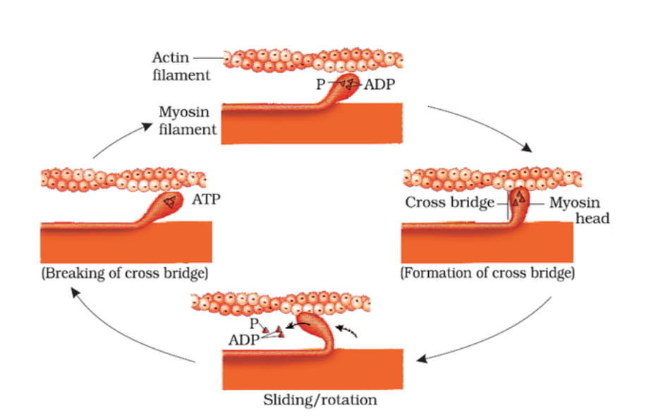
- Increase in Ca++ level leads to the binding of calcium with a subunit of troponin on actin filaments and thereby removing the masking of active sites for myosin.Utilising the energy from ATP hydrolysis, the myosin head now binds to the exposed active sites on actin to form a cross bridge.This pulls the attached actin filaments towards the centre of ‘A’ band. The ‘Z’ line attached to these actins are also pulled inwards thereby causing a shortening of the sarcomere, i.e., contraction. It is clear from the above steps, that during shortening of the muscle, i.e., contraction.
- The ‘I’ bands get reduced, whereas the ‘A’ bands retain the length.
- The myosin, releasing the ADP and P1 goes back to its relaxed state. A new ATP binds and the cross-bridge is broken. The ATP is again hydrolysed by the myosin head and the cycle of cross bridge formation and breakage is repeated causing further sliding.
- The reaction time of the fibres can vary in different muscles.
- During muscle contraction:
- Length of I band gets reduced.
- Length of A band remains the same.
- Length of sarcomere decreases.
- Length of myofibril decreases.
- Henson zone disappears.
- Length of actin and myosin filament remains same.
- Role of ATP:
(i) The 'back & forth' movement of myosin head with in the groove.
(ii) Detachment of myosin head from the actin
ULTRASTRUCTURE OF SKELETAL MUSCLE
- Let us examine a skeletal muscle in detail to understand the structure and mechanism of contraction.
- Each organised skeletal muscle in our body is made of a number of muscle bundles or fascicles held together by a common collagenous connective tissue layer called fascia.Each muscle bundle contains a number of muscle fibres. Each muscle fibre is lined by the plasma membrane called sarcolemma enclosing the sarcoplasm containing many nuclei and numerous mitochondria called sarcomeres and numerous endoplasmic reticulum called sarcoplasmic reticulum.
- The endoplasmic reticulum, i.e., sarcoplasmic reticulum of the muscle fibres is the store house of calcium ions. A characteristic feature of the muscle fibre is the presence of a large number of parallelly arranged filaments in the sarcoplasm called myofilaments of myofibrils.
- The sarcolemma is invaginated to form T-tubules. A triad consists of a t-tubule and the segments of sarcoplasmic reticulum on either side.
- Each myofibril has alternate dark and light bands on it. The striated appearance is due to the distribution pattern of two important proteins – Actin and Myosin.
- The light bands contain actin and are called I-band or Isotropic band, whereas the dark band called ‘A’ or Anisotropic band contains myosin. Both the proteins are arranged as rod-like structures, parallel to each other and also to the longitudinal axis of the myofibrils. Actin filaments are thinner as compared to the myosin filaments, hence are commonly called thin and thick filaments respectively.
- In the centre of each ‘I’ band is an elastic fibre called ‘Z’ line which bisects it. The thin filaments are firmly attached to the ‘Z’ line. The thick filaments in the ‘A’ band are also held together in the middle of this band by a thin fibrous membrane called ‘M’ line. The ‘A’ and ‘I’ bands are arranged alternately throughout the length of the myofibrils.
- The portion of the myofibril between two successive ‘Z’ lines is considered as the functional unit of contraction and is called a sarcomere. In a resting state, the edges of thin filaments on either side of the thick filaments partially overlap the free ends of the thick filaments leaving the central part of the thick filaments. This central part of thick filament, not overlapped by thin filaments, is called the ‘H’ zone.


MUSCLE RELAXATION
- The process continues till the Ca2+ ions are pumped back to the sarcoplasmic cisternae resulting in the masking of actin filaments. This causes the return of ‘Z’ lines back to their original position, i.e., relaxation.
- Energy for muscle contraction:
The main source of energy is ATP which is produced on the oxidative of glucose in the mitochondria abundantly present in the sarcoplasm of skeletal muscles.

STRUCTURE OF CONTRACTILE PROTEINS
- Thick myofilaments
- Each myosin (thick) filament is also a polymerised protein. Many monomeric proteins called Meromyosin ,constitute one thick filament.
- Each meromyosin has two important parts, a globular head with a short arm and a tail, the former being called the heavy meromyosin (HMM) and the latter, the light meromyosin (LMM).
- The HMM component, i.e.; the head and short arm projects outwards at regular distance and angle from each other from the surface of a polymerised myosin filament and is known as cross arm.
- The globular head is an active ATPase enzyme and has binding sites for ATP and active sites for actin.
LMM lacks ATPase activity and does not combine with actin. - The thick filament consists mainly of myosin protein. Myosin contributes 55% of muscle protein by weight.
- Thin myofilaments:
- Each actin (thin) filament is made of two ‘F’ (filamentous) actins helically wound to each other. Each ‘F’ actin is a polymer of monomeric ‘G’ (Globular) actins.
- Tropomyosin also runs close to the ‘F’ actins throughout its length. In the resting state, the tropomyosin molecule lies on top of the active sites of actin strands so that attraction cannot occur between the actin and myosin to cause contraction.
- A complex protein Troponin is distributed at regular intervals on the tropomyosin.
- → TpT binds to tropomyosin as well as to the other two troponin components.
→ TpI inhibits the f-actin myosin interaction and also bonds to other components of troponin.
→ TpC is a calcium binding polypeptide. The strong affinity of the troponin for calcium ions is believed to initiate the contraction process. - In the resting state a subunit of troponin masks the active binding sites for myosin on the actin filaments.
DISORDERS OF MUSCULAR SYSTEM
- Myasthenia gravis: Autoimmune disorder affecting neuromuscular junction leading to fatigue, weakening and paralysis of skeletal muscle.
- Muscular dystrophy: Progressive degeneration of skeletal muscle mostly due to genetic disorder.
- Tetany: Rapid spasm(wild contraction) in muscle due to low calcium ions in body fluid. Decreased calcium leads to opening of sodium gated channels without any stimulus. It leads to muscular twitching, cramps. It can be due to vitamin D deficiency which reduces calcium uptake from the digestive tract or due to hypoparathyroidism due to which there is no mobilisation of calcium from bones.
Sliding Filament Theory
- Yeah theory basically Huxley and Huxley ne muscle contraction and relaxation ko explain karne ke liye di thi. Iss theory ko samajhne ke liye sarcomere ke structure ka yaad karna bahut jaruri hai.
- Thick and thin filaments ki length relaxed and contracted muscle fiber me same rehti hai ; Memorise this point.
- Bilkul ek line me describe kare toh yeah theory yeah kehti hai ki muscle contraction ke dauran, thick filaments ke uper thin filaments slide karte hai and M line ki taraf aate hai jiske karan pura sarcomere shorten hota hai yani ki 2 Z discs ke beech ki length choti ho jati hai. Then muscle relax karti hai toh thin filaments, thick filaments ke uper opposite direction me slide karte hue M line se dur jate hai.
- Neuromuscular junction par jab neurotransmitter release hota hai toh wo action potential generate karta hai jo ki T tubules ki help se pure muscle fibre me lagbhag ussi instant me fail jata hai. Iss action potential ke response me sodium ions ander ghusti hai and membrane ko depolarise karti hai ( yeah action potential and depolarisation vgarah jo terms hai wo Chapter 21 me detail me discuss kari jayengi.) jiske response me sarcoplasmic reticulum se calcium ions sarcoplasm me release kar di jati hai jo ki TpC unit se bind karke tropomyosin ko actin filament ki myosin binding site se hta deti hai jiske karan actin filament pe myosin binding site expose ho jati hai.
- ATP myosin se ATP binding site pe bind karta hai and ATPase enzyme usko ADP + Pi me breakdown kar deti hai lekin ATP hydrolysis ke byproducts abhi bhi myosin head ke sath jude hue hai. ATP hydrolysis se jo energy nikli wo myosin head me store h gyi and ab hum iss myosin head ko energised head kehte hai.
- Energised head ab thick filament ke perpendicular hai and this is known as “cocked” position and yahi position actin filament se bind karne ke liye proper orientation deti hai.
- Ab myosin heads thin filament ke actin me jo myosin binding site thi unpe attach hojata hai and Pi ko release kar deta hai. Yeah myosin and actin ki attachment hi cross-bridge kehlati hai. Ek myosin molecule me 3 myosin head hote hai but ek time par ek hi myosin head ek actin se bind karta hai.
- Ab cross bridge banne ke baad myosin head forward direction me ek stroke ke through thin filament ko M line ki taraf kheenchta hai and ab myosin head and thick filament me 45o ka angle bna hua hai. Myosin me jo ATP hydrolysis ki energy stored thi wo issi power stroke ke generation ke kaam aati hai. Power stroke generate karne ke baad myosin head se ADP ko bhi release kar dia jata hai.
- Fir ek nya ATP molecule firse myosin head se judta hai and cross bridge tut jata hai, thick filaments release thin filaments which slides backwards to their original position and sarcomere gains its original shape and size or ab yeah next contraction cycle ke liye ready hai.
- Ek second me lagbhag 5 aise cycles purey hojate hai.
- Thin filaments thick filaments pe slide karte hai.
- Sarcomere ki length decrease hojati hai.
- I band ka size decrease hota hai.
- H zone chota hojata hai.
- A band and M line constant rehte hai.
- Threshold Stimulus : Yeah wo minimum stimulus hai jo muscle contraction ke liye chahiye hi chahie. Issey kamjor stimulus muscle contract ko initiate nahi kar payega.
- All or None Law : Ek muscle fibre contract karega toh pura karega otherwise nahi hi karega. Iska matlab yeah hai ki ek fibre stimulus milte hi maximally contract karega.
- Isometric Contraction : NO change in muscle length during contraction but tension increases greatly. Ex : pushing a wall, holding a book steady in front of you using an outstretched arm. The tension generated is not enough to exceed the resistance of the object to be moved. These contractions are important for maintaining posture and for supporting objects in a fixed position. Although isometric contractions do not result in body movement, energy is still expended.
- Isotonic Contraction : tension remains constant as muscle length decreases or increases. Ex : lifting an object. Isotonic contractions are used for body movements and for moving objects. The two types of isotonic contractions are concentric (ex : Dumbell curls lgate hue jab aap dumbell ko apne face ke paas lekar aao.) and eccentric (ex : Jab dumbells ko wapis face se dur yani ki unko neeche lekr jao.).
- Muscle twitch : single contraction followed by relaxation period.
- Tetanus ek sustained contraction wala muscle muscle response hai jo ki repeated stimuli se generate hota hai jaise ki lgatar kuch time ke liye likhte hue thumb and finger se pen ko pakadna. Cardiac muscle yeah response show nahi karegi. Repeated stimuli leads to summation os contractions.
- Treppe basically ladder like repeated contraction hai jo ki sub tetanizing stimuli se response me generate hota hai OR it also defined as gradual increase in muscular contraction following rapidly repeated stimulation.
- Refractory period aisa timeperiod hai jisme muscle stimulus ke liye respond karne me ashaksham hoti hai. Skeletal muscles ke liye iski value hai 0.002-0.005 seconds jabki cardiac muscles ke liye value hai 0.1-0.2 seconds.
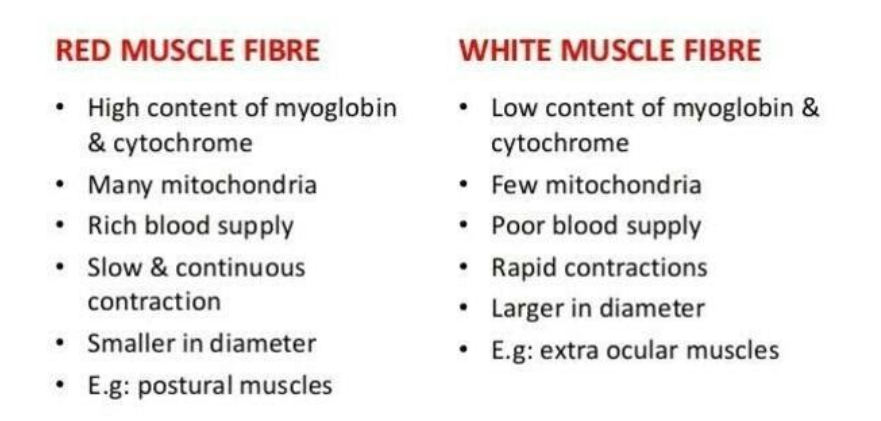
- Red fibers / Type I fibers : inka diameter narrow hota hai, myoglobin ki amount jyada hone ke karan yeah red colour ke dikhte hai and inme high number of mitochondria hota hai, smooth endoplasmic reticulum ka number kam hota hai and yeah slow oxidative fibers hai jinme lactic acid easily accumulate nahi hota toh yeah muscle fibers jaldi se fatigue bhi nahi karte. Ex : Muscle of Back, flight muscles of a kite are red muscle fibers.
- White fibers/ Type II Fibers : yeah basically broad diameter ke hai jinme myoglobin nhi hota toh safed dikhte hai, mitochondria ki amount kam hoti hai lekin smooth endoplasmic reticulum jyada hote hai. Yeah fast glycolytic fibers and inme aasani se lactic acid accumulate hojate hai toh thak bhi jaldi jate hai jaise ki muscles of eyelids ya flight muscles of a sparrow.
- Marathon runners me red fibers jyada developed hote hai toh wo jaldi thakte nahi provided ki wo maximum speed par operate na kare jabki sprinters me white fibers jyada hote hai jo short time me badi jaldi thak jate hai.
Mechanism Of Muscle Contraction :
Changes Occuring During Muscle Contraction :
Red & White Muscle Fibers :
SUMMARY
- Movement is an essential feature of all living beings. Protoplasmic streaming, ciliary movements, movements of fins, limbs, wings, etc., are some forms exhibited by animals.
- A voluntary movement which causes the animal to change its place, is called locomotion.
- The cells of the human body exhibit amoeboid, ciliary and muscular movements.
- Locomotion and many other movements require coordinated muscular activities.
- Three types of muscles are present in our body. Skeletal muscles are attached to skeletal elements. They appear striated and are voluntary in nature. Visceral muscles, present in the inner walls of visceral organs are nonstriated and involuntary. Cardiac muscles are the muscles of the heart. They are striated, branched and involuntary.
- Muscles possess excitability, contractility, extensibility and elasticity. Muscle fibre is the anatomical unit of muscle.
- Each muscle fibre has many parallelly arranged myofibrils. Each myofibril contains many serially arranged units called sarcomeres which are the functional units.
- Each sarcomere has a central ‘A’ band made of thick myosin filaments, and two half ‘I’ bands made of thin actin filaments on either side of it marked by ‘Z’ lines.
- Actin and myosin are polymerised proteins with contractility. The active sites for myosin on resting actin filament are masked by a protein-troponin.
- Myosin head contains ATPase and has ATP binding sites and active sites for actin.
- A motor neuron carries a signal to the muscle fibre which generates an action potential in it. This causes the release of Ca++ from sarcoplasmic reticulum. Ca++ activates actin which binds to the myosin head to form a cross bridge.
- These cross bridges pull the actin filaments causing them to slide over the myosin filaments and thereby causing contraction. Ca2+ are then returned to the sarcoplasmic reticulum which inactivate the actin.
- Cross bridges are broken and the muscles relax.
- Repeated stimulation of muscles leads to fatigue.
- Muscles are classified as Red and White fibres based primarily on the amount of red coloured myoglobin pigment in them.
- Bones and cartilages constitute our skeletal system.
- The skeletal system is divisible into axial and appendicular. Skull, vertebral column, ribs and sternum constitute the axial skeleton. Limb bones and girdles form the appendicular skeleton.
- Three types of joints are formed between bones or between bone and cartilage – fibrous, cartilaginous and synovial. Synovial joints allow considerable movements and therefore, play a significant role in locomotion.
MECHANISM OF MUSCLE CONTRACTION
- Mechanism of muscle contraction is best explained by the sliding filament theory which states that contraction of a muscle fibre takes place by the sliding of the thin filaments over the thick filaments.
- Muscle contraction is initiated by a signal sent by the central nervous system (CNS) via a motor neuron. A motor neuron alongwith the muscle fibres connected to it constitute a motor unit. The junction between a motor neuron and the sarcolemma of the muscle fibre is called the neuromuscular junction or motor-end plate.
- A neural signal reaching this junction releases a neurotransmitter (Acetylcholine) which generates an action potential in the sarcolemma. This spreads through the muscle fibre and causes the release of calcium ions into the sarcoplasm.
- Increase in Ca++ level leads to the binding of calcium with a subunit of troponin on actin filaments and thereby removing the masking of active sites for myosin.Utilising the energy from ATP hydrolysis, the myosin head now binds to the exposed active sites on actin to form a cross bridge.This pulls the attached actin filaments towards the centre of ‘A’ band. The ‘Z’ line attached to these actins are also pulled inwards thereby causing a shortening of the sarcomere, i.e., contraction. It is clear from the above steps, that during shortening of the muscle, i.e., contraction.
- The ‘I’ bands get reduced, whereas the ‘A’ bands retain the length.
- The myosin, releasing the ADP and P1 goes back to its relaxed state. A new ATP binds and the cross-bridge is broken. The ATP is again hydrolysed by the myosin head and the cycle of cross bridge formation and breakage is repeated causing further sliding.
- The reaction time of the fibres can vary in different muscles.
- During muscle contraction:
- Length of I band gets reduced.
- Length of A band remains the same.
- Length of sarcomere decreases.
- Length of myofibril decreases.
- Henson zone disappears.
- Length of actin and myosin filament remains same.
- Role of ATP:
(i) The 'back & forth' movement of myosin head with in the groove.
(ii) Detachment of myosin head from the actin

MUSCLE RELAXATION
- The process continues till the Ca2+ ions are pumped back to the sarcoplasmic cisternae resulting in the masking of actin filaments. This causes the return of ‘Z’ lines back to their original position, i.e., relaxation.
- Energy for muscle contraction:
The main source of energy is ATP which is produced on the oxidative of glucose in the mitochondria abundantly present in the sarcoplasm of skeletal muscles.


DISORDERS OF MUSCULAR SYSTEM
- Myasthenia gravis: Autoimmune disorder affecting neuromuscular junction leading to fatigue, weakening and paralysis of skeletal muscle.
- Muscular dystrophy: Progressive degeneration of skeletal muscle mostly due to genetic disorder.
- Tetany: Rapid spasm(wild contraction) in muscle due to low calcium ions in body fluid. Decreased calcium leads to opening of sodium gated channels without any stimulus. It leads to muscular twitching, cramps. It can be due to vitamin D deficiency which reduces calcium uptake from the digestive tract or due to hypoparathyroidism due to which there is no mobilisation of calcium from bones.
SUMMARY
- Movement is an essential feature of all living beings. Protoplasmic streaming, ciliary movements, movements of fins, limbs, wings, etc., are some forms exhibited by animals.
- A voluntary movement which causes the animal to change its place, is called locomotion.
- The cells of the human body exhibit amoeboid, ciliary and muscular movements.
- Locomotion and many other movements require coordinated muscular activities.
- Three types of muscles are present in our body. Skeletal muscles are attached to skeletal elements. They appear striated and are voluntary in nature. Visceral muscles, present in the inner walls of visceral organs are nonstriated and involuntary. Cardiac muscles are the muscles of the heart. They are striated, branched and involuntary.
- Muscles possess excitability, contractility, extensibility and elasticity. Muscle fibre is the anatomical unit of muscle.
- Each muscle fibre has many parallelly arranged myofibrils. Each myofibril contains many serially arranged units called sarcomeres which are the functional units.
- Each sarcomere has a central ‘A’ band made of thick myosin filaments, and two half ‘I’ bands made of thin actin filaments on either side of it marked by ‘Z’ lines.
- Actin and myosin are polymerised proteins with contractility. The active sites for myosin on resting actin filament are masked by a protein-troponin.
- Myosin head contains ATPase and has ATP binding sites and active sites for actin.
- A motor neuron carries a signal to the muscle fibre which generates an action potential in it. This causes the release of Ca++ from sarcoplasmic reticulum. Ca++ activates actin which binds to the myosin head to form a cross bridge.
- These cross bridges pull the actin filaments causing them to slide over the myosin filaments and thereby causing contraction. Ca2+ are then returned to the sarcoplasmic reticulum which inactivate the actin.
- Cross bridges are broken and the muscles relax.
- Repeated stimulation of muscles leads to fatigue.
- Muscles are classified as Red and White fibres based primarily on the amount of red coloured myoglobin pigment in them.
- Bones and cartilages constitute our skeletal system.
- The skeletal system is divisible into axial and appendicular. Skull, vertebral column, ribs and sternum constitute the axial skeleton. Limb bones and girdles form the appendicular skeleton.
- Three types of joints are formed between bones or between bone and cartilage – fibrous, cartilaginous and synovial. Synovial joints allow considerable movements and therefore, play a significant role in locomotion.

