STRUCTURAL ORGANISATION IN ANIMALS for Class 11 BIOLOGY
INTRODUCTION
- In unicellular organisms, all functions like digestion, respiration and reproduction are performed by a single cell. In the complex body of multicellular animals the same basic functions are carried out by different groups of cells in a well organised manner. The body of a simple organism like Hydra is made of different types of cells and the number of cells in each type can be in thousands. The human body is composed of billions of cells to perform various functions. How do these cells in the body work together? In multicellular animals, a group of similar cells along with intercellular substances perform a specific function. Such an organisation is called tissue. All complex animals consist of only four basic types of tissues (Epithelial, Connective, Muscular And Nervous Tissue). These tissues are organised in specific proportion and pattern to form an organ like stomach, lung, heart and kidney. When two or more organs perform a common function by their physical and/or chemical interaction, they together form an organ system, e.g., digestive system, respiratory system, etc. Cells, tissues, organs and organ systems split up the work in a way that exhibits division of labour and contribute to the survival of the body as a whole.
Chapter Weightage
- Beta yeah puri unit jisme Morphology, Anatomy and third chapter yeah current structural organisation wala hai, yeah puri unit se 12 marks ke question aate hai. Iss chapter se animal tissues ke uper generally short answer questions and cockroach se ek long answer question pucha jata hai.
Epithelial Tissues
- Generally hum epithelial tissue ko define hi karte hai as outermost layer which is skin in case of animals and even plants ki outermost layer bhi epithelium hi hai but apun iss chapter me keval animals ke bare me discuss karenge.
- Epithelium tissue ya toh internal body me body fluids ko face karta hai and agar bahar present hai toh environment ko face karta hai.
- Epithelium tissue is of 2 types : simple and compound epithelium based upon the layers. Simple epithelium me keval ek layer ya sheet me cells present hote hai jabki compound epithelium me multiple layers hoti hai.
- Simple epithelium aapko body cavities ki lining me, ducts and tubes me dekhne ko milta hai. Jarurat ke hisab se yeah tissue khud ko modify kar leta hai and inhi modifications ke basis par NCERT simple epithelium ko 3 types me further divide karti hai :
- Squamous Epithelium : isme patle patle tile ki tarah flat cells ki ek layer hoti hai and cells ki irregular boundaries milengi. Yeah tissue aapko blood vessels me and lungs ki alveoli me dekhne ko milta hai.
- Cuboidal Epithelium : Jaisa ki naama bta rha hai iss case me cube like cells ki ek layer hoti hai jo hume generally glands ki ducts me and nephron ke tubular parts jaise ki PCT, DCT vgarah me dekhne ko milta hai
- Columnar Epithelium : Iss case me column like yani ki pillar like lambe cells hote hai jinme nuclei cell ke base pe dekhne ko milega. Free surface pe microvilli ho sakti hai jo surface area increase karti hai. Generally yeah hume stomach and intestine ki lining me dekhne ko milta hai jha iska main kaam hai secretion and absorption.
- Agar jab columnar ya cuboidal epithelium ke cells par cilia present ho toh hum unko ciliated cuboidal epithelium ya ciliated columnar epithelium bol dete hai. Yeah hume generally hollow organs ke inner surface me dekhne ko milta hai jaise ki bronchioles and fallopian tubules vgarah me. Cilia ka kaam hota hai jo cheez present ho usko move karwana for example fallopian tubes me yeah cilia hi hilte dulte rehte hai jinke karan wo ova ko movement me help karte hai.
- Kuch columnar and cuboidal cells khud ko secretion ke function ke liye specialise kar lete hain yani ki wo different materials secrete karte hai jaise ki mucus, saliva, enzymes etc.
- Yeah secretory cells ya toh unicellular ho sakte hai jaise ki goblet cells of alimentary canal jo mucus secrete karte hain ya fir yeah cells ka ek cluster yani ki bahut sare cells milkar ek gland banate hain which is known as multicellular gland jaise ki salivary glands jo ki mouth me saliva secrete karte hain.
- Ab based upon mode of secretion ki bhyii glands apni secretion kisi special duct ki help se release karte hai ya wo bina duct ke direct blood me apni secretion release karte hai : iske basis pe glands ko further 2 types me banta gya hai : exocrine and endocrine glands.
- Exocrine glands wo hote hai jinke pass duct hai, jasie ki salivary glands and oil glands, milk gland, glands secreting digestive juices in alimentary canal etc.
- Endocrine glands wo hote hai jinke pass duct nahi hoti and wo apni secretion directly blood me release karte hain jaise ki hormone secreting glands.
- Compound epithelium jaisa ka pehle bhi btaya tha, isme ek se jyada cells ki layer hoti hai and inka kaam generally secretion and absorption me ek simit seema tak hi hai. Inka main kaam hai chemical and mechanical stress se protection provide karna. Yeah hume generally salivary glands and pancreatic ducts ki inner lining, buccal cavity ke moist surface and skin ki dry surface pe dekhne ko milti hai. Ab iske bhi bahut sare further types hai jo ki NCERT me given nhi hai so we won’t discuss them here but they are given in detail in the next section.
- Jitne bhi epithelium layer me cells hai wo sab hawa me latke ni hue toh unko apas me jodne ke liye kuch khas junctions hote hai jo ki hume lagbhag har tissue me dekhne ko milenge. Yeah cells me beech me structural (yani ki ek cell ko baki se jod ke rakhega) and functional (ek cell apne cytoplasm se material dusre cell tak bhej sakta hai and kisi or cell se le bhi sakta hai toh it basically interconnects the cytoplasm of various cells.) link bnata ai.
- Cell junction 3 type ke hote hai : tight junctions jo ek cell se kuch bhi dusre tak nhi jane denge toh yeah substances ki leaking ko rokta hai, dusra hai adhering junction jo aas pados ke cells ko ek dusre se adhere yani ki jod ke rakhega and last type ka junction hai gap junction jo ek cell ke cytoplasm ko dusre cell ke cytoplasm se jodta hai taki wo cells aapas me communicate kar sake.
Epithelial Tissues
- Term was coined by Dutch anatomist Ruysch.
- Present in an uniform layer and develops first during embryonic development.
- This also has the power of regeneration. Ye power nervous tissue mein sabse kam hoti h , ya phir absent hoti hai.
- Epithelium is made up of 2 words (epi : upon = thelio :growth ) which means tissue which grows upon another tissue is called Epithelium. It is always present upon connective tissue.
- Cells of this tissue are very closely packed i.e unke beech intercellular space nahi hoti jiski wajah se tissues jese blood vessels, lymph vessels & capillaries unke beech se nahi jaa pate or circulation absent hoti hai . Hence , cells uske neeche wale connective tissue pe dependent hote hain for nutrients.
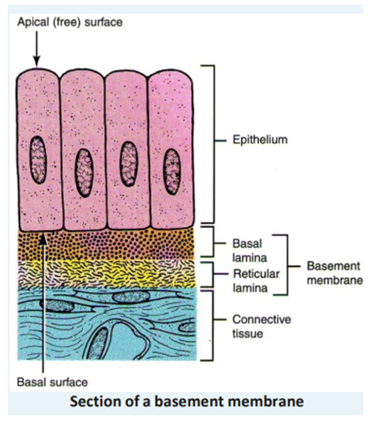
- Between epithelium and connective tissue, a thin layer of nonliving acellular basement membrane is present which is highly permeable. Basement membrane has 2 layers.
- Basal lamina : Present towards epithelium and is made up of glycoproteins. It is secreted by epithelial cells.
- Fibrous lamina : present towards connective tissue, collagen and reticular fibres are suspended in mucopolysaccharide (matrix of connective tissue).
- Hence we can say that, the basement membrane is secreted by both epithelial and connective tissue.
- Mucopolysaccharide is present in the form of Hyaluronic acid which is composed of 2 components – N acetyl D-glucosamine & glucuronic acid. Both these components are found in alternate form. – NAG – GA – NAG –
- For providing mechanical support for the tissue plasma membrane of adjacent epithelium cells are modified to form following structures (Intercellular Junctions) :
- Interdigitation –Finger jese projection hote hai Plasma membrane mei jo adjacent cell ke cytoplasm se interact karte hai . These structures are mainly found in transitional epithelium.
- Desmosomes – (Macula Adherens) these are disc shaped junctions. Location : deep in cell cytoplasm Function :providing mechanical support(stratified epithelium)and they stop substances from leaking across a tissue.
- Tight Junction – kuch jagah pe adjacent cell ki plasma membrane ek sath fuse ho jati hai aur tight junction bana leti hai. Eg. Generally, columnar epithelium
- Gap Junction –these help in cell to cell communication with an adjacent cell’s cytoplasm for rapid movement of ions and molecules.
- Plasma membrane of free end get modified to form 3 types of functional structures.
- Microvilli : minute protoplasmic structures which are non motile, non contractile. Function - absorption, secretion, excretion. They increase the surface more than 20 times. Eg : in the wall of Intestine, Gallbladder, Proximal convoluted tubule etc.
- Cilia : Long cylindrical protoplasmic structures. Motile and contractile. Origin -from basal granule or kinetosome. Movement -uniform direction. e.g. – Fallopian tube ,Trachea, Uterus, Ependymal epithelium (Inner lining of ventricles of brain and central canal of spinal cord. Function of cilia is to conduct substances in CSF.)
- Stereocilia : Long cytoplasmic structure. Non motile, non contractile. Basal granules are absent.Plasma membrane is thick and rigid. Conical in shape. Function: increase in surface area eg. Epididymis , vas deferens.
- Origin of Epithelium Tissue : It is the only tissue originated from all the three primordial germinal layers.
- Ectodermal – Epidermis (stratified squamous Epithelium)
- Mesodermal – Mesothelium (simple squamous Epithelium)
- Endodermal – Endothelium (simple squamous Epithelium)
- Types of epithelial tissue : On the basis of number of layers of cell , It is of two types –
- Simple epithelium – Unilayered (yani ki cells keval ek hi layer present hai)
- Compound epithelium – Multilayered (yani ke cells ki ek se zyada layers present hai)
- SIMPLE EPITHELIUM TISSUE : On the basis of cell shape this is further divided into 4 types :
- Simple Squamous epithelium
- Simple Cuboidal epithelium
- Simple Columnar epithelium
- Pseudostratified epithelium
- Single layered
- Flat cells or scale like
- Flat/round nucleus
- Vertically they appear rectangular because they have more width, less height
- Other names : pavement epithelium ( because of its flat top ), Tessellated epithelium( because of its wavy appearance.)
- Functions : filtration and diffusion.
- Eg. Found in the lining of : Bowman's capsule, Descending limb & thin part of ascending limb of loop of henle, Alveoli of lungs, Rete testis etc.
- Presence of basement membrane
- Cube shaped cells (i.e same length and width ) , round nucleus in centre .
- Function : helps in absorption, secretion & excretion. Also form gametes in gonads.
- Eg :Vesicles of Thyroid gland, Acini of Pancreas, Secretory unit of sweat glands, Iris, Choroid, Ciliary body of eye, Thick part of ascending limb of loop of henle, DCT of nephron.
- Modifications :
- Brush border epithelium : microvilli cuboidal cell ke ek end par present hote hain . Eg: PCT of Nephron .
- Ciliated cuboidal epithelium : have cilia at the free ends . –Eg :Found in certain parts of nephron and in collecting duct.
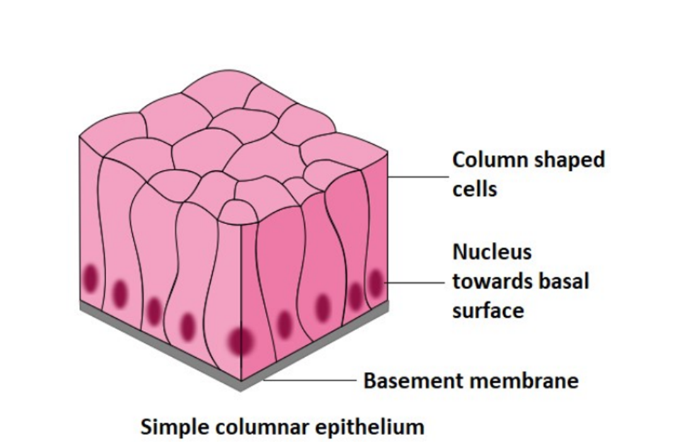
- Characterised by presence of basement membrane and elongated cells .
- Nucleus is present at the base of the cell .
- Cell appear pillar-like ( less width ,more length )
- Function : Absorption and secretion.
- Eg. Bile Duct , Liver
- Modifications:
- Brush Bordered Columnar epithelium : Microvilli cell ke ek end pe present honge. e.g. Gall bladder
- Glandular columnar epithelium : Unicellular mucous secret karne wale goblet cells columnar cells k beech mein present honge . eg. Stomach, Colon , Rectum
- Glandular Brush Border columnar epithelium : Microvilli cell ke ek end par present honge , sath hi goblet cells bhi present honge eg. Duodenum ,Ileum ,Caecum.
- Ciliated Columnar epithelium : in types ke cells mein Cilia present hote hain free end par Eg. Fallopian Tube, Ependymal epithelium.
- Stereo ciliated columnar epithelium : In types ke cells mein stereocilia present hote hain cells ke free end mein Eg. Epididymis ,Vas Deferens.
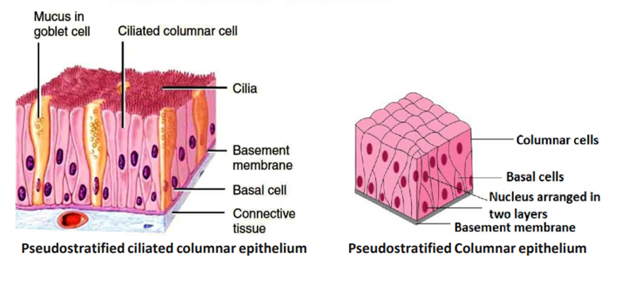
- It has two types of cells : Long and short .
- These are modified columnar epithelium cells.
- Long cells have elongated nuclei towards the base.
- Short cells have rounded nuclei in the center.
- The layer is uniform but gives a false appearance of two layered cells due to differences in the heights of long and small cells, and hence their nuclei are also present on different levels
- Eg. Middle part of male Urethra
- Modification : Pseudostratified ciliated glandular epithelium :Cilia is present at the free end of the epithelium.
- Eg. Trachea, Bronchi, Respiratory epithelium of nasal chambers.
Simple Squamous epithelium
Simple Cuboidal Epithelium
NOTE : It is also also known as Germinal epithelium because in gonads (testis & ovaries) cuboidal cells divide to form egg & sperm. It is found in the peripheral region of ovary & lining of seminiferous tubules in Testis. (Simple cuboidal epithelium)
Simple Columnar Epithelium
PSEUDOSTRATIFIED EPITHELIUM
- Neuro sensory epithelium : In the structure of this epithelium in between pillar shaped supporting cells , modified sensory cells are present. On the free end of the sensory cell sensory hair is present by which they receive sensation. Base of these cells is attached to the sensory nerve. Eg. Gustatory Epithelium ,Covering of the taste buds of tongue receive taste sensation, Olfactory epithelium, Schneidarian membrane receive smell sensation,Statoacoustic, Lining of internal ear, In retina of eye receives optic sensation.
- Myoepithelium :- surrounds mammary and sweat gland (around secretory unit)
- Pigmented epithelium (Cuboidal) :- In Retina of eye.
COMPOUND EPITHELIUM
- Made up of more than one layer : Multilayered.
- Has limited role in secretion and mechanical stresses.
- On the basis of stretching ability, it is of 2 types –
- Transitional epithelium :– which can be stretched.
- Stratified epithelium :– which cannot be stretched.
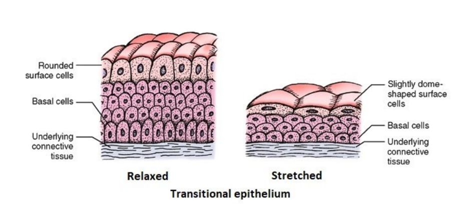
TRANSITIONAL EPITHELIUM (UROTHELIUM)
- Only tissue without a basement membrane so the innermost layer rests directly upon the connective tissue.
- 4-6 layers of cells are present.
- Innermost layer has cube like cells.
- Middle - 2-4 layers have pear shaped or umbrella shaped cells which are known as Polyhedral cells in general.
- Outermost- 1 or 2 layers of oval shaped cells.
- These shapes appear in only the resting stage. When tissue is in stretched condition, the cells become flattened.
- Just after the outermost layer, a thin layer of cuticle is present, making it waterproof.
- Cells are interconnected by interdigitation.
- Eg. Renal Pelvis, Ureter, Urinary Bladder, Proximal part of male urethra.
STRATIFIED EPITHELIUM
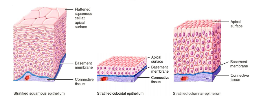
- On the basis of shape of the cells of outermost layer these are of three types :
- Stratified squamous epithelium
- Stratified cuboidal epithelium
- Stratified columnar epithelium
- STRATIFIED SQUAMOUS EPITHELIUM
- Innermost layer of cells are either cuboidal or columnar shaped shaped.
- Dependent upon underlying connective tissue layer for nutrition/food.
- They divide and form stratified layer hence, so also known as germinative epithelium.
- Middle layers are made up of polygonal cells. These cells are interconnected with Desmosomes( jo cells ko rigidity aur mechanical support provide karte hain).
- Cells of outermost layer are scale like flat cells.
- On the basis of presence of keratin protein in the outermost cells this epithelium is of two types :
- Keratinized Stratified squamous epithelium : If keratin protein is present in scaly cells and cells become non nucleated dead cells. eg. Epidermis of skin, Scale, Horns, Nails, Feathers etc.
- Non Keratinized Stratified squamous epithelium : If Keratin protein is absent. Cells are nucleated and Living. eg. Buccal cavity or oral cavity of mammals, Inner lining of cheeks, Inner lining of lips, Lining of hard palate. Lining of tonsils, Lower part of soft palate, Pharynx, Oesophagus, Anal canal, Lining of vagina. Cornea of eye
- STRATIFIED CUBOIDAL EPITHELIUM
- Innermost layer is made up of cuboidal/Columnar cells
- Middle layer is made up of polygonal shaped cells.
- Outermost layer of cells are cube like & cells are nucleated & living.
- Eg. – Secretory duct of sweat glands, mammary glands and sebaceous gland; Secretory duct of sebaceous gland; Secretory duct of salivary glands, Submandibular, Whartson's duct, Stenson's duct Sublingual; Bartholin duct or duct of Rivinus; Pancreatic duct, Female Urethra; Conjunctiva of eye.
- STRATIFIED COLUMNAR EPITHELIUM
- Outermost layer is composed of pillar shaped cells, cells are nucleated.
- On the basis of presence of cilia this epithelium is of 2 types:
- Ciliated stratified columnar epithelium. Cilia are present at the free end. Eg. Buccopharyngeal cavity of Frog ,Upper part of soft palate towards nasopharynx Larynx Uterus
- Non ciliated stratified columnar epithelium.-Cilia absent on free end. Eg. Distal part of male urethra ,Epiglottis.
GLANDS
A cell or a group of cells which secretes chemical substances are called glands. All glands are composed of Epithelium tissue. Glands can originate from all the three germinal layers.
CLASSIFICATION OF GLANDS
- On the basis of presence of secretory duct glands are of 3 types :
- Endocrine glands :- Secretory duct absent.
- Exocrine gland :- Secretory duct present – Liver (largest gland).
- Heterocrine/mixed gland :- Both endocrine & exocrine parts are present. eg. Pancreas, Gonads etc.
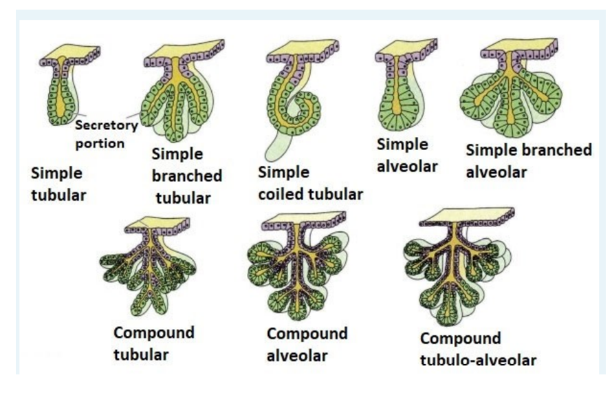
- On the basis of structure :
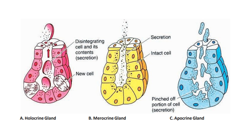
- On the basis of the nature of secretion :- 3 types of glands are there.
- Eccrine/Merocrine gland :- In these glands secretory cells secrete substances by simple diffusion (Exocytosis). No part of cytoplasm is destroyed in secretion. Secretory matter like water. Eg. Maximum sweat glands of humans, claws of rabbit, Goblet cells, Salivary gland, Tear gland, Intestinal glands, Mucous gland.
- Apocrine gland :- In this type of glands secretory products are collected in the apical part of the secretory cell and the apical portion is also shed along with secretory matter. Secretory cells gain their lost part of cytoplasm by process of regeneration. Secretory matter is comparatively concentrated. – Mammary glands. Eg. Sweat gland of arm pit, pubic region, skin around anus, lips, nipples etc. Largest sweat glands of the body are found around nipples - Areola mammae.In Rabbit sweat glands of this type are found on lips & Skin around lips.
- Holocrine glands :- The production or secretion is shed with the whole cell leading to its destruction. i.e Whole cell is shed as secretion (Secretory matter concentrate) Example : Sebaceous, meibomian & Zeis gland.


Connective Tissues
- General features of connective tissue :
- Connective tissue consists of two basic elements: extracellular matrix (aas pass ke cells ke bahar jo material hota hai usso ko extracellular matrix kehte hain and yeah nonliving hota hai) and cells.
- The extracellular matrix consists of protein fibers and ground substance, the material between the cells and the fibers and is usually secreted by the connective tissue cells and determines the tissue's qualities. For instance, in cartilage, the extracellular matrix is firm but flexible. The extracellular matrix of bone, by contrast, is hard and inflexible.
- In contrast to epithelia, connective tissues do not usually occur on body surfaces. Also unlike epithelia, connective tissues usually are highly vascular; that is, they have a rich blood supply. Exceptions include cartilage, which is avascular, and tendons, with a scanty blood supply. Except for cartilage, connective tissues, similar to epithelia, are supplied with nerves.
- In its various forms, connective tissue has a variety of functions, such as follows:
- It binds together, supports, and strengthens other body tissues.
- It protects and insulates internal organs.
- It compartmentalizes structures such as skeletal muscles, etc.
- It serves as the major transport system within the body (blood, a fluid connective tissue).
- It is the primary location of stored energy reserves (adipose, or fat tissue).
- It is the main source of immune responses.
- They range from soft connective tissues to specialised types, which include cartilage, bone, adipose, and blood.
- Ground Substance : ground substance is the material found between the cells and fibers of a tissue and it can be fluid, semifluid, gelatinous, or calcified. The ground substance supports cells, binds them together, stores water, and provides a medium through which substances are exchanged between the blood and cells. It plays an active role in how tissues develop, migrate, proliferate, and change shape, and in how they carry out their metabolic functions.
- Ground substance contains water and an assortment of large organic molecules, many of which are complex combinations of polysaccharides and proteins. The polysaccharides include hyaluronic acid, chondroitin sulfate, dermatan sulfate, and keratan sulfate. Collectively, they are referred to as glycosaminoglycans or GAGs.
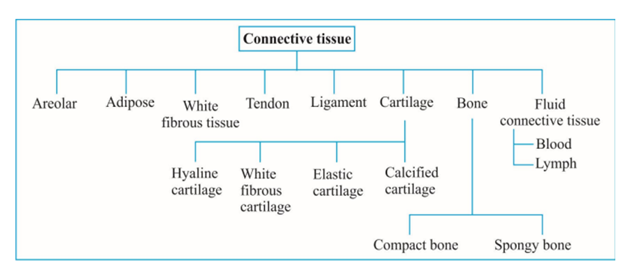
- Connective tissue is classified into 3 types :
- Loose Connective Tissue - Examples are : Areolar and Adipose tissue
- Dense Connective Tissue - further of two types :
- Dense Regular Connective Tissue
- Dense Irregular Connective Tissue
- Specialised Connective Tissue- Example : Cartilage, Blood and Bones
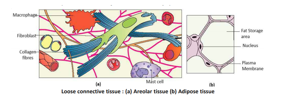
Loose Connective Tissue
- Cells and fibres are loosely arranged in a semi-fluid ground substance.
- Areolar Tissue : present beneath the skin and often it serves as a support framework for epithelium.
- The most widely distributed connective tissue in the body is areolar connective tissue.
- Collagen, elastic, and reticular fibers -are arranged randomly throughout the tissue.
- Macrophages are the phagocytic cells.
- Mast cells play a role in inflammation.
- Adipose Tissue : Iss tissue ke cells fat store karte hain jinko hum adipocytes kehte hain. Adipocytes are derived from fibroblasts. The adipose tissue synthesises, stores and metabolises fat. White fat contains a single large fat globule and hence called Monolocular fat while brown fat has several small fat globules thus, Multilocular fat.
- Fibers in connective tissue :
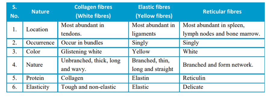
- Collagen Fibers : white in colour so known as white fibers. very strong, flexible and resist pulling forces. Often occur in parallel bundles and are unbranched. Collagen fibers are found in most types of connective tissues, especially bone, cartilage, tendons, and ligaments. When boiled in water, collagen changes into gelatin.
- Elastic fibres (yellow fibres) are smaller in diameter than collagen fibers, branch and join together to form a network within a tissue. Elastic fibers are strong but can be stretched up to 150% of their relaxed length without breaking. Equally important, elastic fibers have the ability to return to their original shape after being stretched. Made up of elastin protein.
- Reticular fibres (reticular ka matlab hai net/jaal like structure ho jiska) are produced by fibroblasts and are much thinner than collagen fibers and form branching networks. Reticular fibers made up of reticulin protein are plentiful in reticular connective tissue, which forms the stroma or supporting framework of many soft organs, such as the spleen and lymph nodes. These fibers also help form the basement membrane.
Dense Connective Tissue
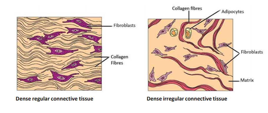
- Dense Connective Tissue : thicker, and denser fibers (packed more closely) but considerably fewer cells than loose connective tissue. If the orientation of fibres show a regular pattern we call it, a dense regular and if orientation is an irregular pattern we call it dense irregular connective tissue.
- Dense regular connective tissue : , bundles of collagen fibers are regularly arranged in parallel patterns. Examples : tendons and most ligaments.
- Tendon :It is a dense, strong fibrous connective tissue with thick, parallel, bundles of collagen fibres (matrix). A few flat and elongated fibroblast cells lie in single rows between the fibre bundles. Tendon forms the strong inextensible attachment of a skeletal muscle to a bone.
- Ligament : It is a dense fibrous connective tissue. Its ground substance or matrix is densely crowded with yellow elastin fibres branched in different directions. A few elongated flat cells (fibroblasts) lie scattered between the fibres. The ligament connects bones at the joints and holds them in position. Because of the fact that ligaments connect the bones at joints, we are able to move and rotate our neck, limbs, fingers, etc. comfortably.
- Dense irregular connective tissue : has fibroblasts and many fibres (mostly collagen) that are oriented differently. Found in the dermis of the skin, fibrous pericardium around the heart.
- Dense regular connective tissue : , bundles of collagen fibers are regularly arranged in parallel patterns. Examples : tendons and most ligaments.
Specialised connective tissue
- Cartilage, bones ,blood and lymph are types of specialised connective tissues.
- Skeletal connective tissues : These connective tissues are a part of the endoskeleton of vertebrates. These include cartilage and bone.
Cartilage
- The intercellular material of cartilage is solid and resists compression.
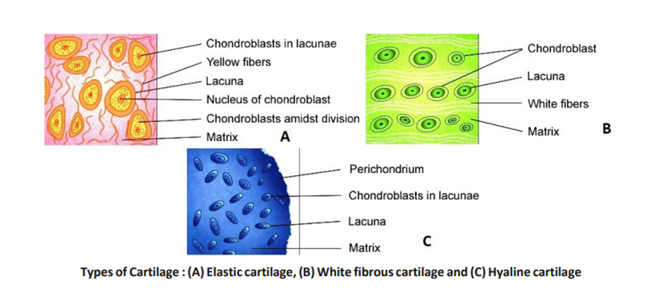
- Young cartilage cells are small, flattened with small surface projections and are known as chondroblasts.
- Chondrocytes are large and bluntly angular cartilage cells lying scattered in the matrix.
- Chondroblasts are metabolically more active than chondrocytes.
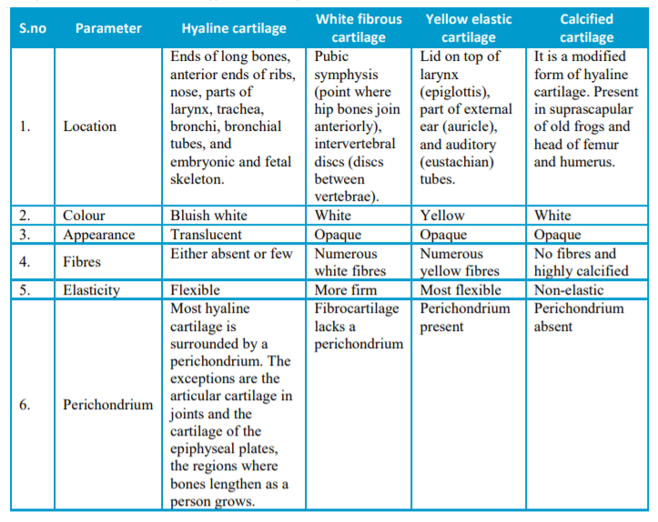
- Cartilage is surrounded by a covering of dense irregular connective tissue known as perichondrium.
Chondrocytes occur in clusters of two or three cells in small fluid filled spaces called lacunae.
NOTE : Mammalian foetus is mainly composed of hyaline cartilage which is gradually replaced by bone. Cartilage is present in the tip of the nose, trachea, bronchi, ends of ribs and the articulating surfaces of joints.
BONE
- Bone is a solid, rigid and strong connective tissue and the hardest tissue of the body.
- Bones have a hard ground substance rich in calcium salts and collagen fibres which give bone its strength
- It makes the basic frame work. Limb bones, such as the long bones of the legs, serve weight-bearing functions. They also interact with skeletal muscles attached to them to bring about movements.
- Support: Skeleton is the basic framework of the body and supports soft tissues and provides attachment for most of the skeletal muscles .
- Protection: The skeleton protects the most important internal organs from injury. For example, cranial bones protect the brain, vertebrae (backbones) protect the spinal cord, and the rib cage protects the heart and lungs.
- Assistance in movement: Most skeletal muscles attach to bones; when they contract, they pull on bones to produce movement.
- MineraI homeostasis (storage and reIease): Bone tissue stores several minerals, especially calcium and phosphorus, which contribute to the strength of bone. Bone tissue stores about 99% of the body's calcium. On demand, bone releases minerals into the blood to maintain critical mineral balances (homeostasis) and to distribute the minerals to other parts of the body.
- Blood cell production: Within certain bones, a connective tissue called red bone marrow produces red blood cells, white blood cells, and platelets, a process called hematopoiesis (hemo- blood; poiesis- making). Red bone marrow consists of developing blood cells, adipocytes, fibroblasts, and macrophages within a network of reticular fibers. It is present in developing bones of the fetus and in some adult bones, such as the hip bones, ribs, breastbone, vertebrae (backbones), skull, and ends of the bones of the arm and thigh.
- Triglyceride storage: Yellow bone marrow consists mainly of adipose cells, which store triglycerides. The stored triglycerides are a potential chemical energy reserve. In a newborn, all bone marrow is red and is involved in hematopoiesis. With increasing age, much of the bone marrow changes from red to yellow.
Functions of a bone
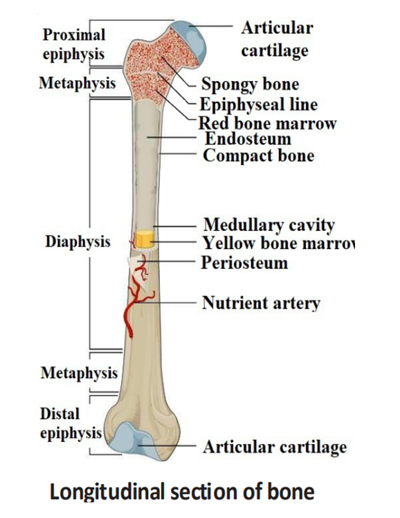
Structure of long bone : Long bone has three regions-
- Epiphysis
- Diaphysis
- Metaphysis
- Epiphysis: Ends of long bone is called Epiphysis.
- This part is composed of spongy tissue. If this part is present at the joint then on the articular surface periosteum is absent & articular cartilage (Hyaline cartilage) is present.
- Cavity is present in the form of trabeculae filled with red bone marrow. It is composed of myeloid tissue which produces blood corpuscles so epiphysis acts as a haemopoietic organ.
- Diaphysis:
- Middle part or shaft of long bone is diaphysis which is composed of compact bone. In this region, a hollow cavity is present called bone marrow cavity filled with yellow bone marrow composed of white fat.
- Function of YBM (yellow bone marrow) is storage of fat. If required or essential in anaemic condition YBM is replaced by RBM.
- At the time of birth RBM is 70 ml. while in adulthood it is about 4 litre.
- Metaphysis
- It formed little part between epiphysis & diaphysis. In this region epiphyseal plate is present which is made up of osteoblast cells.
- They divide to form osteocytes and also synthesize matrices of bone, so the epiphyseal plate is responsible for elongation of bone.
- After complete development of long bone this plate is destroyed. So in a completely developed bone only 2 regions are found while in a developing bone 3 regions are present.
- POINTS TO PONDER:
- Spongy Bones: Bones in which haversian systems are absent. In these bones marrow cavity is present in the form of trabeculae filled with RBM. So all spongy bones of the body are hematopoietic.Eg. Ribs, Pubis, Sternum, Vertebrae, Clavicle, End of long Bones, Scapula
- Compact Bone :In this bone haversian system is present. Eg. Diaphysis of long bone.
- Diploic/Heterotypic: In these bones middle part of bone is composed of spongy bone, in which trabeculae is filled with RBM.While this bone is covered by compact bone on upper & lower surface. Eg. All flat bones of skull.
- Pneumatic Bone In the matrix of these bones air filled spaces are present so bone becomes light in weight. Eg. Bones of birds. (Long Bone).
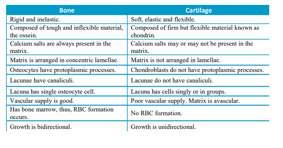
- Difference between BONE and CARTILAGE
Blood :
- Blood is a fluid connective tissue. It is the softest connective tissue. Its cells are quite distinct from other connective tissue cells, both in structure and function.
- There are different types of cells with very specific functions and are free floating in a fluid called plasma. Plasma is equivalent to the matrix of other connective tissues but lacks fibres.
- The matrix of other connective tissues is stationary whereas the matrix of blood is non-stationary and free flowing. That is why blood is also called liquid tissue. It is also called vascular tissue because it is part of the vascular system of many animals.
- Transports oxygen, carbon dioxide, nutrients, hormones, heat, and wastes.
- Regulates pH, body temperature, and water content of cells.
- Protects against blood loss through clotting, and against disease through phagocytic white blood cells and antibodies.
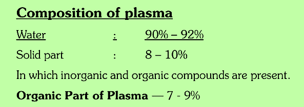
- PIasma :Plasma contains about 92 percent water and 8 per cent solutes most of which are proteins. The solutes present in it include glucose, proteins, amino acids, cholesterol, fatty acids, vitamins, enzymes, hormones, antibodies, oxygen, carbon dioxide and waste products such as urea, uric acid and creatinine, etc.
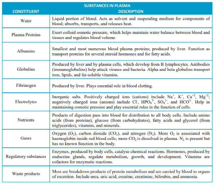
- Plasma contains three major classes of plasma proteins, viz., Albumin, Globulin and Fibrinogen.
- Plasma is faint yellow in colour and slightly alkaline (pH 7.4). It is somewhat viscous and an aqueous solution containing many organic and inorganic substances.
- Plasma proteins serve as a source of proteins for tissue cells and help in transportation and defence mechanism.
- In humans normal blood sugar level is 90-120 mg per 100 ml of blood in an adult person 12 hours after a meal.
- Cholesterol is the main lipid component found in plasma, and in humans it normally ranges from 140 to 260 mg per 100 ml of serum. Inorganic salts present in plasma are ions of sodium, potassium, calcium, magnesium, iron, etc.
- Plasma: It is a straw coloured, slightly alkaline(pH 7.4) viscous fluid constituting nearly 55% of the blood. Plasma mein proteins, lipids, salts, wastes, nutrients, hormones, water etc hota hai.
- Proteins : Albumins - help in osmotic balance. Their deficiency reduces the water content of blood and increases that of tissue fluid, causing oedema. Globulins - primarily are involved in defense mechanisms of the body and transport of substances. Fibrinogens - are needed for clotting or coagulation of blood. Prothrombin- another clotting protein also present in blood. Prothrombin is synthesized in the liver with the help of Vitamin K. Fibrinogens, globulins and albumins are the major proteins.
- Plasma without clotting factors is called serum.
- Nutrients : Plasma includes glucose, amino acids and lipids etc. are always in transit in the body.
- Ions - it contains small amounts of ions like sodium, calcium, magnesium, chloride etc.
- Gases - Nitrogen, oxygen, carbon dioxide occur in a dissolved state.
- Wastes - Wastes like urea, uric acid, etc are present in plasma.
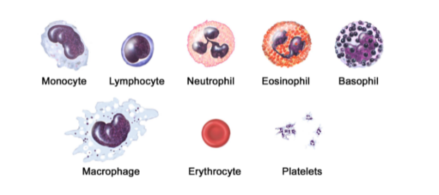
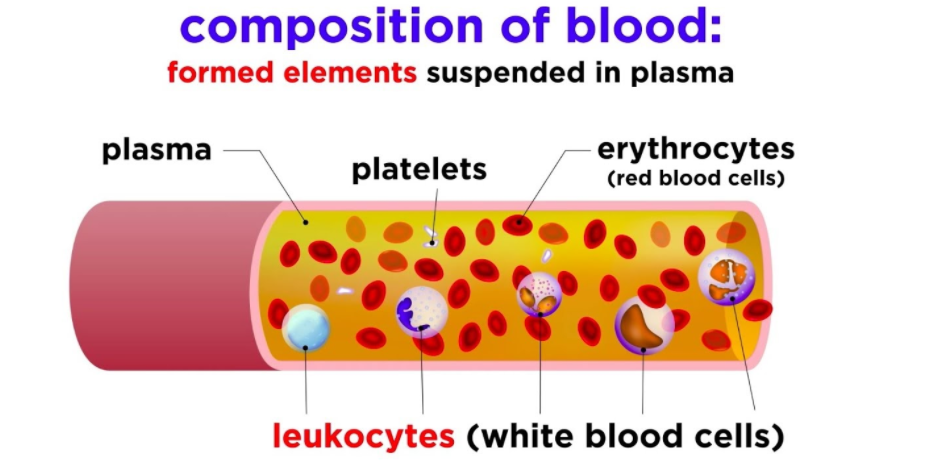
- Blood cells: These are of three types:
Functions of Blood :
Composition of BIood :

Lymph
- Lymph is another fluid connective tissue. It is colourless. It is a translucent alkaline fluid present between capillaries and tissues. It is composed of two parts, namely plasma and leukocytes.
- Plasma of lymph is similar to that of blood except that it has less proteins, calcium and phosphorus.
- The corpuscles present in lymph are leukocytes, mainly floating amoeboid lymphocytes. The composition of the lymph can be expressed as blood minus RBCs, platelets, proteins and some salts.
- Lymph carries materials from tissues into the bloodstream and vice versa. The WBCs present in lymph destroy invading microorganisms.
- Lymph returns blood proteins from tissue fluids to the blood.
- Lymph acts as a "middle man" which transports oxygen, food materials, hormones etc., to the body cells and brings carbon dioxide and other metabolic wastes, from the body cells to blood.
- Destroys the invading microorganisms and foreign particles in the lymph nodes. Maintains the volume of the blood.
Functions :

Connective Tissues
- Naam hi bta rha hai ki iska kaam hai baki tissues ko ek dusre se link yani ki connect karna and support karna toh isliye yeah puri human body me sabse abundant tissue hai.
- Cartilage, bone, adipose tissue and blood sab connective tissue me aate hai.
- Ek bda alag sa feature yeah hai iss tissue ka ki blood ko chor baki jitne bhi connectives tissues hai wo collagen ya elastin naam ke proteinaceous fibers secrete karte hai jo iss tissue ko strength, flexibility and elasticity provide karta hai.
- Iss tissue ke cells kuch modified polysaccharide bhi secrete hai jo ki in sab components ko apas me jod ke rakhta hai and ek medium provide karta hai nutrients and gases ke diffusion ke liye. Issi ko hum matrix ya ground substance bolte hain.
- Connective tissue 3 type ka hai : Loose, Dense and Specialised connective tissue.
- Loose connective tissue me jaise ki naam hi bta rha hai, jo fibres hote hai wo bade loosely arranged hai. Ex : areolar tissue jo ki skin ke neeche present hota hai and epithelium ko support deta hai. Areolar tissue me fibroblast, mast cells and macrophages naam ke cells hote hai. Fibroblasts toh fibers secrete karenge, macrophages phagocytic cells hai jo dead cells and foreign material ko engulf karenge and mast cells generally kuch aise chemicals secrete karte hain jo allergy ko mediate karte hain.
- Khair, issi me dusre type ka tissue hai adipose tissue and yeah bhi skin ke neeche present hota hai but muscle layer ke uper. Main function hai fat droplets ko store karna.
- Dense connective tissue me fibers and fibroblasts kafi compactly packed hote hain. Ab depending upon the orientation of fibers ki fibers kisi regular pattern me arranged hai ya nhi we have further 2 types of dense connective tissue : dense regular and dense irregular tissue.
- Dense regular connective tissue me collagen fibers parallelly arranged hote hai bundles me. Example hai : Tendons jo muscle ko bone se jodte hain and ligaments jo ki bone ko dusri bone se jodte hai.
- Dense irregular me bhi collagen fibers hote hain but kisi ek way me arranged nhi hote wo bas randomly pade hue hai. Location : reticular layer of dermis
- Specialised connective tissue me aate hai : cartilage, blood and bone.
- Cartilage ek solid but pliable yani ki kind of jelly like material se bna hua tissue hai jo compression ko resist karta hai. Cartilage ke cells ko chondrocytes kehte hai jo ki apna matrix khud se secrete karte hain. Yhi matrix cartilage tissue ka ground substance banate hai. Vertebrates me embryo me cartilage ki amount bahut hi jyada hoti hai lekin jaise jaise wo grow karta hai. Bones cartilage ko replace kar deti hai toh adult me cartilage hume nose ki tip and ear me and bones ke joint jaise ki between adjacent bones of vertebral column , limbs and hands in adults.
- Bones ka jo ground material hai wo hard and non pliable yani ki flexible nhi hai. Isme generally calcium, phosphorus and magnesium salts ki amount jyada hoti hai along with more collagen fibers jo bone ko strength provide karte hai. Osteocytes are bone cells. Osteoclasts are bone eating cells. Jis space me osteocytes present hote hai unko lacunae kaha jata hai. Bone ander se spongy hoti hai yani ki bilkul 100% solid rod ki tarah nhi hoti. Bone ke ander bone marrow hote hai jisme hematopoietic stem cells hote hai jinse blood cells jaise ki : RBC, WBC (granulocytes and agranulocytes) and platelets ki formation hoti hai. Ab bone me blood cells ka production choti bones jaisi ki hatho ki ungliyon me jo bones hai unme itna nhi hota lekin badi bones jaise ki humerus, femur, hip bones vgarah jo hai unme actively production hota rehta hai blood cells ka. Bones me kuch canals hoti hai like haversian canals and volkmann canal jinse blood and nerve supply enter and exit karenge. Volksmann canals ka kaam hota hai haversian canals ko aapas me interconnect karna.
- Blood ek fluid connective tissue hai jisme plasma, RBC, WBC, platelets, hormones, proteins like albumin, dissolved gases, nutrients, waste, ions vgarah hote hai. RBC ka kaam hai oxygen ko carry karna. WBC infections se fight karte hain. Platelets and blood coagulating proteins and ions jaise ki calcium ions blood clot and wound heal karne me help karte hain. Blood ke baki function apun human physiology ke chapter me padhenge.
Muscular Tissue
- Muscle tissue is composed of thin and elongated cells that are usually called muscle fibres. They are differentiated cells made up of contractile protein (aise proteins jinmei contract karne ki property ho ).
- Eg -higher animals - found associated with the skeleton, walls of visceral organs, blood vessels and heart, etc.
- Muscular tissue is mesodermal in origin.
- Muscles of the iris of the eye and myoepithelial cells of the salivary, mammary and sweat glands are ectodermal in origin.
- Each muscle is made of many long, cylindrical fibres arranged in parallel arrays. These fibres are composed of numerous fine fibrils, called myofibrils.
- Muscle fibres contract (shorten) in response to stimulation, then relax (lengthen) and return to their uncontracted state in a coordinated fashion. Their action moves the body to adjust to the changes in the environment and to maintain the positions of the various parts of the body. In general, muscles play an active role in all the movements of the body.
- Function of muscle tissue:
- Production of body movements: Movements of the whole body such as walking and running, and localized movements such as grasping a pencil or nodding the head as a result of muscular contractions, rely on the integrated functioning of skeletal muscles, bones, and joints.
- Stabilization of body positions: Skeletal muscle contractions stabilize joints and help maintain body positions, such as standing or sitting. Postural muscles contract continuously when you are awake; for example, sustained contractions of your neck muscles hold your head upright.
- Storage and movement of substances within the body: Storage is accomplished by sustained contractions of ring-like bands of smooth muscle called sphincters, which prevent outflow of the contents of a hollow organ. Eg : Temporary storage of food in stomach , storage of urine , cardiac muscle contraction for blood flow , movement of food in alimentary canal .Skeletal muscle contractions promote the flow of lymph and aid the return of blood to the heart.
- Generating heat:Heat is produced on the contraction of tissue which is called thermogenesis. This is used to maintain normal body temperature. Involuntary contractions of skeletal muscles, which is known as shivering, can increase the rate of heat production.
- According to their structure, location and functions there are three main types :
- Skeletal muscle
- Smooth muscle
- Cardiac muscle
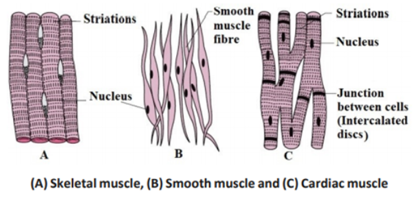
- SKELETAL MUSCLE
- Skeletal muscles are attached to bones by tendons, and consist of bundles of very long (up to 30 cm), cylindrical, multinucleated (syncytial cells). These fibres (cells) can be contracted at desire; therefore, they are also called voluntary muscles.
- The plasma membrane covering each fibre is called sarcolemma. The cytoplasm inside the fibre is called sarcoplasm. The sarcoplasm contains many long, thin, unbranched cross striated cylindrical structures called myofibrils. Each muscle fibre or muscle cell also possesses many oval, flat nuclei at the periphery of the cell under the sarcolemma. They are arranged along the long axis of the fibre.
- Smooth muscle consists of a collection of spindle shaped cells (centre se thick or pointed ends par ) that do not show any striation. They may range in size from 20 m (in small blood vessels) to 500 m (in the pregnant uterus).
- Shorter than the striated fibres with single oval nucleus.
- NO sarcolemma , but fibers have cell membrane.
- The mitochondria are fewer in number, the sarcoplasmic reticulum is less extensive and protein filaments are not regularly arranged to give rise to striations. Their contraction process is slow and not under voluntary control. So they are also called involuntary muscles ( aise muscles jinhe hum apni marzi se control na kar sakein).
- Cell junctions hold them together and they are bundled together in a connective tissue sheath.
- Eg: The wall of internal organs such as the blood vessels, stomach and intestine.
- Cardiac muscle is restricted to the wall of the heart only. These are composed of elongated, branched individual cells that lie parallel to each other.
- Because they need a large amount of energy , they possess numerous mitochondria and glycogen granules.
- They are cross striated (inn par bhi skeletal muscle jaise strips present hain ) and have actin and myosin filaments.
- Cardiac muscle is involuntary, vigorous ,rhythmic and immune to fatigue ( kabhi thakti nahi h )
- Communication junctions or gap junctions (intercalated discs) allow the cells to contract as a unit, i.e., when one cell receives a signal to contract, its neighbours are also stimulated to contract.
SMOOTH MUSCLE
CARDIAC MUSCLE
Unlike multinucleated skeletal muscle, these cells possess only one centrally located nucleus, which lies deep at the centre of the fibre.
NOTE They have specialised junctional complexes at the ends of the adjacent muscle cells called intercalated discs (oval shape k disc).
Function:They hold the cells together ( jo rapid excitation spread mei help karta hai jab heart beat karta hai).
Connective Tissues
- General features of connective tissue :
- Connective tissue consists of two basic elements: extracellular matrix (aas pass ke cells ke bahar jo material hota hai usso ko extracellular matrix kehte hain and yeah nonliving hota hai) and cells.
- The extracellular matrix consists of protein fibers and ground substance, the material between the cells and the fibers and is usually secreted by the connective tissue cells and determines the tissue's qualities. For instance, in cartilage, the extracellular matrix is firm but flexible. The extracellular matrix of bone, by contrast, is hard and inflexible.
- In contrast to epithelia, connective tissues do not usually occur on body surfaces. Also unlike epithelia, connective tissues usually are highly vascular; that is, they have a rich blood supply. Exceptions include cartilage, which is avascular, and tendons, with a scanty blood supply. Except for cartilage, connective tissues, similar to epithelia, are supplied with nerves.
- In its various forms, connective tissue has a variety of functions, such as follows:
- It binds together, supports, and strengthens other body tissues.
- It protects and insulates internal organs.
- It compartmentalizes structures such as skeletal muscles, etc.
- It serves as the major transport system within the body (blood, a fluid connective tissue).
- It is the primary location of stored energy reserves (adipose, or fat tissue).
- It is the main source of immune responses.
- They range from soft connective tissues to specialised types, which include cartilage, bone, adipose, and blood.
- Ground Substance : ground substance is the material found between the cells and fibers of a tissue and it can be fluid, semifluid, gelatinous, or calcified. The ground substance supports cells, binds them together, stores water, and provides a medium through which substances are exchanged between the blood and cells. It plays an active role in how tissues develop, migrate, proliferate, and change shape, and in how they carry out their metabolic functions.
- Ground substance contains water and an assortment of large organic molecules, many of which are complex combinations of polysaccharides and proteins. The polysaccharides include hyaluronic acid, chondroitin sulfate, dermatan sulfate, and keratan sulfate. Collectively, they are referred to as glycosaminoglycans or GAGs.
- Connective tissue is classified into 3 types :
- Loose Connective Tissue - Examples are : Areolar and Adipose tissue
- Dense Connective Tissue - further of two types :
- Dense Regular Connective Tissue
- Dense Irregular Connective Tissue
- Specialised Connective Tissue- Example : Cartilage, Blood and Bones

Loose Connective Tissue
- Cells and fibres are loosely arranged in a semi-fluid ground substance.
- Areolar Tissue : present beneath the skin and often it serves as a support framework for epithelium.
- The most widely distributed connective tissue in the body is areolar connective tissue.
- Collagen, elastic, and reticular fibers -are arranged randomly throughout the tissue.
- Macrophages are the phagocytic cells.
- Mast cells play a role in inflammation.
- Adipose Tissue : Iss tissue ke cells fat store karte hain jinko hum adipocytes kehte hain. Adipocytes are derived from fibroblasts. The adipose tissue synthesises, stores and metabolises fat. White fat contains a single large fat globule and hence called Monolocular fat while brown fat has several small fat globules thus, Multilocular fat.
- Fibers in connective tissue :
- Collagen Fibers : white in colour so known as white fibers. very strong, flexible and resist pulling forces. Often occur in parallel bundles and are unbranched. Collagen fibers are found in most types of connective tissues, especially bone, cartilage, tendons, and ligaments. When boiled in water, collagen changes into gelatin.
- Elastic fibres (yellow fibres) are smaller in diameter than collagen fibers, branch and join together to form a network within a tissue. Elastic fibers are strong but can be stretched up to 150% of their relaxed length without breaking. Equally important, elastic fibers have the ability to return to their original shape after being stretched. Made up of elastin protein.
- Reticular fibres (reticular ka matlab hai net/jaal like structure ho jiska) are produced by fibroblasts and are much thinner than collagen fibers and form branching networks. Reticular fibers made up of reticulin protein are plentiful in reticular connective tissue, which forms the stroma or supporting framework of many soft organs, such as the spleen and lymph nodes. These fibers also help form the basement membrane.
Dense Connective Tissue
- Dense Connective Tissue : thicker, and denser fibers (packed more closely) but considerably fewer cells than loose connective tissue. If the orientation of fibres show a regular pattern we call it, a dense regular and if orientation is an irregular pattern we call it dense irregular connective tissue.
- Dense regular connective tissue : , bundles of collagen fibers are regularly arranged in parallel patterns. Examples : tendons and most ligaments.
- Tendon :It is a dense, strong fibrous connective tissue with thick, parallel, bundles of collagen fibres (matrix). A few flat and elongated fibroblast cells lie in single rows between the fibre bundles. Tendon forms the strong inextensible attachment of a skeletal muscle to a bone.
- Ligament : It is a dense fibrous connective tissue. Its ground substance or matrix is densely crowded with yellow elastin fibres branched in different directions. A few elongated flat cells (fibroblasts) lie scattered between the fibres. The ligament connects bones at the joints and holds them in position. Because of the fact that ligaments connect the bones at joints, we are able to move and rotate our neck, limbs, fingers, etc. comfortably.
- Dense irregular connective tissue : has fibroblasts and many fibres (mostly collagen) that are oriented differently. Found in the dermis of the skin, fibrous pericardium around the heart.
- Dense regular connective tissue : , bundles of collagen fibers are regularly arranged in parallel patterns. Examples : tendons and most ligaments.
Specialised connective tissue
- Cartilage, bones ,blood and lymph are types of specialised connective tissues.
- Skeletal connective tissues : These connective tissues are a part of the endoskeleton of vertebrates. These include cartilage and bone.
Cartilage
- The intercellular material of cartilage is solid and resists compression.
- Young cartilage cells are small, flattened with small surface projections and are known as chondroblasts.
- Chondrocytes are large and bluntly angular cartilage cells lying scattered in the matrix.
- Chondroblasts are metabolically more active than chondrocytes.
- Cartilage is surrounded by a covering of dense irregular connective tissue known as perichondrium.
Chondrocytes occur in clusters of two or three cells in small fluid filled spaces called lacunae.
NOTE : Mammalian foetus is mainly composed of hyaline cartilage which is gradually replaced by bone. Cartilage is present in the tip of the nose, trachea, bronchi, ends of ribs and the articulating surfaces of joints.


BONE
- Bone is a solid, rigid and strong connective tissue and the hardest tissue of the body.
- Bones have a hard ground substance rich in calcium salts and collagen fibres which give bone its strength
- It makes the basic frame work. Limb bones, such as the long bones of the legs, serve weight-bearing functions. They also interact with skeletal muscles attached to them to bring about movements.
- Support: Skeleton is the basic framework of the body and supports soft tissues and provides attachment for most of the skeletal muscles .
- Protection: The skeleton protects the most important internal organs from injury. For example, cranial bones protect the brain, vertebrae (backbones) protect the spinal cord, and the rib cage protects the heart and lungs.
- Assistance in movement: Most skeletal muscles attach to bones; when they contract, they pull on bones to produce movement.
- MineraI homeostasis (storage and reIease): Bone tissue stores several minerals, especially calcium and phosphorus, which contribute to the strength of bone. Bone tissue stores about 99% of the body's calcium. On demand, bone releases minerals into the blood to maintain critical mineral balances (homeostasis) and to distribute the minerals to other parts of the body.
- Blood cell production: Within certain bones, a connective tissue called red bone marrow produces red blood cells, white blood cells, and platelets, a process called hematopoiesis (hemo- blood; poiesis- making). Red bone marrow consists of developing blood cells, adipocytes, fibroblasts, and macrophages within a network of reticular fibers. It is present in developing bones of the fetus and in some adult bones, such as the hip bones, ribs, breastbone, vertebrae (backbones), skull, and ends of the bones of the arm and thigh.
- Triglyceride storage: Yellow bone marrow consists mainly of adipose cells, which store triglycerides. The stored triglycerides are a potential chemical energy reserve. In a newborn, all bone marrow is red and is involved in hematopoiesis. With increasing age, much of the bone marrow changes from red to yellow.
Functions of a bone
Structure of long bone : Long bone has three regions-
- Epiphysis
- Diaphysis
- Metaphysis
- Epiphysis: Ends of long bone is called Epiphysis.
- This part is composed of spongy tissue. If this part is present at the joint then on the articular surface periosteum is absent & articular cartilage (Hyaline cartilage) is present.
- Cavity is present in the form of trabeculae filled with red bone marrow. It is composed of myeloid tissue which produces blood corpuscles so epiphysis acts as a haemopoietic organ.
- Diaphysis:
- Middle part or shaft of long bone is diaphysis which is composed of compact bone. In this region, a hollow cavity is present called bone marrow cavity filled with yellow bone marrow composed of white fat.
- Function of YBM (yellow bone marrow) is storage of fat. If required or essential in anaemic condition YBM is replaced by RBM.
- At the time of birth RBM is 70 ml. while in adulthood it is about 4 litre.
- Metaphysis
- It formed little part between epiphysis & diaphysis. In this region epiphyseal plate is present which is made up of osteoblast cells.
- They divide to form osteocytes and also synthesize matrices of bone, so the epiphyseal plate is responsible for elongation of bone.
- After complete development of long bone this plate is destroyed. So in a completely developed bone only 2 regions are found while in a developing bone 3 regions are present.
- POINTS TO PONDER:
- Spongy Bones: Bones in which haversian systems are absent. In these bones marrow cavity is present in the form of trabeculae filled with RBM. So all spongy bones of the body are hematopoietic.Eg. Ribs, Pubis, Sternum, Vertebrae, Clavicle, End of long Bones, Scapula
- Compact Bone :In this bone haversian system is present. Eg. Diaphysis of long bone.
- Diploic/Heterotypic: In these bones middle part of bone is composed of spongy bone, in which trabeculae is filled with RBM.While this bone is covered by compact bone on upper & lower surface. Eg. All flat bones of skull.
- Pneumatic Bone In the matrix of these bones air filled spaces are present so bone becomes light in weight. Eg. Bones of birds. (Long Bone).
- Difference between BONE and CARTILAGE
Blood :
- Blood is a fluid connective tissue. It is the softest connective tissue. Its cells are quite distinct from other connective tissue cells, both in structure and function.
- There are different types of cells with very specific functions and are free floating in a fluid called plasma. Plasma is equivalent to the matrix of other connective tissues but lacks fibres.
- The matrix of other connective tissues is stationary whereas the matrix of blood is non-stationary and free flowing. That is why blood is also called liquid tissue. It is also called vascular tissue because it is part of the vascular system of many animals.
- Transports oxygen, carbon dioxide, nutrients, hormones, heat, and wastes.
- Regulates pH, body temperature, and water content of cells.
- Protects against blood loss through clotting, and against disease through phagocytic white blood cells and antibodies.
- PIasma :Plasma contains about 92 percent water and 8 per cent solutes most of which are proteins. The solutes present in it include glucose, proteins, amino acids, cholesterol, fatty acids, vitamins, enzymes, hormones, antibodies, oxygen, carbon dioxide and waste products such as urea, uric acid and creatinine, etc.
- Plasma contains three major classes of plasma proteins, viz., Albumin, Globulin and Fibrinogen.
- Plasma is faint yellow in colour and slightly alkaline (pH 7.4). It is somewhat viscous and an aqueous solution containing many organic and inorganic substances.
- Plasma proteins serve as a source of proteins for tissue cells and help in transportation and defence mechanism.
- In humans normal blood sugar level is 90-120 mg per 100 ml of blood in an adult person 12 hours after a meal.
- Cholesterol is the main lipid component found in plasma, and in humans it normally ranges from 140 to 260 mg per 100 ml of serum. Inorganic salts present in plasma are ions of sodium, potassium, calcium, magnesium, iron, etc.
- Plasma: It is a straw coloured, slightly alkaline(pH 7.4) viscous fluid constituting nearly 55% of the blood. Plasma mein proteins, lipids, salts, wastes, nutrients, hormones, water etc hota hai.
- Proteins : Albumins - help in osmotic balance. Their deficiency reduces the water content of blood and increases that of tissue fluid, causing oedema. Globulins - primarily are involved in defense mechanisms of the body and transport of substances. Fibrinogens - are needed for clotting or coagulation of blood. Prothrombin- another clotting protein also present in blood. Prothrombin is synthesized in the liver with the help of Vitamin K. Fibrinogens, globulins and albumins are the major proteins.
- Plasma without clotting factors is called serum.
- Nutrients : Plasma includes glucose, amino acids and lipids etc. are always in transit in the body.
- Ions - it contains small amounts of ions like sodium, calcium, magnesium, chloride etc.
- Gases - Nitrogen, oxygen, carbon dioxide occur in a dissolved state.
- Wastes - Wastes like urea, uric acid, etc are present in plasma.
- Blood cells: These are of three types:

Functions of Blood :
Composition of BIood :


Lymph
- Lymph is another fluid connective tissue. It is colourless. It is a translucent alkaline fluid present between capillaries and tissues. It is composed of two parts, namely plasma and leukocytes.
- Plasma of lymph is similar to that of blood except that it has less proteins, calcium and phosphorus.
- The corpuscles present in lymph are leukocytes, mainly floating amoeboid lymphocytes. The composition of the lymph can be expressed as blood minus RBCs, platelets, proteins and some salts.
- Lymph carries materials from tissues into the bloodstream and vice versa. The WBCs present in lymph destroy invading microorganisms.
- Lymph returns blood proteins from tissue fluids to the blood.
- Lymph acts as a "middle man" which transports oxygen, food materials, hormones etc., to the body cells and brings carbon dioxide and other metabolic wastes, from the body cells to blood.
- Destroys the invading microorganisms and foreign particles in the lymph nodes. Maintains the volume of the blood.
Functions :

Nervous Tissue
- Composed of specialized cells capable of reacting to external and internal stimuli and conducting impulses to various organs in the body.
- Makes up the various organs of the nervous system namely the brain, the spinal cord and peripheral nerves.
- Neurons provide most of the unique functions of the nervous system, such as sensing, thinking, remembering, controlling muscle activity, and regulating glandular secretions.
- Neuroglia support, nourish, and protect the neurons and maintain homeostasis in the interstitial fluid that bathes them.
- Properties of neural tissue
- Excitability: It is the ability to initiate nerve impulse in response to stimuli.
- Conductivity: It is the ability to transmit a nerve impulse.
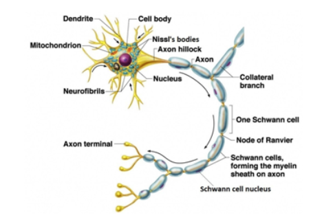
- A neuron has a large cell body with two or more thin protoplasmic processes extending from it. The cell body called cyton or perikaryon or soma is the part of a neuron that contains the nucleus and the cytoplasm. The nucleus is spherical and large with a prominent nucleus.
- The cytoplasm contains a rough endoplasmic reticulum and free ribosomes, which is a properly stained cell that appears as granular areas called Nissl bodies which are involved in the synthesis of proteins of the neurofibrils.
- The processes of the neuron are known as Neurites. One of the processes, called the axon, is long and conducts nerve impulses away from the cell body, thus, also known as the Efferent process. It ends in several small branches on muscle fibers, gland cells, or other neurons. The remaining processes (one or more) conduct nerve impulses towards the cell body and are called dendrites or dendrites or afferent processes.
- The plasma membrane and cytoplasm of an axon are known as Axolemma and Axoplasm respectively.
- The extended axon or dendrite of a neuron is called a nerve fiber. Nerve fibers are collected into bundles. Each nerve fiber is covered with a continuous sheath, called the neurolemma.
- The neurolemma sheath is made up of a single layer of flat expanded Schwann cells. Neurolemma is absent in the brain and spinal cord.
- The neurons are of five types based on the number of nerve processes:
- Apolar neurons: Neurons of hydra and amacrine cells of the retina have no definite cell process. These types of neurons are known as apolar neurons.
- Unipolar neurons:Which have only axon but no dendron and are found only in early embryos of invertebrates and vertebrates.
- Pseudo-unipolar neurons: resemble unipolar neurons because each of them has an axon, but no true dendrites. However, pseudo-unipolar neurons are variants of bipolar neurons.The reason for this is that the single axon attached to the cell body proceeds to two opposite "poles" or directions - one towards the muscle, joints, and skin, and the other towards the spinal cord. They are present in the dorsal root ganglion of the spinal cord.
- Bipolar neurons which have two processes, one axon, and another dendron, and are found in the olfactory epithelium and retina of the eye, vestibular and cochlear parts of the membranous labyrinth of the internal ear.
- Multipolar neurons, which have many processes arising from the cell body; out of them, one (longer) acts as an axon and the remaining as dendrons. Multipolar neurons are found in the brain and spinal cord.
- Neurons are classified into three types according to their function:
-(i) Sensory or afferent neurons conduct impulses from the sensory organs to the central nervous system. Most sensory neurons are unipolar in structure.
-(ii) Motor or efferent neurons conduct impulses from the central nervous system to the effector organs (muscles and glands). Most motor neurons are multipolar in structure.
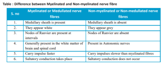
-(iii) Interneurons (connector/association neurons) connect sensory neurons to motor neurons. Interneurons integrate (process) incoming sensory information from sensory neurons and then elicit a motor response by activating the appropriate motor neurons. Most interneurons are multipolar in structure. - MyeIinated or meduIIated nerve fibres : Some nerve fibres are covered by a lipid-rich insulating layer, called the myelin sheath. Such nerve fibres are called myelinated or medullated nerve fibres. Each myelinated nerve fibre shows constrictions at regular intervals called nodes of Ranvier; these result from interruptions in the myelin sheath at those places. These are found in brain and spinal cord.When an impulse travels along the medullated nerve fibre, it does not proceed uniformly along the length but jumps from one node of Ranvier to the other. This type of conduction is known as Saltatory conduction.
- Non-myelinated or non-medullated nerve fibres: Some nerve fibres are not covered by myelin sheath. They are called non-myelinated or non-medullated nerve fibres. They do not possess nodes of Ranvier. Myelinated fibres are generally thicker than non-myelinated ones. These are found in the autonomic nervous system. It conducts message either to other neurons or to muscles and glands. The response elicited may be sensation such as pain or an action like muscle contraction or glandular secretion.
STRUCTURE OF NEURON
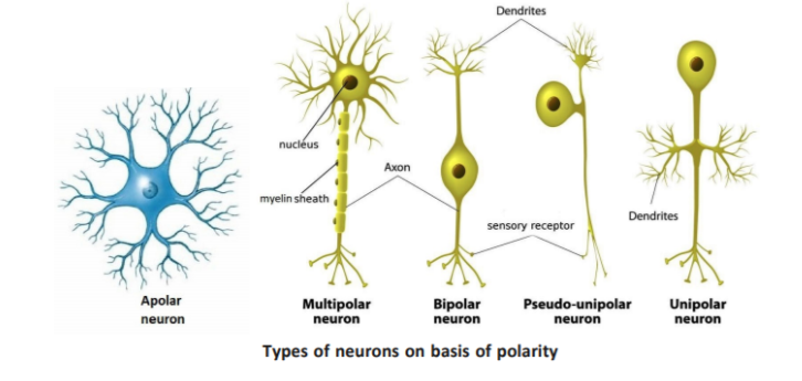
Classification of Neurons: Nerve fibres are of two types:
Muscular Tissue
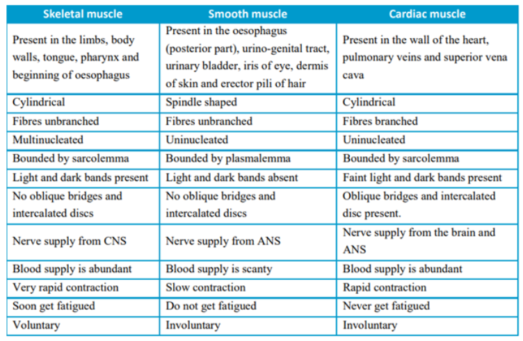
- Har ek muscle me bahut sare lambe lambe patle cylindrical fibres parallelly arranged hote hain. Ab har ek fibre me bahut sare myofibrils hote hain jo ki contractile proteins : myosin and actin se bante hai. Muscle contract and relax kar sakti hai. Kuch muscles ki contraction ko hum control kar sakte hain like sans lena ya na lena, bicep muscles, legs and hands ki movement ke liye responsible muscles vgarah, jinko hum voluntary muscles kehte hain. Jin muscle ko hum control nhi kar sakte unko involuntary muscle bolte hai. Generally muscles ke neeche diye gaye 3 types hote hai :
Differences between different muscles
| Skeletal Muscle | Smooth Muscle | Cardiac Muscles |
|---|---|---|
| Yea voluntary muscle hai jiski appearance striated hai yani ki microscope me dekhte hue isme dark and light colour se alternate bands dekhne ko milte hain. | involuntary muscle jisme straitions nahi hote. | Involuntary with striations. |
| Hatho or legs me milti hai jaise ki bicep muscles, triceps, hamstrings, calf muscles and back muscles. | Yea hume oesophagus ke posterior part me, blood vessels me, urino-genital tract and alimentary tract me milti hai. | Cardiac muscles milti hai keval heart tissue me along with pulmonary veins and superior vena cava me. |
| cylindrical shape | spindle shaped hoti hai jiske dono end middle portion ke comparison me patle and pointed hote hai. | cylindrical shape |
| Unbranched | Unbranched | Branched |
| Multinucleated | Uninucleated | Uninucleated |
| Oblique bridges and intercalated discs are absent | Oblique bridges and intercalated discs are absent | Oblique bridges and intercalated discs are present. |
| Nerve supply inhe CNS se milti hai. | Nerve supply inhe ANS se milti hai. | Nerve supply inhe ANS and brain se milti hai. |
| very rapid contractions but gets fatigued soon due to accumulation of lactic acid. | Slow contraction rate so doesn't get fatigued much | Rapid contraction and doesn't get fatigued till the life time. |
Cockroach
- Classification :
- During day time, they remain inactive and hiding and during the night, they show much activity and run here and there in search of food showing their nocturnal habit. Their narrow and flattened body is adapted to slip into narrow creeks and crevices.
- Four species of cockroaches found in India are :
- Blattella germanica (the German or Croton cockroach)
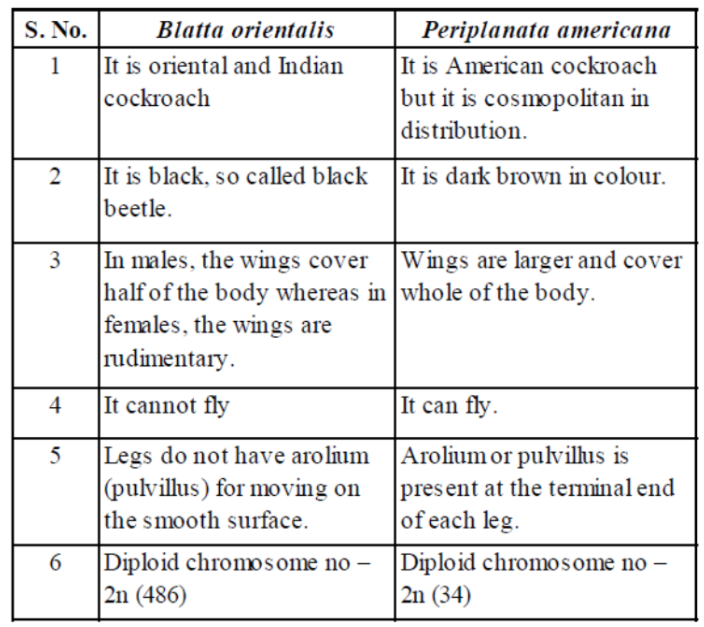
- Blatta orientalis (the Oriental or Indian Cockroach) : It is also called black beetle.
- Periplaneta americana (the American cockroach or ship cockroach) : the biggest cockroach
- Periplaneta australasiae (the Australian cockroach)
- Differences between Blatta orientalis and Periplaneta americana are as follows :
- Cockroach shows metameric segmentation.
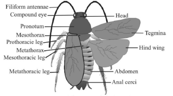
- The body of a cockroach is divisible into three parts – head, thorax and abdomen.
- Head
- Made by the fusion of 6 embryonic segments.
- Triangular and pear shaped.Head is covered by an exoskeleton called head capsule.
- It is made by six sclerites.
- Two compound eyes are present on dorso-lateral sides which are kidney shaped.
- At the base of the antennae, 2 white spots which function as photoreceptors are present which are known as fenestra or ocellar spots.
- Antennae are olfactory and tangoreceptors.
- Cockroach locates food with the help of antennae.
- Thorax
- Thorax is the middle part of the body and consists of three parts – prothorax, mesothorax and metathorax.
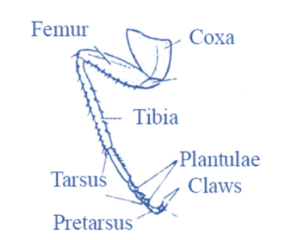
- 3 pairs of legs on the ventral side and two pairs of wings on the dorsolateral side.Each leg is made by 5 podomeres –
- The first podomere is coxa. It is broad and attached to the thorax. It is the most swollen segment.
- The second podomere is small triangular trochanter. It gives viability for leg movement. Trochanter is freely movable on coxa but fixed to the femur.
- The third podomere is femur. Femur is the strongest part of the leg. It shows bristles.
- The fourth podomere is tibia. Tibia is the longest part of the leg. It also bears bristles.
- The fifth podomere is tarsus. Tarsus contains 5 tarsomeres. The tarsomeres have plantulae on their ventral side. The last tarsomere is known as pretarsus which terminally contains a pair of claws. Between the two claws, a soft hairy pad is present called arolium or pulvillus. Arolium and plantulae serve as organs of attachment and provide a firm grip on smooth surfaces. When arolium and plantae are removed, cockroach cannot move on smooth surface.
- The first pair of wings arises from mesothorax and the second pair from metathorax. Forewings (mesothoracic), called tegmina or elytra, are opaque dark and leathery and cover the hind wings when at rest. The hind wings are transparent, membranous and are used in flight. They are folded like a fan and covered by elytra while at rest.
- Abdomen
- Broader than thorax and consists of 10 segments while embryo has 11 segments.
- 10th segment in both sexes bears a pair of small, filamentous and sensory anal cerci. Each anal cercus is made of 15 segment. In male, in addition of anal cerci, 9th sternum bears anal style (unjointed thread like) which are absent in females. In female cockroach, abdomen is broader than in male.
- The body of a cockroach is covered by chitinous exoskeleton. It is in the form of sclerites. The dorsal sclerite is called tergal plate. The ventral sclerite is called sternal plate. Two sclerites are connected by arthrodial membrane.
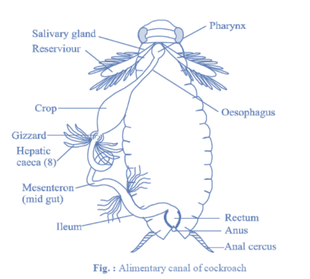
- Alimentary canal is divided into three regions – foregut, midgut and hindgut. Foregut includes mouth cavity, pharynx, oesophagus, crop & gizzard. Crop is the largest part of foregut & serve as a reservoir for storing food. Next segment is gizzard or proventriculus which has outer muscular layer and chitinous teeth on the internal side which help in grinding of the food. A ring of 6-8 blind tubules called hepatic or gastric caeca is present at the junction of foregut and midgut, which secrete digestive juice. Midgut (mesenteron or ventriculus) is short, tubular lined with glandular endoderm, so the function is to secrete digestive juices. The anterior part of midgut consists of 4 parts of hepatic caeca. Midgut forms the true stomach whose main functions are digestion and absorption. At the junction of midgut and hindgut is present another ring of 100-150 yellow coloured thin filamentous Malphigian tubules. They help in removal of excretory products from haemolymph. The rectum opens out through anus.
CIRCULATORY SYSTEM
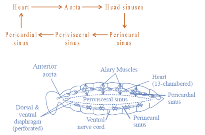
- Open type with poorly developed blood vessels which open into haemocoel and the fluid flowing through it is known as haemolymph which is basically colourless plasma + haemocytes. Neurogenic heart has 13 muscular chambers(with alary muscles at the junction of two chambers) with ostia on either side . Blood from sinuses enter the heart through ostia and is pumped anteriorly to sinuses again.
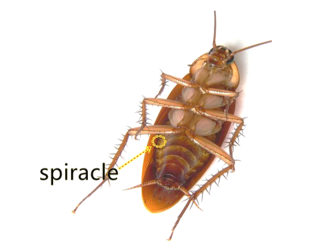
RESPIRATORY SYSTEM - The respiratory system of cockroach consists of a network of trachea, that open through 10 pairs of small holes called spiracles (two pairs thoracic and eight pairs of abdominal) present on the lateral side of the body. Thin branching tubes (tracheal tubes subdivided into tracheoles) carry oxygen from the air to all the parts. Opening of the spiracles is regulated by the sphincters. Exchange of gases : by diffusion. Muscles involved in tracheal ventilation are : abdominal muscles namely, tergo-sternal muscles.
EXCRETORY SYSTEM - Excretion is performed by Malpighian tubules. Uricotelic in nature. the fat body, nephrocytes and uricose glands also help in excretion.
- The nervous system of cockroach consists of a series of fused, segmentally arranged ganglia (total 9) joined by paired longitudinal connectives on the ventral side. Three ganglia lie in the thorax, and six in the abdomen.Central nervous system consists of cerebral or supraesophageal ganglion (brain), suboesophageal ganglion, paired circumesophageal connectives and double ventral nerve cord (with three thoracic and six abdominal compound segmental ganglia). Generally insects with compound eyes have two types of vision in insects – mosaic vision (or apposition image) during the day time and superposition (or dull image) in dim light but but in cockroach, pigment sheath of ommatidia is non-contractile so capable of only mosaic vision even during the night.
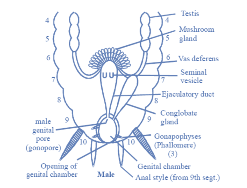
- Male organs consist of testes, vasa deferentia, ejaculatory duct, mushroom or utricular gland, phallic or conglobate gland and male gonapophysis.
- Testes of cockroach are located in the abdominal segments 4, 5 and 6. From each testis arises, a thin vas deferens, which opens into ejaculatory duct through seminal vesicle.
- The ejaculatory duct opens into male gonopore situated ventral to anus.
- A characteristic mushroom shaped gland is present in the 6th-7th abdominal segments which functions as an accessory reproductive glands.
- Mushroom gland consists of two types of tubules, the long slender tubules, the utriculi majores or peripheral tubules; and short tubules, the utriculi breviores, making up of the major part of the gland.
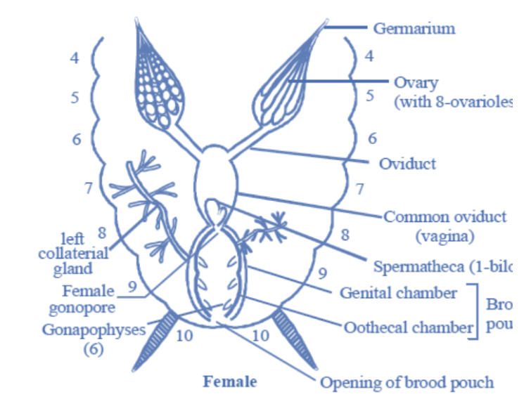
- The female reproductive system consists of two large ovaries, lying laterally in the 2nd – 6th abdominal segments.
- Each ovary is formed of a group of eight ovarian tubules or ovarioles, containing a chain of developing ova.
- A pair of spermatheca is present in the 6th segment which opens into the genital chamber and receives sperms from spermatophores. Their fertilized eggs are encased in capsules called ootheca. Ootheca is a dark reddish to blackish brown capsule, about 3/8" (8 mm) long.
- On an average, females produce 9-10 ootheca, each containing 14-16 eggs.
- The development of P. americana is paurometabolous, meaning there is development through nymphal stage. The nymphs look very much like adults. The nymph grows by moulting about 13 times to reach the adult form. The next to last nymphal stage has wing pads but only adult cockroaches have wings.
- Metamorphosis is regulated by two hormones ecdysone (secreted by prothoracic glands) and juvenile hormone or neotenin (secreted by corpora allata).
Phylum – Arthropoda
Class – Insecta
Subclass – Pterygota
Order – Orthoptera
Genus – Periplaneta
Species – americana
DIGESTIVE SYSTEM
NERVOUS SYSTEM
REPRODUCTIVE SYSTEM
Muscular Tissue
- Muscle tissue is composed of thin and elongated cells that are usually called muscle fibres. They are differentiated cells made up of contractile protein (aise proteins jinmei contract karne ki property ho ).
- Eg -higher animals - found associated with the skeleton, walls of visceral organs, blood vessels and heart, etc.
- Muscular tissue is mesodermal in origin.
- Muscles of the iris of the eye and myoepithelial cells of the salivary, mammary and sweat glands are ectodermal in origin.
- Each muscle is made of many long, cylindrical fibres arranged in parallel arrays. These fibres are composed of numerous fine fibrils, called myofibrils.
- Muscle fibres contract (shorten) in response to stimulation, then relax (lengthen) and return to their uncontracted state in a coordinated fashion. Their action moves the body to adjust to the changes in the environment and to maintain the positions of the various parts of the body. In general, muscles play an active role in all the movements of the body.
- Function of muscle tissue:
- Production of body movements: Movements of the whole body such as walking and running, and localized movements such as grasping a pencil or nodding the head as a result of muscular contractions, rely on the integrated functioning of skeletal muscles, bones, and joints.
- Stabilization of body positions: Skeletal muscle contractions stabilize joints and help maintain body positions, such as standing or sitting. Postural muscles contract continuously when you are awake; for example, sustained contractions of your neck muscles hold your head upright.
- Storage and movement of substances within the body: Storage is accomplished by sustained contractions of ring-like bands of smooth muscle called sphincters, which prevent outflow of the contents of a hollow organ. Eg : Temporary storage of food in stomach , storage of urine , cardiac muscle contraction for blood flow , movement of food in alimentary canal .Skeletal muscle contractions promote the flow of lymph and aid the return of blood to the heart.
- Generating heat:Heat is produced on the contraction of tissue which is called thermogenesis. This is used to maintain normal body temperature. Involuntary contractions of skeletal muscles, which is known as shivering, can increase the rate of heat production.
- According to their structure, location and functions there are three main types :
- Skeletal muscle
- Smooth muscle
- Cardiac muscle

- SKELETAL MUSCLE
- Skeletal muscles are attached to bones by tendons, and consist of bundles of very long (up to 30 cm), cylindrical, multinucleated (syncytial cells). These fibres (cells) can be contracted at desire; therefore, they are also called voluntary muscles.
- The plasma membrane covering each fibre is called sarcolemma. The cytoplasm inside the fibre is called sarcoplasm. The sarcoplasm contains many long, thin, unbranched cross striated cylindrical structures called myofibrils. Each muscle fibre or muscle cell also possesses many oval, flat nuclei at the periphery of the cell under the sarcolemma. They are arranged along the long axis of the fibre.
- Smooth muscle consists of a collection of spindle shaped cells (centre se thick or pointed ends par ) that do not show any striation. They may range in size from 20 m (in small blood vessels) to 500 m (in the pregnant uterus).
- Shorter than the striated fibres with single oval nucleus.
- NO sarcolemma , but fibers have cell membrane.
- The mitochondria are fewer in number, the sarcoplasmic reticulum is less extensive and protein filaments are not regularly arranged to give rise to striations. Their contraction process is slow and not under voluntary control. So they are also called involuntary muscles ( aise muscles jinhe hum apni marzi se control na kar sakein).
- Cell junctions hold them together and they are bundled together in a connective tissue sheath.
- Eg: The wall of internal organs such as the blood vessels, stomach and intestine.
- Cardiac muscle is restricted to the wall of the heart only. These are composed of elongated, branched individual cells that lie parallel to each other.
- Because they need a large amount of energy , they possess numerous mitochondria and glycogen granules.
- They are cross striated (inn par bhi skeletal muscle jaise strips present hain ) and have actin and myosin filaments.
- Cardiac muscle is involuntary, vigorous ,rhythmic and immune to fatigue ( kabhi thakti nahi h )
- Communication junctions or gap junctions (intercalated discs) allow the cells to contract as a unit, i.e., when one cell receives a signal to contract, its neighbours are also stimulated to contract.
SMOOTH MUSCLE
CARDIAC MUSCLE
Unlike multinucleated skeletal muscle, these cells possess only one centrally located nucleus, which lies deep at the centre of the fibre.
NOTE They have specialised junctional complexes at the ends of the adjacent muscle cells called intercalated discs (oval shape k disc).
Function:They hold the cells together ( jo rapid excitation spread mei help karta hai jab heart beat karta hai).

Summary
- Cells, tissues, organs and organ systems split up the work in a way that ensures the survival of the body as a whole and exhibit division of labour. A tissue is defined as group of cells along with intercellular substances performing one or more functions in the body. Epithelia are sheet like tissues lining the body’s surface and its cavities, ducts and tubes. Epithelia have one free surface facing a body fluid or the outside environment. Their cells are structurally and functionally connected at junctions.
- Diverse types of connective tissues bind together, support, strengthen, protect and insulate other tissue in the body. Soft connective tissues consist of protein fibres as well as a variety of cells arranged in a ground substance. Cartilage, bone, blood, and adipose tissue are specialised connective tissues. Cartilage and bone are both structural materials. Blood is a fluid tissue with transport functions. Adipose tissue is a reservoir of stored energy. Muscle tissue, which can contract (shorten) in response to stimulation, helps in movement of the body and specific body parts. Skeletal muscle is the muscle tissue attached to bones. Smooth muscle is a component of internal organs. Cardiac muscle makes up the contractile walls of the heart. Connective tissue covers all three types of tissues. Nervous tissue exerts greatest control over the response of body. Neurons are the basic units of nervous tissue.
- The body of Cockroach (Periplaneta americana) is covered by chitinous exoskeleton. It is divided into head, thorax and abdomen. Segments bear jointed appendages. There are three segments of thorax, each bearing a pair of walking legs. Two pairs of wings are present, one pair each on 2nd and 3rd segment. There are ten segments in abdomen. Alimentary canal is well developed with a mouth surrounded by mouth parts, a pharynx, oesophagus, crop, gizzard, midgut, hindgut and anus. Hepatic caecae are present at the junction of foregut and midgut. Malpighian tubules are present at the junction of midgut and hindgut and help in excretion. A pair of salivary gland is present near crop. The blood vascular system is of open type. Respiration takes place by network of tracheae. Trachea opens outside with spiracles. Nervous system is represented by segmentally arranged ganglia and ventral nerve cord. A pair of testes is present in 4th-6th segments and ovaries in 2nd-6th segments. Fertilisation is internal. Female produces 9-10 ootheca bearing developing embryos. After rupturing of single ootheca sixteen young ones, called nymphs come out.
Nervous Tissue
- Nervous tissue aisa tissue hai jo excite ho sakta hai and it produces and conducts electric current. Halakni current ki voltage bahut kam hoti hai but issi ke jariye nervous tissue ke cells yani ki neurons information ko conduct karte hain. Toh 2 main property nervous tissue ki hui : excitability and conductibility.
- Structure : Ek neuron ke 3 main part hai : soma/cell body, axon and axon terminals.
- Soma ya cell body me cytoplasm and nucleus hai or sath hi me kuch projections bahar ki taraf nikle hue hai jinko hum dendrites kehte hain. Dendrites baki ke neurons ke sath connection banate hai take ek neuron se information dusre ko conduct ki ja sake. Soma me kuch granules dikhte hain jinko hum nissl's granules kehte hain and unka main kaam hai protein synthesis ka.
- Axon basically ek elongated tube like structure hai jo cell body se originate hota hai and iski length kabhi kabhi meters tak bhi ho jati hai. Axon ke uper myelin sheath jo ki ek lipid rich insulating layer hai, wo present hoti hai toh myelinated nerve fibres keh dete hai otherwise non myelinated nerve fibers kehte hain.
- Nerve impulse myelinated nerve fibers me jyada speed se travel karti hai as compared to non myelinated neurons.
- Myelin sheath beech beech me absent hoti hai nerve fibre ya axon se toh uss jagah ko hum Nodes of Ranvier kehte hain. Yeah bhi nerve impulse conduction me help karti hai ab kaisey kya kyu wo sab human physiology ke last second chapter ka part hai. Uss pure chapter me hume bas nervous tissue hi padhna hai. Abhi toh bas yahaan overview dekh rahe hai neuron ka.
- Wo neurons jo body organs se information brain tak lekr jata hai jaise ki aapne kuch garam chua toh skin se neuron uss information ko brain ko dega ki kuch garam cheez hai. Aisi information ko sensory information kehte hai and jo neurons isko conduct karte hain usko hum sensory neurons kehte hai.
- Ab brain uss information ko process karke kuch command behjege muscle tissue ko jiske karan muscle contract karegi and aap apna hath uss garam cheez se hta loge. Yeah brain ne jo muscle tissue ko information di usko hum motor information kehte hain and jo neuron isko conduct krte hai usko motor neuron kehte hain.
- Last part axon ka branched hota hai jiski ending mein bulb like structures hote hain. Yeah part nerve ending kehlata hai. And yhi wo part hai jo next neuron ke dendrites ke sath ek connection establish karega jisko synapse bolte hain. Ab axon terminals ke through information kuch chemical messengers jinko hum neurotransmitters kehte hai wo synapse se travel hokr next neuron ko information dete hai ki ab us neuron ko koi specific function karna hai ya kisi dusre neuron ko information pass karni hai. Aise hi pure body me connection and circuits bane hue hain.
Nervous Tissue
- Composed of specialized cells capable of reacting to external and internal stimuli and conducting impulses to various organs in the body.
- Makes up the various organs of the nervous system namely the brain, the spinal cord and peripheral nerves.
- Neurons provide most of the unique functions of the nervous system, such as sensing, thinking, remembering, controlling muscle activity, and regulating glandular secretions.
- Neuroglia support, nourish, and protect the neurons and maintain homeostasis in the interstitial fluid that bathes them.
- Properties of neural tissue
- Excitability: It is the ability to initiate nerve impulse in response to stimuli.
- Conductivity: It is the ability to transmit a nerve impulse.
- A neuron has a large cell body with two or more thin protoplasmic processes extending from it. The cell body called cyton or perikaryon or soma is the part of a neuron that contains the nucleus and the cytoplasm. The nucleus is spherical and large with a prominent nucleus.
- The cytoplasm contains a rough endoplasmic reticulum and free ribosomes, which is a properly stained cell that appears as granular areas called Nissl bodies which are involved in the synthesis of proteins of the neurofibrils.
- The processes of the neuron are known as Neurites. One of the processes, called the axon, is long and conducts nerve impulses away from the cell body, thus, also known as the Efferent process. It ends in several small branches on muscle fibers, gland cells, or other neurons. The remaining processes (one or more) conduct nerve impulses towards the cell body and are called dendrites or dendrites or afferent processes.
- The plasma membrane and cytoplasm of an axon are known as Axolemma and Axoplasm respectively.
- The extended axon or dendrite of a neuron is called a nerve fiber. Nerve fibers are collected into bundles. Each nerve fiber is covered with a continuous sheath, called the neurolemma.
- The neurolemma sheath is made up of a single layer of flat expanded Schwann cells. Neurolemma is absent in the brain and spinal cord.
- The neurons are of five types based on the number of nerve processes:
- Apolar neurons: Neurons of hydra and amacrine cells of the retina have no definite cell process. These types of neurons are known as apolar neurons.
- Unipolar neurons:Which have only axon but no dendron and are found only in early embryos of invertebrates and vertebrates.
- Pseudo-unipolar neurons: resemble unipolar neurons because each of them has an axon, but no true dendrites. However, pseudo-unipolar neurons are variants of bipolar neurons.The reason for this is that the single axon attached to the cell body proceeds to two opposite "poles" or directions - one towards the muscle, joints, and skin, and the other towards the spinal cord. They are present in the dorsal root ganglion of the spinal cord.
- Bipolar neurons which have two processes, one axon, and another dendron, and are found in the olfactory epithelium and retina of the eye, vestibular and cochlear parts of the membranous labyrinth of the internal ear.
- Multipolar neurons, which have many processes arising from the cell body; out of them, one (longer) acts as an axon and the remaining as dendrons. Multipolar neurons are found in the brain and spinal cord.
- Neurons are classified into three types according to their function:
-(i) Sensory or afferent neurons conduct impulses from the sensory organs to the central nervous system. Most sensory neurons are unipolar in structure.
-(ii) Motor or efferent neurons conduct impulses from the central nervous system to the effector organs (muscles and glands). Most motor neurons are multipolar in structure.
-(iii) Interneurons (connector/association neurons) connect sensory neurons to motor neurons. Interneurons integrate (process) incoming sensory information from sensory neurons and then elicit a motor response by activating the appropriate motor neurons. Most interneurons are multipolar in structure. - MyeIinated or meduIIated nerve fibres : Some nerve fibres are covered by a lipid-rich insulating layer, called the myelin sheath. Such nerve fibres are called myelinated or medullated nerve fibres. Each myelinated nerve fibre shows constrictions at regular intervals called nodes of Ranvier; these result from interruptions in the myelin sheath at those places. These are found in brain and spinal cord.When an impulse travels along the medullated nerve fibre, it does not proceed uniformly along the length but jumps from one node of Ranvier to the other. This type of conduction is known as Saltatory conduction.
- Non-myelinated or non-medullated nerve fibres: Some nerve fibres are not covered by myelin sheath. They are called non-myelinated or non-medullated nerve fibres. They do not possess nodes of Ranvier. Myelinated fibres are generally thicker than non-myelinated ones. These are found in the autonomic nervous system. It conducts message either to other neurons or to muscles and glands. The response elicited may be sensation such as pain or an action like muscle contraction or glandular secretion.
STRUCTURE OF NEURON


Classification of Neurons: Nerve fibres are of two types:

Cockroach
- Cockroach dekha toh sabne haina? Right. Ab iss chapter me isko detail me padhna hai. Toh classification se shuru karte hain ki bhyii cockroach ka kingdom ? Animalia. Phylum arthropoda hai, class insects, order orthoptera, genus periplaneta and species hai americana. Normal colour adult ka dark brown hota hai. Kuch species udd sakti hai and kuch nahi. India me jo cockroach dekhne ko milta hai wo hai Blatta orientalis jo udd ni sakta.
- Segmented body hai jo 3 segments : head, thorax and abdomen me divided hoti hai.
- Pura shareer inka exoskeleton se covered hai jo chitinous plates jinko sclerites kehte hai unse bni hui hai. Inhi plates ko dorsal surface pe tergites kehte hai and ventral surface ki plates ko sternites kehte hai. Yeah plates ek dusre se ek patli or flexible membrane se joined hoti hai jisko arthrodial membrane kehte hai.
- Triangular shape ka head hai jo body ki longitudinal axis ke 90 degrees par situated hai. Yeah 6 segments ke fusion se bnta hai and neck kafi flexible hoti hai. Head me compound eyes ka ek pair hoti hai jinse mosaic vision me inko cheezein dikhti hain. Antennae sensory structures hote hain jinke karan cockroach apna khana vgarah dhundte hain. Mouth parts inke biting and chewing type ke hai. Although details itni NCERT me nhi hai although neeche wale section me given hai jo ki keval unhi students ke liye hai jinko aage competitive exams ki preparation karni hai ya jinke respective Board me padhaya jata hai.
- Thorax ko 3 parts me divide kar sakte hai : prothorax, mesothorax and metathorax. Neck head ko prothorax se connect karti hai. Har thoracic segment me legs ka ek pair hota hai. Wings ke 2 pair hote hai. Pehla pair jinko forewings kehte hain wo mesothorax se arise hote hain and tegmina kehlate hai. Yeah dark brown colour ke opaque non functional wings hai jo rest ki situation me hind wings ko cover karte hain. Transparent, Membranous wings jo udaan me help karte hai wo metathorax se arise hote hain.
- Males and females dono ke abdomen me 10 segments hote hain. Halanki difference yeah hai ki females me 7th sternum boat shaped hote hain and aathve or novey sternum ke sath milkar yeah brood pouch ya genital pouch form karte hain jiske anterior part me yani ki head ki taraf gonopore, spermathecal pores and collateral glands hote hain. Males me genital pouch abdomen ke hind part me hota hai jo ki dorsally ninth and tenth terga and ventrally ninth sternum se covered hote hain. Jisme dorsal anus, ventral surface pe male genital pore and gonapophysis hote hain. Males me anal styles hote hai jo ki females me absent hoti hai. Dono sexes me tenth segment me anal cerci present hoti hai.
- Alimentary canal 3 parts me divided hai : foregut, midgut and hind gut. Mouth pharynx me open hota hai jo ki oesophagus me open hoti hai. Oesophagus aage chal kar crop me khulta hai. Uska next part hai gizzard ya proventriculus jo khana grind karne me help karta hai. Hepatic caeca ya gastric caeca hoti hai foregut and midgut ke junction pe jo ki gastric juice secrete karti hai. Midgut or hindgut ke junction par malpighian tubules hoti hai wo excretion me help karti hai. Hindgut ke 3 part hai : ileum, colon and rectum and rectum anus ke through bahar ke environment me open hoti hai taki excreta ko bahar nikal sake.
- Circulatory system open type ka hai, koi respiratory pigment nhi hota, neurogenic heart hai jiske 13 chambers hote hai. Blood ko haemolymph bola jata hai. Heart se haemolymph jata hai aorta me, aorta se head sinuses me, vhan se perineural sinuses me, then to perivisceral sinuses, fir jayega pericardial sinus se and vhan se blood/hemolymph fir heart me enter karke yahi pura cycle dohraye ga.
- Respiratory system trachea ka network hai jo ki sharir ke bahar spiracales ke jariye khulte hai. Spiracles 10 pair hote hai and yeah lateral side pe khulte hai. Trachea fir tracheal tubes me khulti hai fir wo tracheoles me subdivided hoti hai and sare parts tak aise oxygen pahuchti hai. Sphincters aisi muscles hoti hai jo spiracles ki opening and closing ko regulate hoti hai.
- Excretion toh bta hi dia ki Malpighian tubules se hota hai jo nitrogenous waste ko uric acid me convert karke excrete karti hai isiliye cockroaches ko uricotelic kaha jata hai.
- Nervous system. Segmentally arranged ganglia hai jo ki paired longitudinal connectives se joined hote hain on the ventral side. 3 ganglia thorax me, 6 abdomen me toh 9 ganglia hogye. Head me thoda bahut part hai nervous system ka baki ka rest of the body me, isiliye jab aap cockroach ka keval head separate kardenge toh bhi cockroach ki death ekdum nhi hogi, wo death uski dheere dheere starvation ke karan hogi kyunki bina head khayega kaise? Right.
- Male ke case me testes ka ek pair hota hai jo 4-6 segment ki lateral side pe situated hai. From each testis arises a thin vas deferens, which opens into the ejaculatory duct through a seminal vesicle. The ejaculatory duct opens into male gonopore situated ventral to anus. Ek unique feature males ka yeah hai ki inki abdomen ki sixth-seventh segment me ek mushroom gland hota hai jo as an accessory reproductive gland function karta hai. Asymmetrical chitinous structures jinko phallomere kehte hai wo phallus ko represent karte hain.
- Female me ovaries, 2-6 segment ki lateral sides pe hoti hai. Har ek ovary 8 ovarioles se bnti hai jinme developing ova ki ek chain present hoti hai. Dono ovaries se aane wali oviducts ek single median oviduct me fuse karti hai jisko hum vagina kehte hain. Spermatheca sixth segment me present hota hai.
- Spermatophores ke jariye sperms transfer hote hain. Fertilised eggs ko capsules jinhe ootheca bola jata hai, unse cover kar dia jata hai. Ootheca dark red se blackish brown colour ka 8mm ka cover hai jo ki fertilised egg ke charo taraf present hota hai. Inhi encased eggs ko ek jagah pe drop kr dete hain.
- Aamtaur par ek female, 9-10 oothecae produce karti hai and har ootheca me 14-16 eggs hote hain.
- Development is paurometabolous type yani ki nymph stage involved hai. Nymph 13 baar moulting kar karke finally adult cockroach me badal jata hai. But yaad rakhna ki keval adult cockroach ke pass hi wings hote hain.
Anatomy
Cockroach
- Classification :
- During day time, they remain inactive and hiding and during the night, they show much activity and run here and there in search of food showing their nocturnal habit. Their narrow and flattened body is adapted to slip into narrow creeks and crevices.
- Four species of cockroaches found in India are :
- Blattella germanica (the German or Croton cockroach)
- Blatta orientalis (the Oriental or Indian Cockroach) : It is also called black beetle.
- Periplaneta americana (the American cockroach or ship cockroach) : the biggest cockroach
- Periplaneta australasiae (the Australian cockroach)
- Differences between Blatta orientalis and Periplaneta americana are as follows :
- Cockroach shows metameric segmentation.
- The body of a cockroach is divisible into three parts – head, thorax and abdomen.
- Head
- Made by the fusion of 6 embryonic segments.
- Triangular and pear shaped.Head is covered by an exoskeleton called head capsule.
- It is made by six sclerites.
- Two compound eyes are present on dorso-lateral sides which are kidney shaped.
- At the base of the antennae, 2 white spots which function as photoreceptors are present which are known as fenestra or ocellar spots.
- Antennae are olfactory and tangoreceptors.
- Cockroach locates food with the help of antennae.
- Thorax
- Thorax is the middle part of the body and consists of three parts – prothorax, mesothorax and metathorax.
- 3 pairs of legs on the ventral side and two pairs of wings on the dorsolateral side.Each leg is made by 5 podomeres –
- The first podomere is coxa. It is broad and attached to the thorax. It is the most swollen segment.
- The second podomere is small triangular trochanter. It gives viability for leg movement. Trochanter is freely movable on coxa but fixed to the femur.
- The third podomere is femur. Femur is the strongest part of the leg. It shows bristles.
- The fourth podomere is tibia. Tibia is the longest part of the leg. It also bears bristles.
- The fifth podomere is tarsus. Tarsus contains 5 tarsomeres. The tarsomeres have plantulae on their ventral side. The last tarsomere is known as pretarsus which terminally contains a pair of claws. Between the two claws, a soft hairy pad is present called arolium or pulvillus. Arolium and plantulae serve as organs of attachment and provide a firm grip on smooth surfaces. When arolium and plantae are removed, cockroach cannot move on smooth surface.
- The first pair of wings arises from mesothorax and the second pair from metathorax. Forewings (mesothoracic), called tegmina or elytra, are opaque dark and leathery and cover the hind wings when at rest. The hind wings are transparent, membranous and are used in flight. They are folded like a fan and covered by elytra while at rest.
- Abdomen
- Broader than thorax and consists of 10 segments while embryo has 11 segments.
- 10th segment in both sexes bears a pair of small, filamentous and sensory anal cerci. Each anal cercus is made of 15 segment. In male, in addition of anal cerci, 9th sternum bears anal style (unjointed thread like) which are absent in females. In female cockroach, abdomen is broader than in male.
- The body of a cockroach is covered by chitinous exoskeleton. It is in the form of sclerites. The dorsal sclerite is called tergal plate. The ventral sclerite is called sternal plate. Two sclerites are connected by arthrodial membrane.
- Alimentary canal is divided into three regions – foregut, midgut and hindgut. Foregut includes mouth cavity, pharynx, oesophagus, crop & gizzard. Crop is the largest part of foregut & serve as a reservoir for storing food. Next segment is gizzard or proventriculus which has outer muscular layer and chitinous teeth on the internal side which help in grinding of the food. A ring of 6-8 blind tubules called hepatic or gastric caeca is present at the junction of foregut and midgut, which secrete digestive juice. Midgut (mesenteron or ventriculus) is short, tubular lined with glandular endoderm, so the function is to secrete digestive juices. The anterior part of midgut consists of 4 parts of hepatic caeca. Midgut forms the true stomach whose main functions are digestion and absorption. At the junction of midgut and hindgut is present another ring of 100-150 yellow coloured thin filamentous Malphigian tubules. They help in removal of excretory products from haemolymph. The rectum opens out through anus.
CIRCULATORY SYSTEM
- Open type with poorly developed blood vessels which open into haemocoel and the fluid flowing through it is known as haemolymph which is basically colourless plasma + haemocytes. Neurogenic heart has 13 muscular chambers(with alary muscles at the junction of two chambers) with ostia on either side . Blood from sinuses enter the heart through ostia and is pumped anteriorly to sinuses again.
RESPIRATORY SYSTEM - The respiratory system of cockroach consists of a network of trachea, that open through 10 pairs of small holes called spiracles (two pairs thoracic and eight pairs of abdominal) present on the lateral side of the body. Thin branching tubes (tracheal tubes subdivided into tracheoles) carry oxygen from the air to all the parts. Opening of the spiracles is regulated by the sphincters. Exchange of gases : by diffusion. Muscles involved in tracheal ventilation are : abdominal muscles namely, tergo-sternal muscles.
EXCRETORY SYSTEM - Excretion is performed by Malpighian tubules. Uricotelic in nature. the fat body, nephrocytes and uricose glands also help in excretion.
- The nervous system of cockroach consists of a series of fused, segmentally arranged ganglia (total 9) joined by paired longitudinal connectives on the ventral side. Three ganglia lie in the thorax, and six in the abdomen.Central nervous system consists of cerebral or supraesophageal ganglion (brain), suboesophageal ganglion, paired circumesophageal connectives and double ventral nerve cord (with three thoracic and six abdominal compound segmental ganglia). Generally insects with compound eyes have two types of vision in insects – mosaic vision (or apposition image) during the day time and superposition (or dull image) in dim light but but in cockroach, pigment sheath of ommatidia is non-contractile so capable of only mosaic vision even during the night.
- Male organs consist of testes, vasa deferentia, ejaculatory duct, mushroom or utricular gland, phallic or conglobate gland and male gonapophysis.
- Testes of cockroach are located in the abdominal segments 4, 5 and 6. From each testis arises, a thin vas deferens, which opens into ejaculatory duct through seminal vesicle.
- The ejaculatory duct opens into male gonopore situated ventral to anus.
- A characteristic mushroom shaped gland is present in the 6th-7th abdominal segments which functions as an accessory reproductive glands.
- Mushroom gland consists of two types of tubules, the long slender tubules, the utriculi majores or peripheral tubules; and short tubules, the utriculi breviores, making up of the major part of the gland.
- The female reproductive system consists of two large ovaries, lying laterally in the 2nd – 6th abdominal segments.
- Each ovary is formed of a group of eight ovarian tubules or ovarioles, containing a chain of developing ova.
- A pair of spermatheca is present in the 6th segment which opens into the genital chamber and receives sperms from spermatophores. Their fertilized eggs are encased in capsules called ootheca. Ootheca is a dark reddish to blackish brown capsule, about 3/8" (8 mm) long.
- On an average, females produce 9-10 ootheca, each containing 14-16 eggs.
- The development of P. americana is paurometabolous, meaning there is development through nymphal stage. The nymphs look very much like adults. The nymph grows by moulting about 13 times to reach the adult form. The next to last nymphal stage has wing pads but only adult cockroaches have wings.
- Metamorphosis is regulated by two hormones ecdysone (secreted by prothoracic glands) and juvenile hormone or neotenin (secreted by corpora allata).
Phylum – Arthropoda
Class – Insecta
Subclass – Pterygota
Order – Orthoptera
Genus – Periplaneta
Species – americana

DIGESTIVE SYSTEM
NERVOUS SYSTEM
REPRODUCTIVE SYSTEM


Summary
- Cells, tissues, organs and organ systems split up the work in a way that ensures the survival of the body as a whole and exhibit division of labour. A tissue is defined as group of cells along with intercellular substances performing one or more functions in the body. Epithelia are sheet like tissues lining the body’s surface and its cavities, ducts and tubes. Epithelia have one free surface facing a body fluid or the outside environment. Their cells are structurally and functionally connected at junctions.
- Diverse types of connective tissues bind together, support, strengthen, protect and insulate other tissue in the body. Soft connective tissues consist of protein fibres as well as a variety of cells arranged in a ground substance. Cartilage, bone, blood, and adipose tissue are specialised connective tissues. Cartilage and bone are both structural materials. Blood is a fluid tissue with transport functions. Adipose tissue is a reservoir of stored energy. Muscle tissue, which can contract (shorten) in response to stimulation, helps in movement of the body and specific body parts. Skeletal muscle is the muscle tissue attached to bones. Smooth muscle is a component of internal organs. Cardiac muscle makes up the contractile walls of the heart. Connective tissue covers all three types of tissues. Nervous tissue exerts greatest control over the response of body. Neurons are the basic units of nervous tissue.
- The body of Cockroach (Periplaneta americana) is covered by chitinous exoskeleton. It is divided into head, thorax and abdomen. Segments bear jointed appendages. There are three segments of thorax, each bearing a pair of walking legs. Two pairs of wings are present, one pair each on 2nd and 3rd segment. There are ten segments in abdomen. Alimentary canal is well developed with a mouth surrounded by mouth parts, a pharynx, oesophagus, crop, gizzard, midgut, hindgut and anus. Hepatic caecae are present at the junction of foregut and midgut. Malpighian tubules are present at the junction of midgut and hindgut and help in excretion. A pair of salivary gland is present near crop. The blood vascular system is of open type. Respiration takes place by network of tracheae. Trachea opens outside with spiracles. Nervous system is represented by segmentally arranged ganglia and ventral nerve cord. A pair of testes is present in 4th-6th segments and ovaries in 2nd-6th segments. Fertilisation is internal. Female produces 9-10 ootheca bearing developing embryos. After rupturing of single ootheca sixteen young ones, called nymphs come out.

