BIOTECHNOLOGY : PRINCIPLES AND PROCESSES for Class 12 BIOLOGY
INTRODUCTION
- This is a whole new unit in its own and very important in current days. Biotechnology basically means using techniques so that we can use live organisms or their products for the welfare of human beings. The very basic form of using microbes to prepare curd, bread, wine etc can be viewed as a basic form of technology but biotechnology in its true potential can do much much more. The current ongoing research to synthesize corona vaccine or new treatments for cancer are all possible only because of biotechnology. Biotechnology is what has made biology much more diverse and useful to us in the past years so in this chapter we will study about different techniques of how and what to use and we will see what biotechnology is. In the next chapter we will study the applications of all the processes and techniques that we will cover in this chapter.
CHAPTER WEIGHTAGE
- The whole biotechnology unit comprises two chapters which are incomplete without each other and the unit carries 10 marks in the final exam. Important topics include: Cloning Vector, Restriction Enzyme, Recombinant Techology Process, Bioreactors and Downstream Processing.
CHAPTER WEIGHTAGE
- The whole biotechnology unit comprises two chapters which are incomplete without each other and the unit carries 12 marks in the exam. Important topics include: Cloning Vector, Restriction Enzyme, Recombinant Techology Process, Bioreactors and Downstream Processing.
CHAPTER WEIGHTAGE
- The whole biotechnology unit comprises two chapters which are incomplete without each other and the unit carries 12 marks in the exam. Important topics include: Cloning Vector, Restriction Enzyme, Recombinant Techology Process, Bioreactors and Downstream Processing.
BACKGROUND & BASICS
BACKGROUND TERMS
- Old/Traditional Biotechnology
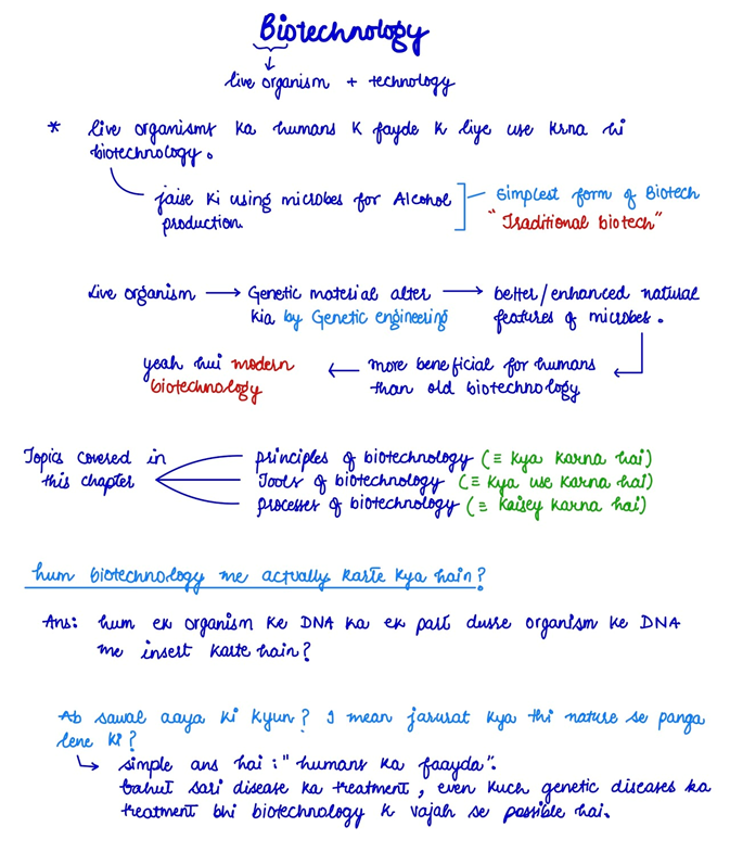
- The basic use of microorganism to produce citric acid or penicillin or wine, curd etc. are obtained from natural strains of microorganisms and often the yield is low. This use of microorganism in their natural form without any genetic modification comes under old biotechnology.
- The use of microorganism beyond their natural capabilities like production of human insulin from E.coli bacteria (which we will study in this unit later on) or increased production of antibiotics from microorganisms after their genetic modifications or using genetic engineering for green revolution or to produce golden rice, all comes under modern biotechnology where some kind of genetic modification is involved to enhance the natural capabilities of these microorganisms.
- Biotechnology is the integrated use of biochemistry, microbiology and engineering sciences in order to achieve technological or industrial application of the capabilities of organisms, cultured tissues/cells and parts thereof.
Modern Biotechnology
Definition Of Biotechnology
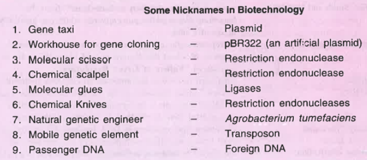
The image below shows some nicknames that are quite common and we will also be using them through our unit so as of now we just need to memorise them and slowly they’ll start to make some sense.

PRINCIPLES OF BIOTECHNOLOGY
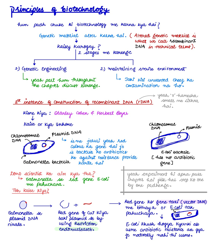
- Genetic engineering : the technique to alter the genetic material (DNA/RNA) and maintenance of sterile environment : to provide an environment free from microbial contamination to alter genetic material.
- In genetic engineering the genetic material of an organism is altered which means either some genes are added or deleted or both and this new altered DNA is what is known as recombinant DNA .
- The new gene or DNA to be inserted into genetic material of other organism is known as gene of interest which is always inserted near the origin of replication or ori point because the newly inserted segment need to be replicate and multiply itself so that this gene of interest can maintain itself in the progenies. This multiplication of gene of interest in the host organism is known as gene cloning
- In 1972 Stanley Cohen & Herbert Boyer isolated an antibiotic resistance gene segment from plasmid of Salmonella typhimurium by using restriction enzymes (or molecular scissors) and this gene segment coded for an antibiotic resistance protein. They then ligated (joined) this gene segment into the plasmid DNA of Salmonella typhimurium. This plasmid DNA now has a foreign gene segment which was naturally not its own so this plasmid DNA along with the antibiotic resistance gene is now known as a rDNA.
- This plasmid will now be used to transfer this antibiotic gene to another bacteria like Escherichia coli so that E.coli which originally did not have this antibiotic resistance gene, now has it. In this process the plasmid DNA from Salmonella is used to carry that antibiotic resistance gene to E.coli that is why the plasmid DNA is generally known as gene taxi or vector in technical terms.
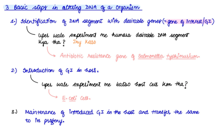
- Identification of DNA with desirable genes or identification of gene of interest (in above case that would be antibiotic resistance gene)
- Introduction of the gene of interest into the host (in above case that would be transferring the antibiotic resistance gene into E.coli using plasmid DNA of Salmonella).
- Maintenance of introduced DNA in the host and transfer of the DNA to its progeny (this basically means that we introduced our gene of interest in plasmid near to the ori point and then antibiotic resistance would replicate itself using DNA polymerase of the E.coli bacteria).
are the two basic techniques and the very core of modern biotechnology. These techniques made modern biotechnology modern in a true sense.
These were the basic terms and before advancing further make sure that you know about terms like, gene transfer, gene cloning, rDNA/recombinant DNA, ori point and some basics from the genetics unit like DNA multiplication, transcription etc.
First Instance Of Construction Of An Artificial rDNA Molecule
So, there are 3 basic steps that happened above in modifying E.coli bacteria are :
PRINCIPLES OF BIOTECHNOLOGY
TOOLS OF rDNA TECHNOLOGY
- Just like any other technology, biotechnology also requires its own tools to successfully alter the genome of a cell. At the very basic core of biotechnology we have three basic tools :
- Enzymes : like restriction enzymes, polymerase enzymes and ligase enzymes etc.
- Vectors : to carry our gene of interest into the host cell.
- Competent host : a host is the organism in which we insert our gene of interest. But looking from the host point of view, that gene of interest is a foreign molecule and the host won’t take it up willingly so we have to make our host able to pick up the vector (along with gene of interest) and this willing host which can take up that foreign gene of interest is known as a competent host.
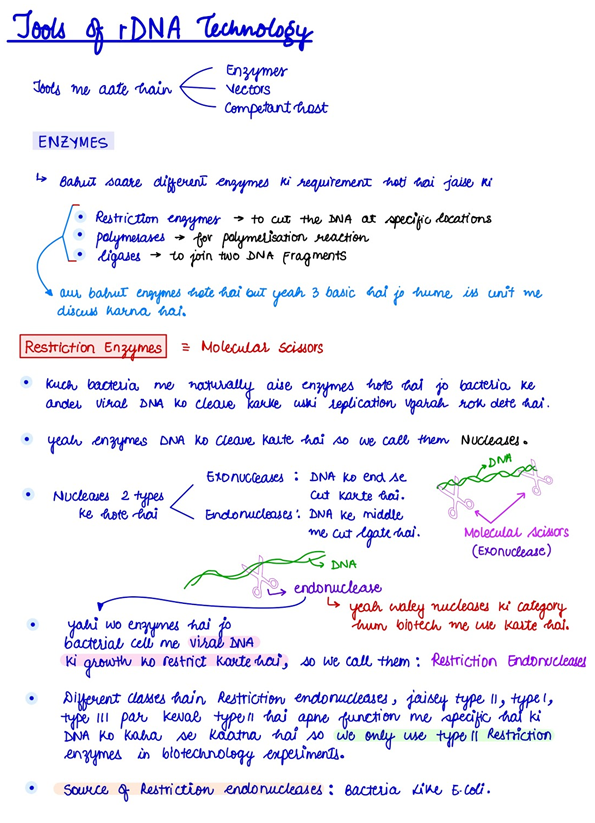
Now, we will discuss each of the three tools one by one in detail. - These enzymes are used to cut the DNA and different types of restriction enzymes are used to cut DNA at specific locations. The enzymes that cut or break the DNA are known as nucleases because they degrade the DNA(nucleic acid), hence the name nuclease.
- These enzymes can either cut the DNA from the middle and in that case they are known as endonuclease or they can cut the DNA from either of the termini/ends and in that case they will be known as exonucleases.
- Now these endonucleases were naturally found in some microbes like E.coli where they degrade the viral DNA to restrict the growth of viral DNA. Due to their ability to restrict the growth of viral DNA, these specific endonucleases which recognized and cut a specific sequence of 6-8 base pairs in the viral DNA were named as Restriction Endonucleases which we use in rDNA technology.
- Exonucleases are not used because they tend to degrade the DNA and render it non useful to us. While restriction endonucleases are of different types : some are more specific as to where they make a cut in the DNA, some are less accurate and some cut the DNA at a random site. So the class of restriction endonucleases that we use in the rDNA technology are the Type II Restriction Endonucleasewhich have a high accuracy of cutting the DNA at a given specific site.
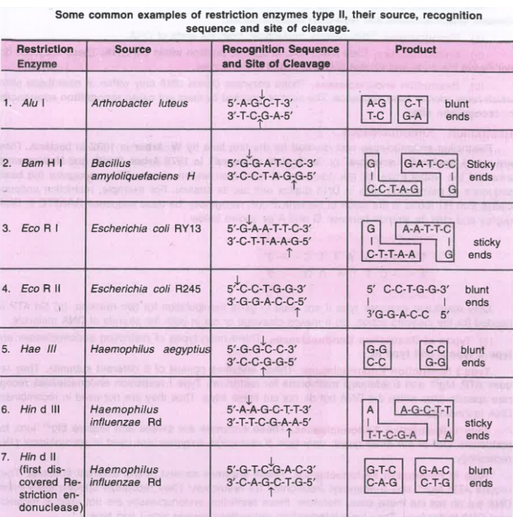
- Hind II was the first restriction enzyme and besides it we have isolated over 900 restriction enzymes from over 230 strains of bacteria and each of which recognize different sites.
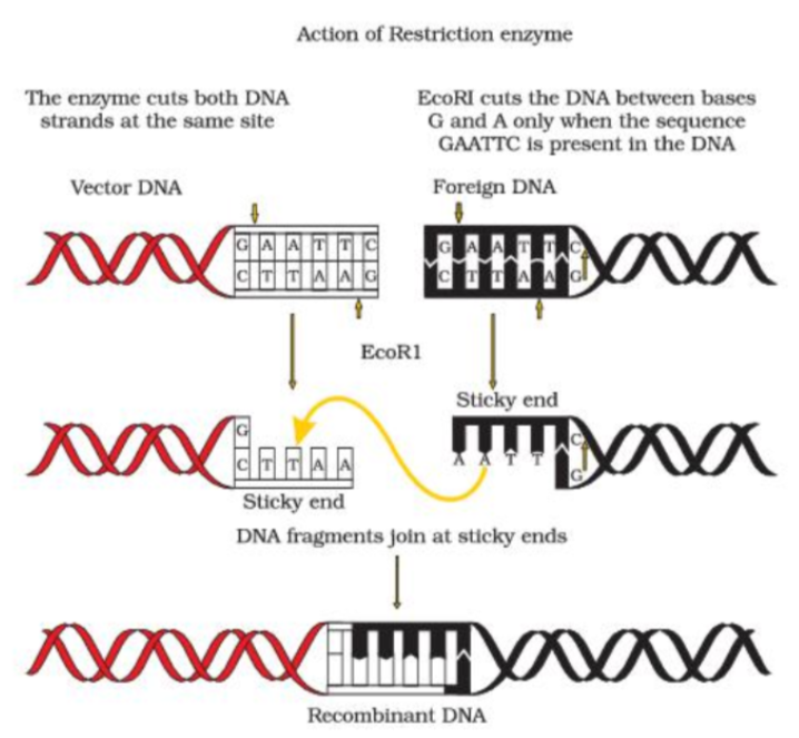
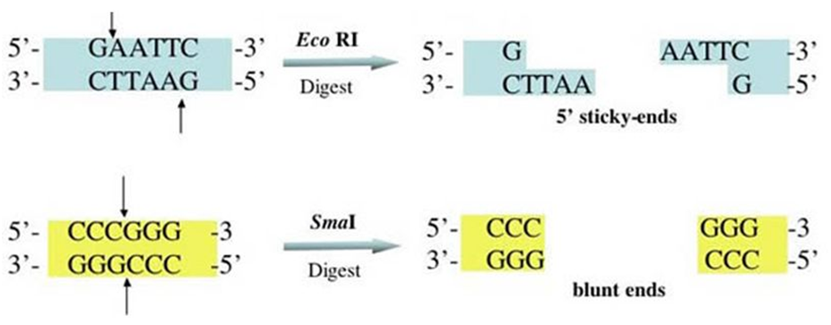
- Now each sequence recognized by RE is a palindromic sequence which means that the sequence will read the same from both sides like the word ‘MALAYALAM’. For example the RE EcoRI will always cut the DNA between the bases G and A only when the sequence GAATTC is present in the DNA.
- The first letter of the name of RE comes from the genus and is written in capital. The next two letters come from the species of the microbe from which that RE was isolated and written in small. The next capital word comes from the strain of the microbe and after that we write the roman number indicating the order in which the enzymes were isolated from the strain of bacteria.
- For example the EcoRI comes from Escherichia coli RY 13. E from genus, co from species, R from strain (which is RY in this case ) and I represents that it was the first RE to be isolated from this particular strain of E.coli
- The above picture shows the sequence GAATTC recognized by the EcoRI enzyme and this RE will always cut in between G and A of the sequence (which you can see in second part of this image)thus leaving a overhang/stick end. This sticky end helps in easy ligation of red and black DNA segments which represents the vector and foreign DNA respectively.
- Now there are some other REs which produce blunt ends after cutting the DNA like shown in the picture below this where the enzyme SmaIalways cuts in between the G and C base pair of the recognition sequence. Recognition sequence in the part highlighted in yellow and blue colour while the cut is made either in exactly in the middle of the sequence like by SmaI thus producing the blunt ends or the cut is made a little away from so that a overhang is left like in the case of EcoRI an overhang/sticky end is left at the 5’ end of the sequence.
- These sticky ends will be joined easily by the enzyme : DNA ligase. Ligation of blunt ends is a bit tricky but anyhow it is not a part of our discussion at this point.
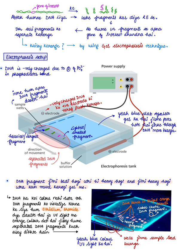
- We know that DNA is negatively charged due to the negative charge on phosphate bonds. So under the influence of the electric field the DNA fragments will move from the negative electrode to the positive electrode because the current from the negative electrode will repel the negatively charged DNA towards the positive electrode. So the DNA will move from negative to positive electrode in a media of agarose gel.
- Agarose gel has pores which offer resistance to the free movement of the DNA fragments. DNA fragments will move through these pores towards the positive electrode but this movement will depend on the size of the fragment.
- Higher the number of base pairs in a fragment, more will be its weight and it will travel less distance towards the positive electrode. On the other hand, the less number of base pairs a fragment has, less will be its weight and it farther it moves.
- So, the different fragments in the test tube are now separated in the agarose gel according to their weight.
- Now see, DNA doesn’t have a colour, the agarose gel is also colourless so how do we know the DNA is travelling in the agarose media ?
Well the answer is simple, we add a dye to the DNA fragment solution in our tube so that we can track the dye going from the negative to the positive electrode and just behind the dye will be our DNA. - See, even after adding the dye we can’t actually visualize the DNA fragments because the dye travels ahead of the fragments and the only purpose of the dye is to tell us when to stop the current and in what time our electrophoresis will be done.
- So, to actually visualize the DNA we use staining agent or another dye named Ethidium Bromide (EtBr) which intercalated itself between the DNA base pair and this dye is bright orange colored under UV light and no color under normal visible light conditions.
- So also add EtBr and under the UV light our DNA fragments will be of bright orange colored bands separated according to their weight.
- Now we know which our fragment of interest is and that particular fragment is cut out of the gel to use it for ligation with the vector DNA. The extraction of DNA fragments from agarose is called elution.
- Vectors are the DNA molecules that can carry and replicate our gene of interest into the host. Vector could a plasmid DNA of a bacteria or a bacteriophage (a virus which has innate ability to inject its genetic material into the bacterial cells), Yeast artificial chromosomes (YAC) or bacterial artificial chromosome (BAC), transposons or even other viruses like influenza virus. Nowadays we are also successful in using disarmed HIV for targeted gene delivery but till date the most common vectors are plasmids and bacteriophages.
- There a few features that are required to facilitate cloning/replication into a vector which are discussed below :
1.) Origin Of Replication (Ori) - Ori point is required for replication because we insert our gene of interest in the vector so that whenever this vector replicates itself, our gene of interest also gets replicated and this is how the gene of interest will be maintained in the progenies of the host.
- Selectable markers basically help us to select transformants and non - transformants. Transformants are the vectors in which our gene of interest is successfully ligated. This ligation again occurs in the test tube under aqueous conditions so we need some markers which will allow us to differentiate whether the gene of interest has been successfully ligated within the vector (transformant vector) or not.
- These selectable markers are generally the genes responsible for providing resistance to a particular antibiotic like resistance against ampicillin, chloramphenicol, tetracycline or kanamycin.
- Normal E.coli cells do not carry resistance against any antibiotic.
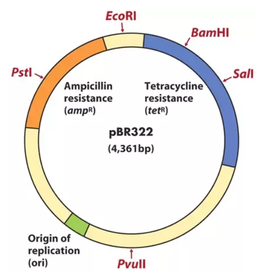
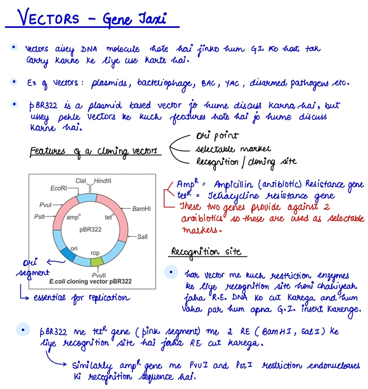
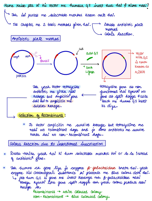
- Ideally a vector should have only one recognition site / cloning site for a single restriction endonuclease enzyme. The given figure below this section is of a vector DNA which is generated in lab by Boliver & Rodrigez from a plasmid DNA and this circular vector is named pBR322 where p stand for plasmid. This plasmid has 2 antibiotic resistance genes named tetR & ampR which provide resistance against tetracycline and ampicillin antibiotics respectively.
- These antibiotic resistance genes have recognition sequence for restriction endonucleases like PstI, BamHI, EcoRI and SalI.
- our gene of interest will be inserted either in between the orange segment (ampicillin resistance gene) or in between the blue region which provide resistance against tetracycline antibiotic.
- Now when we will insert our gene of interest in either of the orange or blue segments of this vector, that particular segment will lose its resistance against that antibiotic.
- This way selecting recombinants from non recombinant become easy because non recombinants will survive in both of the antibiotic mediums but the recombinants will grow only in the ampicillin media but not in the media containing tetracycline.
- An alternate selectable marker is the gene coding for enzyme beta-galactosidase which produces a blue color in presence of a chromogenic substance. So when a gene of interest is inserted within the DNA sequence for the enzyme beta-galactosidase, the enzyme (beta-galactosidase) won’t be produced because its gene is being split into 2 by insertion of foreign DNA in between this gene.
- So the recombinants won’t produce blue color with the chromogenic substance so they will produce white colored colonies while the non recombinants will be producing a functional beta - galactosidase which will produce blue colored colonies in presence of the chromogenic substance.
- The plasmid vector pBR322 is a plasmid based vector so this vector is useful only for cloning gene in bacterial cells like E.coli but for cloning the genes in plants or animal cells this vector won’t work efficiently so we need some new cloning vectors.
- The tumor inducing Ti Plasmid of Agrobacterium tumefaciens is an excellent vector for plant cells and and several disarmed retroviruses work efficiently for cloning genes in the animal cells.
- we will be studying about the Ti Plasmid in detail in the next chapter.
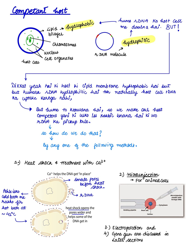
- DNA is hydrophilic for the host cell to pick up the vector molecule, the vector DNA should pass through the lipid bilayer membrane which is hydrophobic so any host cell would not have a inbuilt ability to pick up the vector DNA so we have to make the host cell competent. There are several ways to make a host cell competent which are discussed below :
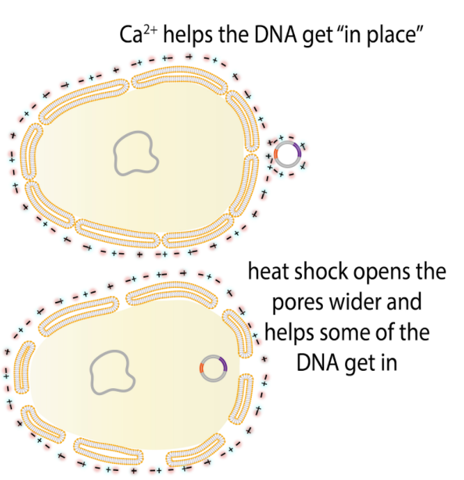
- Treatment with divalent cations such as Ca2+ which increase the efficiency with which DNA enters bacteria through pores in its cell wall.
- Heat shock
Host cells are incubated with rDNA on ice and then they are placed at 42oC. This sudden change from cold to hot temperature is known as the heat shock method.,
Restriction Enzymes - The Molecular Scissors
From now onwards we will use RE as an abbreviation for restriction endonucleases.
Nomenclature of Restriction Endonucleases
Description Of The Picture Given Above
Separation And Isolation Of Dna Fragments
Till now we have generated the gene of interest by cutting DNA with the help of RE but remember that we are doing all this in a test tube where many fragments will be found because because the DNA from a single cell may have many restriction sites (where RE makes a cut) for a single RE and we generally start these experiments by taking DNA from many cells so many fragments will be produced in that test tube after the treatment of DNA with RE. So do we know and isolate our fragment of interest from the other DNA fragments of that test tube ?
The simple answer would be : GEL ELECTROPHORESIS which is a technique that separates different DNA fragments based on their size under the influence of an electric field.
GEL ELECTROPHORESIS
Cloning Vectors - Vehicles For Cloning
2.) Selectable Marker
3.) Cloning Sites
→ This method of selection of recombinants from non recombinants is usually known as double plate antibiotic based selection which is cumbersome so we have another method which is known as Insertional inactivation
Insertional Inactivation / Color Reaction
Vectors for cloning genes in plants & animals
Competent Host (For Transformation With rDNA)
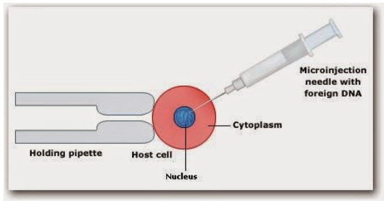
3.) Microinjection
In this method the Recombinant DNA is directly injected into the cell using a micro needle and a micropipette.
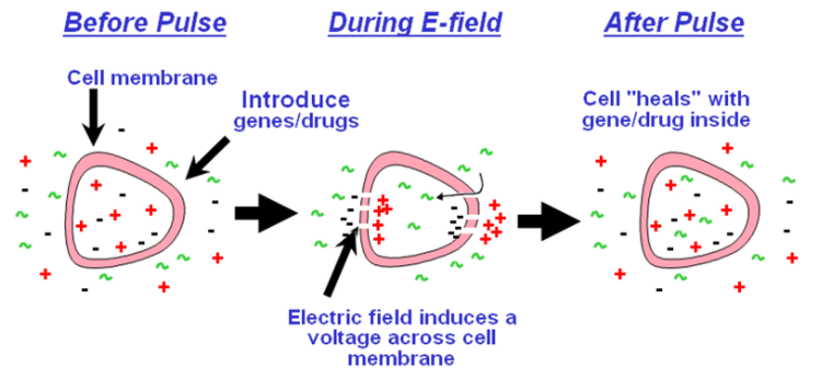
4.) Electroporation
The electrical shock makes temporary pulse into the plasma membrane through which DNA molecules enter the cell.
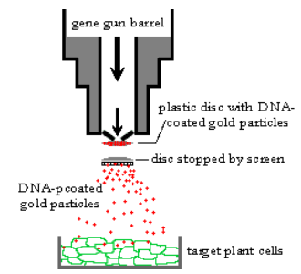
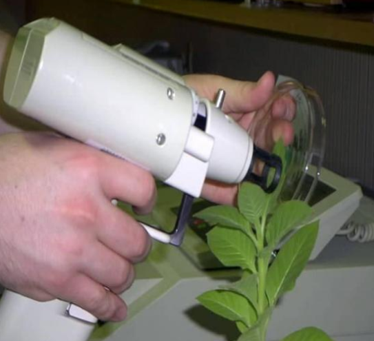
5.) Gene Gun / Biolistics
This method is suitable for plant cells. The cells are bombarded with the high velocity microparticles of gold or tungsten which are coated with foreign DNA so as to directly inject the DNA into the cell.
6.) Disarmed Pathogens
pathogens they have a natural ability to enter inside any cell so we use this property of these agents to deliver our gene of interest but before that these vectors have to be disarmed which makes that their disease causing part of the DNA is ripped off so that this pathogen vector would not cause any disease in the host cell. An example of such a case would be using disarmed retroviruses, disarmed HIV for transfection of animal cells and the use of disarmed agrobacterium tumefaciens tumor plasmid for transformation of plant cells.
TOOLS OF rDNA TECHNOLOGY
- Just like any other technology, biotechnology also requires its own tools to successfully alter the genome of a cell. At the very basic core of biotechnology we have three basic tools :
- Enzymes : like restriction enzymes, polymerase enzymes and ligase enzymes etc.
- Vectors : to carry our gene of interest into the host cell.
- Competent host : a host is the organism in which we insert our gene of interest. But looking from the host point of view, that gene of interest is a foreign molecule and the host won’t take it up willingly so we have to make our host able to pick up the vector (along with gene of interest) and this willing host which can take up that foreign gene of interest is known as a competent host.
Now, we will discuss each of the three tools one by one in detail. - These enzymes are used to cut the DNA and different types of restriction enzymes are used to cut DNA at specific locations. The enzymes that cut or break the DNA are known as nucleases because they degrade the DNA(nucleic acid), hence the name nuclease.
- These enzymes can either cut the DNA from the middle and in that case they are known as endonuclease or they can cut the DNA from either of the termini/ends and in that case they will be known as exonucleases.
- Now these endonucleases were naturally found in some microbes like E.coli where they degrade the viral DNA to restrict the growth of viral DNA. Due to their ability to restrict the growth of viral DNA, these specific endonucleases which recognized and cut a specific sequence of 6-8 base pairs in the viral DNA were named as Restriction Endonucleases which we use in rDNA technology.
- Exonucleases are not used because they tend to degrade the DNA and render it non useful to us. While restriction endonucleases are of different types : some are more specific as to where they make a cut in the DNA, some are less accurate and some cut the DNA at a random site. So the class of restriction endonucleases that we use in the rDNA technology are the Type II Restriction Endonucleasewhich have a high accuracy of cutting the DNA at a given specific site.
- Hind II was the first restriction enzyme and besides it we have isolated over 900 restriction enzymes from over 230 strains of bacteria and each of which recognize different sites.
- Now each sequence recognized by RE is a palindromic sequence which means that the sequence will read the same from both sides like the word ‘MALAYALAM’. For example the RE EcoRI will always cut the DNA between the bases G and A only when the sequence GAATTC is present in the DNA.
- The first letter of the name of RE comes from the genus and is written in capital. The next two letters come from the species of the microbe from which that RE was isolated and written in small. The next capital word comes from the strain of the microbe and after that we write the roman number indicating the order in which the enzymes were isolated from the strain of bacteria.
- For example the EcoRI comes from Escherichia coli RY 13. E from genus, co from species, R from strain (which is RY in this case ) and I represents that it was the first RE to be isolated from this particular strain of E.coli
- The above picture shows the sequence GAATTC recognized by the EcoRI enzyme and this RE will always cut in between G and A of the sequence (which you can see in second part of this image)thus leaving a overhang/stick end. This sticky end helps in easy ligation of red and black DNA segments which represents the vector and foreign DNA respectively.
- Now there are some other REs which produce blunt ends after cutting the DNA like shown in the picture below this where the enzyme SmaIalways cuts in between the G and C base pair of the recognition sequence. Recognition sequence in the part highlighted in yellow and blue colour while the cut is made either in exactly in the middle of the sequence like by SmaI thus producing the blunt ends or the cut is made a little away from so that a overhang is left like in the case of EcoRI an overhang/sticky end is left at the 5’ end of the sequence.
- These sticky ends will be joined easily by the enzyme : DNA ligase. Ligation of blunt ends is a bit tricky but anyhow it is not a part of our discussion at this point.
- We know that DNA is negatively charged due to the negative charge on phosphate bonds. So under the influence of the electric field the DNA fragments will move from the negative electrode to the positive electrode because the current from the negative electrode will repel the negatively charged DNA towards the positive electrode. So the DNA will move from negative to positive electrode in a media of agarose gel.
- Agarose gel has pores which offer resistance to the free movement of the DNA fragments. DNA fragments will move through these pores towards the positive electrode but this movement will depend on the size of the fragment.
- Higher the number of base pairs in a fragment, more will be its weight and it will travel less distance towards the positive electrode. On the other hand, the less number of base pairs a fragment has, less will be its weight and it farther it moves.
- So, the different fragments in the test tube are now separated in the agarose gel according to their weight.
- Now see, DNA doesn’t have a colour, the agarose gel is also colourless so how do we know the DNA is travelling in the agarose media ?
Well the answer is simple, we add a dye to the DNA fragment solution in our tube so that we can track the dye going from the negative to the positive electrode and just behind the dye will be our DNA. - See, even after adding the dye we can’t actually visualize the DNA fragments because the dye travels ahead of the fragments and the only purpose of the dye is to tell us when to stop the current and in what time our electrophoresis will be done.
- So, to actually visualize the DNA we use staining agent or another dye named Ethidium Bromide (EtBr) which intercalated itself between the DNA base pair and this dye is bright orange colored under UV light and no color under normal visible light conditions.
- So also add EtBr and under the UV light our DNA fragments will be of bright orange colored bands separated according to their weight.
- Now we know which our fragment of interest is and that particular fragment is cut out of the gel to use it for ligation with the vector DNA. The extraction of DNA fragments from agarose is called elution.
- Vectors are the DNA molecules that can carry and replicate our gene of interest into the host. Vector could a plasmid DNA of a bacteria or a bacteriophage (a virus which has innate ability to inject its genetic material into the bacterial cells), Yeast artificial chromosomes (YAC) or bacterial artificial chromosome (BAC), transposons or even other viruses like influenza virus. Nowadays we are also successful in using disarmed HIV for targeted gene delivery but till date the most common vectors are plasmids and bacteriophages.
- There a few features that are required to facilitate cloning/replication into a vector which are discussed below :
1.) Origin Of Replication (Ori) - Ori point is required for replication because we insert our gene of interest in the vector so that whenever this vector replicates itself, our gene of interest also gets replicated and this is how the gene of interest will be maintained in the progenies of the host.
- Selectable markers basically help us to select transformants and non - transformants. Transformants are the vectors in which our gene of interest is successfully ligated. This ligation again occurs in the test tube under aqueous conditions so we need some markers which will allow us to differentiate whether the gene of interest has been successfully ligated within the vector (transformant vector) or not.
- These selectable markers are generally the genes responsible for providing resistance to a particular antibiotic like resistance against ampicillin, chloramphenicol, tetracycline or kanamycin.
- Normal E.coli cells do not carry resistance against any antibiotic.
- Ideally a vector should have only one recognition site / cloning site for a single restriction endonuclease enzyme. The given figure below this section is of a vector DNA which is generated in lab by Boliver & Rodrigez from a plasmid DNA and this circular vector is named pBR322 where p stand for plasmid. This plasmid has 2 antibiotic resistance genes named tetR & ampR which provide resistance against tetracycline and ampicillin antibiotics respectively.
- These antibiotic resistance genes have recognition sequence for restriction endonucleases like PstI, BamHI, EcoRI and SalI.
- our gene of interest will be inserted either in between the orange segment (ampicillin resistance gene) or in between the blue region which provide resistance against tetracycline antibiotic.
- Now when we will insert our gene of interest in either of the orange or blue segments of this vector, that particular segment will lose its resistance against that antibiotic.
- This way selecting recombinants from non recombinant become easy because non recombinants will survive in both of the antibiotic mediums but the recombinants will grow only in the ampicillin media but not in the media containing tetracycline.
- An alternate selectable marker is the gene coding for enzyme beta-galactosidase which produces a blue color in presence of a chromogenic substance. So when a gene of interest is inserted within the DNA sequence for the enzyme beta-galactosidase, the enzyme (beta-galactosidase) won’t be produced because its gene is being split into 2 by insertion of foreign DNA in between this gene.
- So the recombinants won’t produce blue color with the chromogenic substance so they will produce white colored colonies while the non recombinants will be producing a functional beta - galactosidase which will produce blue colored colonies in presence of the chromogenic substance.
- The plasmid vector pBR322 is a plasmid based vector so this vector is useful only for cloning gene in bacterial cells like E.coli but for cloning the genes in plants or animal cells this vector won’t work efficiently so we need some new cloning vectors.
- The tumor inducing Ti Plasmid of Agrobacterium tumefaciens is an excellent vector for plant cells and and several disarmed retroviruses work efficiently for cloning genes in the animal cells.
- we will be studying about the Ti Plasmid in detail in the next chapter.
- DNA is hydrophilic for the host cell to pick up the vector molecule, the vector DNA should pass through the lipid bilayer membrane which is hydrophobic so any host cell would not have a inbuilt ability to pick up the vector DNA so we have to make the host cell competent. There are several ways to make a host cell competent which are discussed below :
- Treatment with divalent cations such as Ca2+ which increase the efficiency with which DNA enters bacteria through pores in its cell wall.
- Heat shock
Host cells are incubated with rDNA on ice and then they are placed at 42oC. This sudden change from cold to hot temperature is known as the heat shock method.,
Restriction Enzymes - The Molecular Scissors
From now onwards we will use RE as an abbreviation for restriction endonucleases.
Nomenclature of Restriction Endonucleases

Description Of The Picture Given Above


Separation And Isolation Of Dna Fragments
Till now we have generated the gene of interest by cutting DNA with the help of RE but remember that we are doing all this in a test tube where many fragments will be found because because the DNA from a single cell may have many restriction sites (where RE makes a cut) for a single RE and we generally start these experiments by taking DNA from many cells so many fragments will be produced in that test tube after the treatment of DNA with RE. So do we know and isolate our fragment of interest from the other DNA fragments of that test tube ?
The simple answer would be : GEL ELECTROPHORESIS which is a technique that separates different DNA fragments based on their size under the influence of an electric field.
GEL ELECTROPHORESIS
Cloning Vectors - Vehicles For Cloning
2.) Selectable Marker
3.) Cloning Sites
→ This method of selection of recombinants from non recombinants is usually known as double plate antibiotic based selection which is cumbersome so we have another method which is known as Insertional inactivation
Insertional Inactivation / Color Reaction
Vectors for cloning genes in plants & animals

Competent Host (For Transformation With rDNA)

3.) Microinjection
In this method the Recombinant DNA is directly injected into the cell using a micro needle and a micropipette.

4.) Electroporation
The electrical shock makes temporary pulse into the plasma membrane through which DNA molecules enter the cell.

5.) Gene Gun / Biolistics
This method is suitable for plant cells. The cells are bombarded with the high velocity microparticles of gold or tungsten which are coated with foreign DNA so as to directly inject the DNA into the cell.


6.) Disarmed Pathogens
pathogens they have a natural ability to enter inside any cell so we use this property of these agents to deliver our gene of interest but before that these vectors have to be disarmed which makes that their disease causing part of the DNA is ripped off so that this pathogen vector would not cause any disease in the host cell. An example of such a case would be using disarmed retroviruses, disarmed HIV for transfection of animal cells and the use of disarmed agrobacterium tumefaciens tumor plasmid for transformation of plant cells.
PRINCIPLES OF BIOTECHNOLOGY
- Genetic engineering : the technique to alter the genetic material (DNA/RNA) and maintenance of sterile environment : to provide an environment free from microbial contamination to alter genetic material.
- In genetic engineering the genetic material of an organism is altered which means either some genes are added or deleted or both and this new altered DNA is what is known as recombinant DNA .
- The new gene or DNA to be inserted into genetic material of other organism is known as gene of interest which is always inserted near the origin of replication or ori point because the newly inserted segment need to be replicate and multiply itself so that this gene of interest can maintain itself in the progenies. This multiplication of gene of interest in the host organism is known as gene cloning
- In 1972 Stanley Cohen & Herbert Boyer isolated an antibiotic resistance gene segment from plasmid of Salmonella typhimurium by using restriction enzymes (or molecular scissors) and this gene segment coded for an antibiotic resistance protein. They then ligated (joined) this gene segment into the plasmid DNA of Salmonella typhimurium. This plasmid DNA now has a foreign gene segment which was naturally not its own so this plasmid DNA along with the antibiotic resistance gene is now known as a rDNA.
- This plasmid will now be used to transfer this antibiotic gene to another bacteria like Escherichia coli so that E.coli which originally did not have this antibiotic resistance gene, now has it. In this process the plasmid DNA from Salmonella is used to carry that antibiotic resistance gene to E.coli that is why the plasmid DNA is generally known as gene taxi or vector in technical terms.
- Identification of DNA with desirable genes or identification of gene of interest (in above case that would be antibiotic resistance gene)
- Introduction of the gene of interest into the host (in above case that would be transferring the antibiotic resistance gene into E.coli using plasmid DNA of Salmonella).
- Maintenance of introduced DNA in the host and transfer of the DNA to its progeny (this basically means that we introduced our gene of interest in plasmid near to the ori point and then antibiotic resistance would replicate itself using DNA polymerase of the E.coli bacteria).
are the two basic techniques and the very core of modern biotechnology. These techniques made modern biotechnology modern in a true sense.
These were the basic terms and before advancing further make sure that you know about terms like, gene transfer, gene cloning, rDNA/recombinant DNA, ori point and some basics from the genetics unit like DNA multiplication, transcription etc.
First Instance Of Construction Of An Artificial rDNA Molecule
So, there are 3 basic steps that happened above in modifying E.coli bacteria are :
IMPORTANT POINT
- We have to cut the vector DNA and the source DNA (from which we are taking our gene of interest) with the same restriction enzyme of that complementary ends could be generated which can be ligated/joined by the ligase enzymes. If we cut the two DNA with different RE, we will have two sliced DNA fragments with no complementary bases to ligate them to produce a rDNA.
- The dye generally used to track the progress of electrophoresis is bromophenol blue.
TOOLS OF rDNA TECHNOLOGY
PROCESSES OF RECOMBINANT DNA TECHNOLOGY
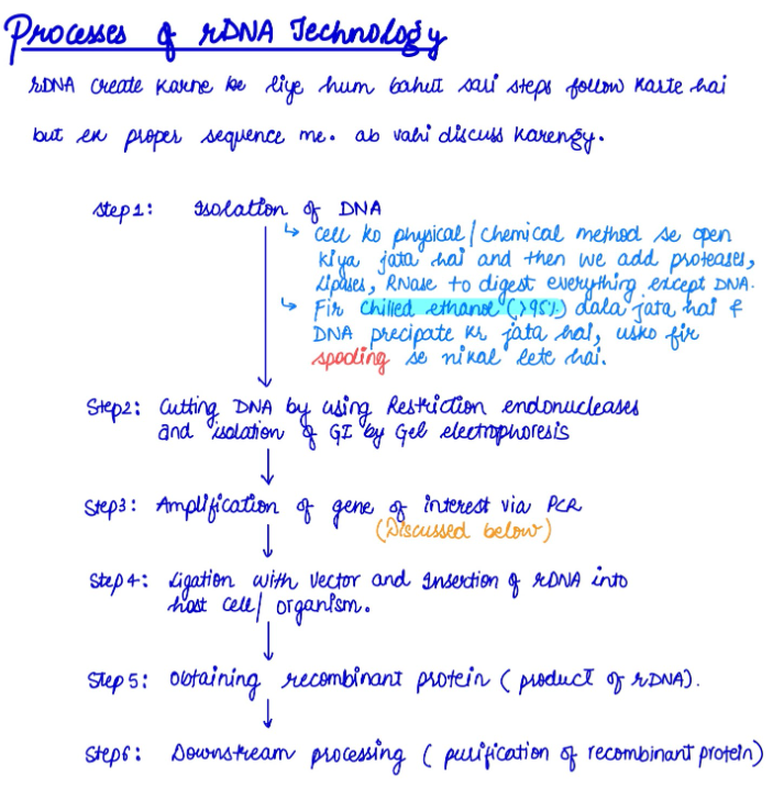
- rDNA technology involves several steps in specific sequence which are mentioned below:
- A cell has various macromolecules like proteins RNA, lipids and of course DNA But which specifically required in DNA for biotechnology process so we need to digest all the other bio macromolecules but first of all we need to digest the cell wall and cell membrane to open the cell up which we do by using enzymes such as lysosomes to degrade the cell wall for bacteria cell and release to the digest the cell wall of plant cells and chitin is in same to digest the cell wall of fungus after this we had other in enzymes such as RNases, lipases and proteases so that now we are only left with DNA.
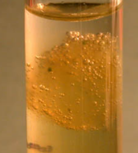
- Now in our solution we only have DNA which we can precipitate by addition of chilled ethanol and after doing all this our test tube will look something like this :
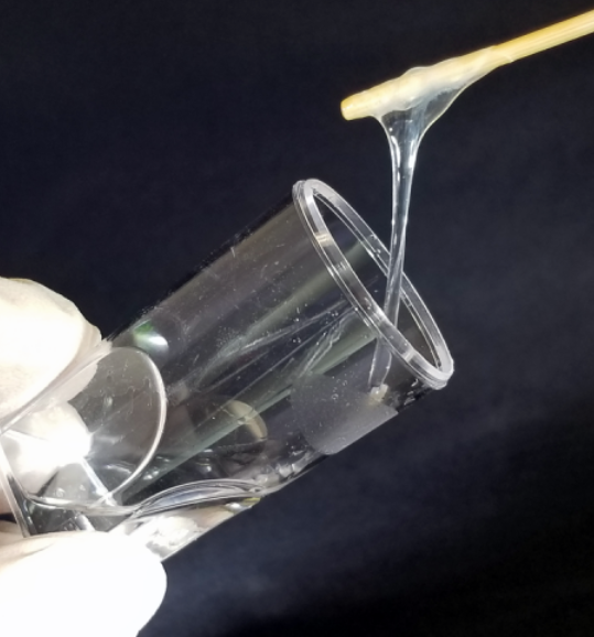
- Extraction of this DNA from the test tube using a glass rod or something else is known as Spooling which is picturised below : (The slimy thing you are seeing below hanging from the chopstick like tool is actually precipitated DNA)
- After the isolation of the DNA we have to cut the DNA at specific position by using restriction endonuclease and we already know about this step.
- After cutting, the fragments have to be separated using gel electrophoresis. The above two steps are repeated with vector DNA also and finally foreign DNA segment is ligated with a vector with the help of ligases after which the final result is recombinant DNA.
- PCR stands for Polymerase Chain Reaction which means a chain reaction is carried out using DNA Polymerase enzyme and our gene of interest in presence of deoxynucleotides and some RNA primers. In simpler terms we can say that PCR is used to create multiple copies (amplification) of our gene of interest.
- This is the first step where two strands of gene of interest/DNA are separated using heat and so this step is carried out 3-5oC below the melting point of the RNA primers. So normally the temperature for this step is generally 92oC.
Annealing - In this second step temperature is lowered to 50−55oC so that the RNA primers (red thing in above diagram) can attach themselves to the 3' end of the separated DNA strands (annealing).
Extension - In this step temperature is again raised to 72oC and DNA dependent taq DNA Polymerase (from Thermus aquaticus)starts polymerization reaction by extending the primers in 5’ to 3’ end so that now after this step we have twice the amount of DNA we had initially.
- These 3 steps make one cycle so we run PCR multiple time and after 30 cycle we can get upto a billion fragments from a single DNA molecule.
- For insertion of our gene of interest we use several vectors as described above. So the rDNA is inserted into the selectable marker gene of the vector and then we select transformants (with rDNA) from non transformants.
Obtaining the Foreign Gene Product - The vector carries our gene of interest into a host cell where it will replicate and will be transcribed after which a recombinant protein will be translated from its mRNA.
- Collecting that protein formed in the host cell (from our gene of interest) is the ultimate aim.
- After inserting the foreign gene let’s say in bacterial cells, the transformants will be grown first in lab and if everything goes alright then we can even go for large scale production (by using bioreactors) of the recombinant protein in cases where the recombinant protein has application like in medical field. Such an example would be large scale industrial production of Human insulin from bacteria which we will discuss in the next chapter.
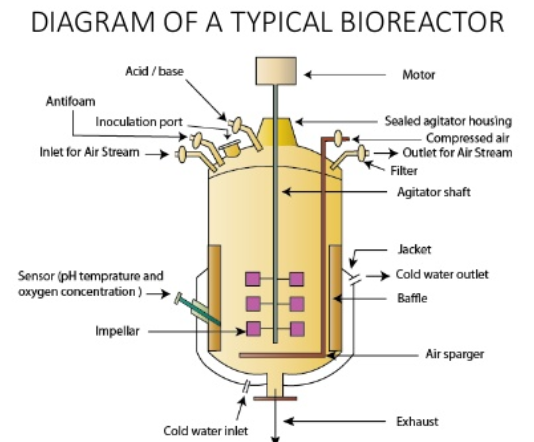
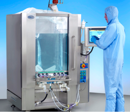
- Bioreactors are the large vessels which are used to culture transformants in high quantities ranging from 100 -1000L. A bioreactors basically provides the optimum conditions for the growth of transformants so we put things like vitamins, substrate,salts,oxygen and create an optimum temperature and pH for the optimal growth of the transformants. This is the biosynthetic phase where desired products are produced but we can’t use them directly. First we have to purify them through a number of series and this purification phase is known as downstream processing.
- This step includes separation and purification of the desired product and we add suitable preservatives to preserve the product so that it can be used later on.
- If the desired product happens to be a new drug then the drug goes for clinical trials in various phases to see its efficiency, accuracy and associated side effects. But the protocol varies from product to product.
1.) Isolation of DNA
2.) Cutting of DNA at specific locations using restriction enzymes
3.) Amplification of the gene of interest using PCR
4.) Insertion of recombinant DNA into the host cell or organism
5.) Collecting the foreign gene product
6.) Downstream processing.
Isolation of DNA
Cutting of DNA at Specific Locations
Amplification of Gene of Interest Using PCR
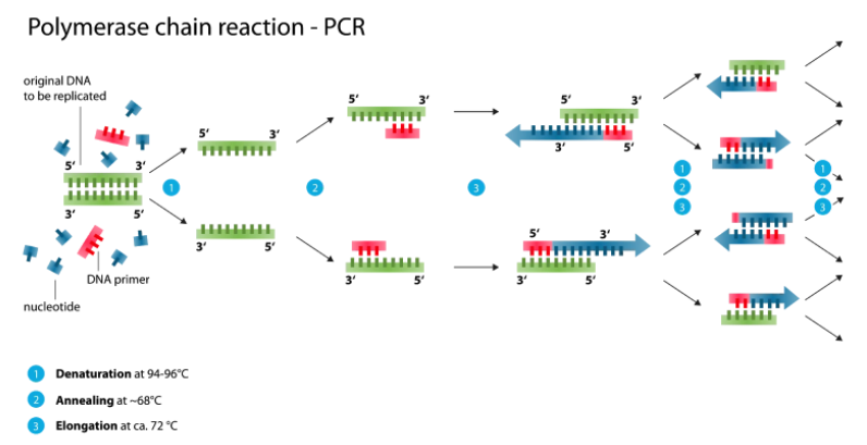
PCR has three basic steps which are described below:
Denaturation
Insertion of rDNA Into The Host Cell/Organism
Bioreactors
Downstream Processing
PROCESSES OF RECOMBINANT DNA TECHNOLOGY
- rDNA technology involves several steps in specific sequence which are mentioned below:
- A cell has various macromolecules like proteins RNA, lipids and of course DNA But which specifically required in DNA for biotechnology process so we need to digest all the other bio macromolecules but first of all we need to digest the cell wall and cell membrane to open the cell up which we do by using enzymes such as lysosomes to degrade the cell wall for bacteria cell and release to the digest the cell wall of plant cells and chitin is in same to digest the cell wall of fungus after this we had other in enzymes such as RNases, lipases and proteases so that now we are only left with DNA.
- Now in our solution we only have DNA which we can precipitate by addition of chilled ethanol and after doing all this our test tube will look something like this :
- Extraction of this DNA from the test tube using a glass rod or something else is known as Spooling which is picturised below : (The slimy thing you are seeing below hanging from the chopstick like tool is actually precipitated DNA)
- After the isolation of the DNA we have to cut the DNA at specific position by using restriction endonuclease and we already know about this step.
- After cutting, the fragments have to be separated using gel electrophoresis. The above two steps are repeated with vector DNA also and finally foreign DNA segment is ligated with a vector with the help of ligases after which the final result is recombinant DNA.
- PCR stands for Polymerase Chain Reaction which means a chain reaction is carried out using DNA Polymerase enzyme and our gene of interest in presence of deoxynucleotides and some RNA primers. In simpler terms we can say that PCR is used to create multiple copies (amplification) of our gene of interest.
- This is the first step where two strands of gene of interest/DNA are separated using heat and so this step is carried out 3-5oC below the melting point of the RNA primers. So normally the temperature for this step is generally 92oC.
Annealing - In this second step temperature is lowered to 50−55oC so that the RNA primers (red thing in above diagram) can attach themselves to the 3' end of the separated DNA strands (annealing).
Extension - In this step temperature is again raised to 72oC and DNA dependent taq DNA Polymerase (from Thermus aquaticus)starts polymerization reaction by extending the primers in 5’ to 3’ end so that now after this step we have twice the amount of DNA we had initially.
- These 3 steps make one cycle so we run PCR multiple time and after 30 cycle we can get upto a billion fragments from a single DNA molecule.
- For insertion of our gene of interest we use several vectors as described above. So the rDNA is inserted into the selectable marker gene of the vector and then we select transformants (with rDNA) from non transformants.
Obtaining the Foreign Gene Product - The vector carries our gene of interest into a host cell where it will replicate and will be transcribed after which a recombinant protein will be translated from its mRNA.
- Collecting that protein formed in the host cell (from our gene of interest) is the ultimate aim.
- After inserting the foreign gene let’s say in bacterial cells, the transformants will be grown first in lab and if everything goes alright then we can even go for large scale production (by using bioreactors) of the recombinant protein in cases where the recombinant protein has application like in medical field. Such an example would be large scale industrial production of Human insulin from bacteria which we will discuss in the next chapter.
- Bioreactors are the large vessels which are used to culture transformants in high quantities ranging from 100 -1000L. A bioreactors basically provides the optimum conditions for the growth of transformants so we put things like vitamins, substrate,salts,oxygen and create an optimum temperature and pH for the optimal growth of the transformants. This is the biosynthetic phase where desired products are produced but we can’t use them directly. First we have to purify them through a number of series and this purification phase is known as downstream processing.
- This step includes separation and purification of the desired product and we add suitable preservatives to preserve the product so that it can be used later on.
- If the desired product happens to be a new drug then the drug goes for clinical trials in various phases to see its efficiency, accuracy and associated side effects. But the protocol varies from product to product.
1.) Isolation of DNA
2.) Cutting of DNA at specific locations using restriction enzymes
3.) Amplification of the gene of interest using PCR
4.) Insertion of recombinant DNA into the host cell or organism
5.) Collecting the foreign gene product
6.) Downstream processing.
Isolation of DNA


Cutting of DNA at Specific Locations
Amplification of Gene of Interest Using PCR

PCR has three basic steps which are described below:
Denaturation
Insertion of rDNA Into The Host Cell/Organism
Bioreactors


Downstream Processing
TOOLS OF rDNA TECHNOLOGY
- Just like any other technology, biotechnology also requires its own tools to successfully alter the genome of a cell. At the very basic core of biotechnology we have three basic tools :
- Enzymes : like restriction enzymes, polymerase enzymes and ligase enzymes etc.
- Vectors : to carry our gene of interest into the host cell.
- Competent host : a host is the organism in which we insert our gene of interest. But looking from the host point of view, that gene of interest is a foreign molecule and the host won’t take it up willingly so we have to make our host able to pick up the vector (along with gene of interest) and this willing host which can take up that foreign gene of interest is known as a competent host.
Now, we will discuss each of the three tools one by one in detail. - These enzymes are used to cut the DNA and different types of restriction enzymes are used to cut DNA at specific locations. The enzymes that cut or break the DNA are known as nucleases because they degrade the DNA(nucleic acid), hence the name nuclease.
- These enzymes can either cut the DNA from the middle and in that case they are known as endonuclease or they can cut the DNA from either of the termini/ends and in that case they will be known as exonucleases.
- Now these endonucleases were naturally found in some microbes like E.coli where they degrade the viral DNA to restrict the growth of viral DNA. Due to their ability to restrict the growth of viral DNA, these specific endonucleases which recognized and cut a specific sequence of 6-8 base pairs in the viral DNA were named as Restriction Endonucleases which we use in rDNA technology.
- Exonucleases are not used because they tend to degrade the DNA and render it non useful to us. While restriction endonucleases are of different types : some are more specific as to where they make a cut in the DNA, some are less accurate and some cut the DNA at a random site. So the class of restriction endonucleases that we use in the rDNA technology are the Type II Restriction Endonucleasewhich have a high accuracy of cutting the DNA at a given specific site.
- Hind II was the first restriction enzyme and besides it we have isolated over 900 restriction enzymes from over 230 strains of bacteria and each of which recognize different sites.
- Now each sequence recognized by RE is a palindromic sequence which means that the sequence will read the same from both sides like the word ‘MALAYALAM’. For example the RE EcoRI will always cut the DNA between the bases G and A only when the sequence GAATTC is present in the DNA.
- The first letter of the name of RE comes from the genus and is written in capital. The next two letters come from the species of the microbe from which that RE was isolated and written in small. The next capital word comes from the strain of the microbe and after that we write the roman number indicating the order in which the enzymes were isolated from the strain of bacteria.
- For example the EcoRI comes from Escherichia coli RY 13. E from genus, co from species, R from strain (which is RY in this case ) and I represents that it was the first RE to be isolated from this particular strain of E.coli
- The above picture shows the sequence GAATTC recognized by the EcoRI enzyme and this RE will always cut in between G and A of the sequence (which you can see in second part of this image)thus leaving a overhang/stick end. This sticky end helps in easy ligation of red and black DNA segments which represents the vector and foreign DNA respectively.
- Now there are some other REs which produce blunt ends after cutting the DNA like shown in the picture below this where the enzyme SmaIalways cuts in between the G and C base pair of the recognition sequence. Recognition sequence in the part highlighted in yellow and blue colour while the cut is made either in exactly in the middle of the sequence like by SmaI thus producing the blunt ends or the cut is made a little away from so that a overhang is left like in the case of EcoRI an overhang/sticky end is left at the 5’ end of the sequence.
- These sticky ends will be joined easily by the enzyme : DNA ligase. Ligation of blunt ends is a bit tricky but anyhow it is not a part of our discussion at this point.
- We know that DNA is negatively charged due to the negative charge on phosphate bonds. So under the influence of the electric field the DNA fragments will move from the negative electrode to the positive electrode because the current from the negative electrode will repel the negatively charged DNA towards the positive electrode. So the DNA will move from negative to positive electrode in a media of agarose gel.
- Agarose gel has pores which offer resistance to the free movement of the DNA fragments. DNA fragments will move through these pores towards the positive electrode but this movement will depend on the size of the fragment.
- Higher the number of base pairs in a fragment, more will be its weight and it will travel less distance towards the positive electrode. On the other hand, the less number of base pairs a fragment has, less will be its weight and it farther it moves.
- So, the different fragments in the test tube are now separated in the agarose gel according to their weight.
- Now see, DNA doesn’t have a colour, the agarose gel is also colourless so how do we know the DNA is travelling in the agarose media ?
Well the answer is simple, we add a dye to the DNA fragment solution in our tube so that we can track the dye going from the negative to the positive electrode and just behind the dye will be our DNA. - See, even after adding the dye we can’t actually visualize the DNA fragments because the dye travels ahead of the fragments and the only purpose of the dye is to tell us when to stop the current and in what time our electrophoresis will be done.
- So, to actually visualize the DNA we use staining agent or another dye named Ethidium Bromide (EtBr) which intercalated itself between the DNA base pair and this dye is bright orange colored under UV light and no color under normal visible light conditions.
- So also add EtBr and under the UV light our DNA fragments will be of bright orange colored bands separated according to their weight.
- Now we know which our fragment of interest is and that particular fragment is cut out of the gel to use it for ligation with the vector DNA. The extraction of DNA fragments from agarose is called elution.
- Vectors are the DNA molecules that can carry and replicate our gene of interest into the host. Vector could a plasmid DNA of a bacteria or a bacteriophage (a virus which has innate ability to inject its genetic material into the bacterial cells), Yeast artificial chromosomes (YAC) or bacterial artificial chromosome (BAC), transposons or even other viruses like influenza virus. Nowadays we are also successful in using disarmed HIV for targeted gene delivery but till date the most common vectors are plasmids and bacteriophages.
- There a few features that are required to facilitate cloning/replication into a vector which are discussed below :
1.) Origin Of Replication (Ori) - Ori point is required for replication because we insert our gene of interest in the vector so that whenever this vector replicates itself, our gene of interest also gets replicated and this is how the gene of interest will be maintained in the progenies of the host.
- Selectable markers basically help us to select transformants and non - transformants. Transformants are the vectors in which our gene of interest is successfully ligated. This ligation again occurs in the test tube under aqueous conditions so we need some markers which will allow us to differentiate whether the gene of interest has been successfully ligated within the vector (transformant vector) or not.
- These selectable markers are generally the genes responsible for providing resistance to a particular antibiotic like resistance against ampicillin, chloramphenicol, tetracycline or kanamycin.
- Normal E.coli cells do not carry resistance against any antibiotic.
- Ideally a vector should have only one recognition site / cloning site for a single restriction endonuclease enzyme. The given figure below this section is of a vector DNA which is generated in lab by Boliver & Rodrigez from a plasmid DNA and this circular vector is named pBR322 where p stand for plasmid. This plasmid has 2 antibiotic resistance genes named tetR & ampR which provide resistance against tetracycline and ampicillin antibiotics respectively.
- These antibiotic resistance genes have recognition sequence for restriction endonucleases like PstI, BamHI, EcoRI and SalI.
- our gene of interest will be inserted either in between the orange segment (ampicillin resistance gene) or in between the blue region which provide resistance against tetracycline antibiotic.
- Now when we will insert our gene of interest in either of the orange or blue segments of this vector, that particular segment will lose its resistance against that antibiotic.
- This way selecting recombinants from non recombinant become easy because non recombinants will survive in both of the antibiotic mediums but the recombinants will grow only in the ampicillin media but not in the media containing tetracycline.
- An alternate selectable marker is the gene coding for enzyme beta-galactosidase which produces a blue color in presence of a chromogenic substance. So when a gene of interest is inserted within the DNA sequence for the enzyme beta-galactosidase, the enzyme (beta-galactosidase) won’t be produced because its gene is being split into 2 by insertion of foreign DNA in between this gene.
- So the recombinants won’t produce blue color with the chromogenic substance so they will produce white colored colonies while the non recombinants will be producing a functional beta - galactosidase which will produce blue colored colonies in presence of the chromogenic substance.
- The plasmid vector pBR322 is a plasmid based vector so this vector is useful only for cloning gene in bacterial cells like E.coli but for cloning the genes in plants or animal cells this vector won’t work efficiently so we need some new cloning vectors.
- The tumor inducing Ti Plasmid of Agrobacterium tumefaciens is an excellent vector for plant cells and and several disarmed retroviruses work efficiently for cloning genes in the animal cells.
- we will be studying about the Ti Plasmid in detail in the next chapter.
- DNA is hydrophilic for the host cell to pick up the vector molecule, the vector DNA should pass through the lipid bilayer membrane which is hydrophobic so any host cell would not have a inbuilt ability to pick up the vector DNA so we have to make the host cell competent. There are several ways to make a host cell competent which are discussed below :
- Treatment with divalent cations such as Ca2+ which increase the efficiency with which DNA enters bacteria through pores in its cell wall.
- Heat shock
Host cells are incubated with rDNA on ice and then they are placed at 42oC. This sudden change from cold to hot temperature is known as the heat shock method.,
Restriction Enzymes - The Molecular Scissors
From now onwards we will use RE as an abbreviation for restriction endonucleases.
Nomenclature of Restriction Endonucleases

Description Of The Picture Given Above


Separation And Isolation Of Dna Fragments
Till now we have generated the gene of interest by cutting DNA with the help of RE but remember that we are doing all this in a test tube where many fragments will be found because because the DNA from a single cell may have many restriction sites (where RE makes a cut) for a single RE and we generally start these experiments by taking DNA from many cells so many fragments will be produced in that test tube after the treatment of DNA with RE. So do we know and isolate our fragment of interest from the other DNA fragments of that test tube ?
The simple answer would be : GEL ELECTROPHORESIS which is a technique that separates different DNA fragments based on their size under the influence of an electric field.
GEL ELECTROPHORESIS
Cloning Vectors - Vehicles For Cloning
2.) Selectable Marker
3.) Cloning Sites
→ This method of selection of recombinants from non recombinants is usually known as double plate antibiotic based selection which is cumbersome so we have another method which is known as Insertional inactivation
Insertional Inactivation / Color Reaction
Vectors for cloning genes in plants & animals

Competent Host (For Transformation With rDNA)

3.) Microinjection
In this method the Recombinant DNA is directly injected into the cell using a micro needle and a micropipette.

4.) Electroporation
The electrical shock makes temporary pulse into the plasma membrane through which DNA molecules enter the cell.

5.) Gene Gun / Biolistics
This method is suitable for plant cells. The cells are bombarded with the high velocity microparticles of gold or tungsten which are coated with foreign DNA so as to directly inject the DNA into the cell.


6.) Disarmed Pathogens
pathogens they have a natural ability to enter inside any cell so we use this property of these agents to deliver our gene of interest but before that these vectors have to be disarmed which makes that their disease causing part of the DNA is ripped off so that this pathogen vector would not cause any disease in the host cell. An example of such a case would be using disarmed retroviruses, disarmed HIV for transfection of animal cells and the use of disarmed agrobacterium tumefaciens tumor plasmid for transformation of plant cells.
SUMMARY
- Biotechnology deals with large scale production and marketing of products and processes using live organisms, cells or enzymes. Modern biotechnology using genetically modified organisms was made possible only when man learnt to alter the chemistry of DNA and construct recombinant DNA. This key process is called recombinant DNA technology or genetic engineering. This process involves the use of restriction endonucleases, DNA ligase, appropriate plasmid or viral vectors to isolate and ferry the foreign DNA into host organisms, expression of the foreign gene, purification of the gene product, i.e., the functional protein and finally making a suitable formulation for marketing. Large scale production involves use of bioreactors.
IMPORTANT POINT
- We have to cut the vector DNA and the source DNA (from which we are taking our gene of interest) with the same restriction enzyme of that complementary ends could be generated which can be ligated/joined by the ligase enzymes. If we cut the two DNA with different RE, we will have two sliced DNA fragments with no complementary bases to ligate them to produce a rDNA.
- The dye generally used to track the progress of electrophoresis is bromophenol blue.
PROCESSES OF RECOMBINANT DNA TECHNOLOGY
PROCESSES OF RECOMBINANT DNA TECHNOLOGY
- rDNA technology involves several steps in specific sequence which are mentioned below:
- A cell has various macromolecules like proteins RNA, lipids and of course DNA But which specifically required in DNA for biotechnology process so we need to digest all the other bio macromolecules but first of all we need to digest the cell wall and cell membrane to open the cell up which we do by using enzymes such as lysosomes to degrade the cell wall for bacteria cell and release to the digest the cell wall of plant cells and chitin is in same to digest the cell wall of fungus after this we had other in enzymes such as RNases, lipases and proteases so that now we are only left with DNA.
- Now in our solution we only have DNA which we can precipitate by addition of chilled ethanol and after doing all this our test tube will look something like this :
- Extraction of this DNA from the test tube using a glass rod or something else is known as Spooling which is picturised below : (The slimy thing you are seeing below hanging from the chopstick like tool is actually precipitated DNA)
- After the isolation of the DNA we have to cut the DNA at specific position by using restriction endonuclease and we already know about this step.
- After cutting, the fragments have to be separated using gel electrophoresis. The above two steps are repeated with vector DNA also and finally foreign DNA segment is ligated with a vector with the help of ligases after which the final result is recombinant DNA.
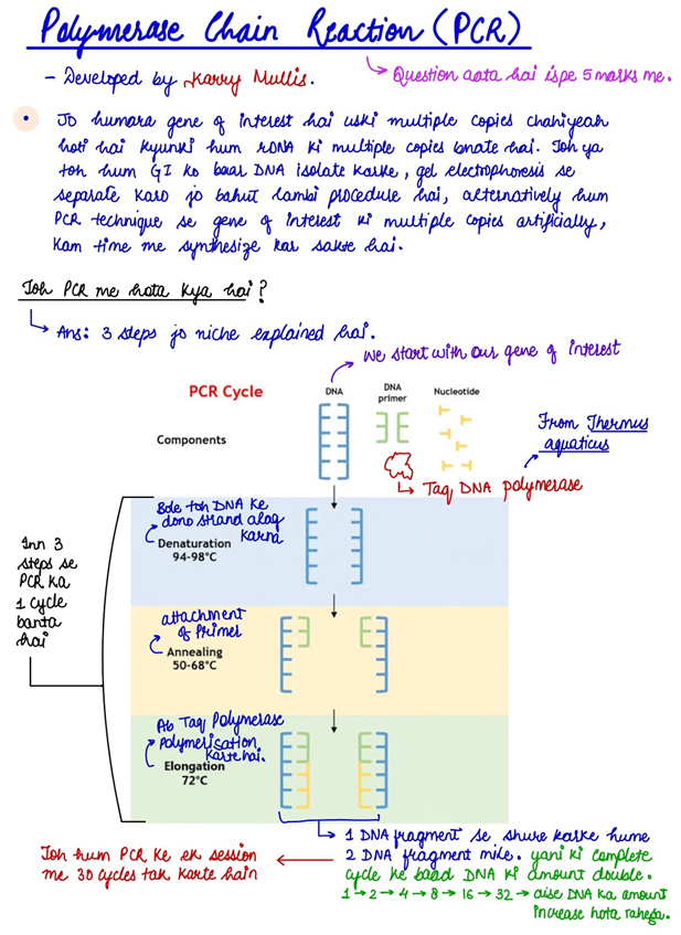
- PCR stands for Polymerase Chain Reaction which means a chain reaction is carried out using DNA Polymerase enzyme and our gene of interest in presence of deoxynucleotides and some RNA primers. In simpler terms we can say that PCR is used to create multiple copies (amplification) of our gene of interest.
- This is the first step where two strands of gene of interest/DNA are separated using heat and so this step is carried out 3-5oC below the melting point of the RNA primers. So normally the temperature for this step is generally 92oC.
Annealing - In this second step temperature is lowered to 50−55oC so that the RNA primers (red thing in above diagram) can attach themselves to the 3' end of the separated DNA strands (annealing).
Extension - In this step temperature is again raised to 72oC and DNA dependent taq DNA Polymerase (from Thermus aquaticus)starts polymerization reaction by extending the primers in 5’ to 3’ end so that now after this step we have twice the amount of DNA we had initially.
- These 3 steps make one cycle so we run PCR multiple time and after 30 cycle we can get upto a billion fragments from a single DNA molecule.
- For insertion of our gene of interest we use several vectors as described above. So the rDNA is inserted into the selectable marker gene of the vector and then we select transformants (with rDNA) from non transformants.
Obtaining the Foreign Gene Product - The vector carries our gene of interest into a host cell where it will replicate and will be transcribed after which a recombinant protein will be translated from its mRNA.
- Collecting that protein formed in the host cell (from our gene of interest) is the ultimate aim.
- After inserting the foreign gene let’s say in bacterial cells, the transformants will be grown first in lab and if everything goes alright then we can even go for large scale production (by using bioreactors) of the recombinant protein in cases where the recombinant protein has application like in medical field. Such an example would be large scale industrial production of Human insulin from bacteria which we will discuss in the next chapter.
- Bioreactors are the large vessels which are used to culture transformants in high quantities ranging from 100 -1000L. A bioreactors basically provides the optimum conditions for the growth of transformants so we put things like vitamins, substrate,salts,oxygen and create an optimum temperature and pH for the optimal growth of the transformants. This is the biosynthetic phase where desired products are produced but we can’t use them directly. First we have to purify them through a number of series and this purification phase is known as downstream processing.
- This step includes separation and purification of the desired product and we add suitable preservatives to preserve the product so that it can be used later on.
- If the desired product happens to be a new drug then the drug goes for clinical trials in various phases to see its efficiency, accuracy and associated side effects. But the protocol varies from product to product.
1.) Isolation of DNA
2.) Cutting of DNA at specific locations using restriction enzymes
3.) Amplification of the gene of interest using PCR
4.) Insertion of recombinant DNA into the host cell or organism
5.) Collecting the foreign gene product
6.) Downstream processing.
Isolation of DNA


Cutting of DNA at Specific Locations
Amplification of Gene of Interest Using PCR
Generally a 5 marks question is asked on PCR. Explain all 3 steps along with the diagram.

PCR has three basic steps which are described below:
Denaturation
Insertion of rDNA Into The Host Cell/Organism
Bioreactors


Downstream Processing
SUMMARY
- Biotechnology deals with large scale production and marketing of products and processes using live organisms, cells or enzymes. Modern biotechnology using genetically modified organisms was made possible only when man learnt to alter the chemistry of DNA and construct recombinant DNA. This key process is called recombinant DNA technology or genetic engineering. This process involves the use of restriction endonucleases, DNA ligase, appropriate plasmid or viral vectors to isolate and ferry the foreign DNA into host organisms, expression of the foreign gene, purification of the gene product, i.e., the functional protein and finally making a suitable formulation for marketing. Large scale production involves use of bioreactors.