MOLECULAR BASIS OF INHERITANCE for Class 12 BIOLOGY
INTRODUCTION
- Last chapter was all about the pattern of inheritance of genetic material. In this chapter we will study about how exactly the genetic material was discovered, the experiments which helped us conclude that DNA is in fact the genetic material, how this genetic matic material is passed on to daughter cells and how exactly genes behave to control a particular trait. Mendel told us that some ”factors'' are passed on from one generation to another. This chapter will focus on chemical nature on those “factors”.
INTRODUCTION
- In this chapter we will study about how exactly the genetic material was discovered, the experiments which helped us conclude that DNA is in fact the genetic material, how this genetic material is passed on to daughter cells and how exactly genes behave to control a particular trait. Mendel told us that some ”factors'' are passed on from one generation to another. This chapter will focus on chemical nature on those “factors”.
CHAPTER WEIGHTAGE
- Genetics and Evolution chapter ka weightage is 20 marks. Evolution part is deducted. So expected marks is around 8-10 M. Important topics include:
- Structure of DNA
- Replication
- Transcription and Translation ( including differences among eukaryotes and prokaryotes)
- Gene expression and Regulation and Lac Operon
- DNA Fingerprinting
- Human Genome Project
CHAPTER WEIGHTAGE
- Genetics and Evolution chapters are very important. Expected questions are usually around 8-10 . Important topics include:
- Structure of DNA
- Replication
- Transcription and Translation ( including differences among eukaryotes and prokaryotes)
- Gene expression and Regulation and Lac Operon
- DNA Fingerprinting
- Human Genome Project
THE DISCOVERY OF DNA
HISTORICAL BACKGROUND
- In 1902, Sutton and Boveri gave the chromosomal theory of inheritance. They said that genetic material of each and every cell is contained in the chromosomes but at that time we didn’t know what chromosomes were made of. So studies were conducted to determine the chemical nature of chromosomes. Recall that in class 11, chapter : Biomolecules you studied about some macromolecules like proteins, fats and Nucleic acids (DNA, RNA). The chemical studies on the chromosomes showed that they had 2 biomacromolecules : Nucleic acid (DNA) along with the proteins.
- So until the 1940s it was thought that the proteins were the actual genetic material because they show so much variety due to the presence of 20 different amino acids. This somehow explained the genetic diversity and variety shown by sexually reproducing organisms like humans. On the other hand DNA is made up of only 4 nitrogenous base pairs along with a pentose sugar and a phosphate group. So it seemed reasonable that 20 amino acids accounted for more genetic diversity than 4 nitrogenous base pairs ever could.
- During the 1940s different experiments indeed proved that DNA is the genetic material instead of protein. We will discuss these experiments in the coming section but first we need to know what are the essential requirements of genetic material and whether DNA has them or not?
- It should be present in every cell
- The amount should be same in all the cells.
- It should be able to replicate precisely.
- it should be able to faithfully pass its copies into the progeny.
- Should have the capacity to show heritable changes (mutations).
- Should be able to express itself in the progeny.
- should be stable enough but at the same time it should have a capacity to show mutation.
Essential Requirements of Genetic Material
Now we will see some of the features of DNA to understand why DNA is the genetic material and how conclusions were drawn from the experiments that showed that DNA is indeed the genetic material we need to familiarize ourselves with the basics of DNA like what is it made up of, type of chemical bonds, its structure, how it's packed inside tiny nucleus of a tiny cell. And then we will study about the experiments leading to the discovery of the DNA as genetic material.
THE DISCOVERY & STRUCTURE OF DNA
STRUCTURE & PACKAGING OF DNA
DEOXYRIBONUCLEIC ACID (DNA)
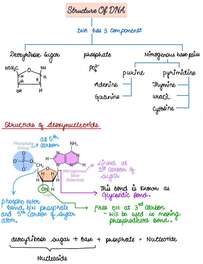
- DNA is a long chain of polynucleotides. A single nucleotide is made up of 3 components :
- Deoxyribose Sugar
- Nitrogenous Base Pair (either purine or pyrimidine) Recall class 11, chapter : Biomolecules.
- Phosphate Bond
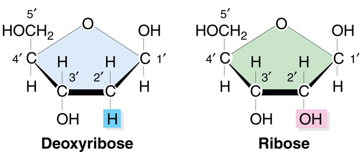
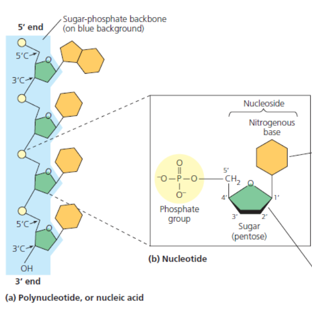
- The above picture shows two types of pentose sugar (sugar having 5 carbon atoms) : Deoxyribose and Ribose. Deoxy in Deoxyribose means without oxygen (at second carbon atom highlighted in blue) and ribose is a 5C sugar represented by the green coloured structure.
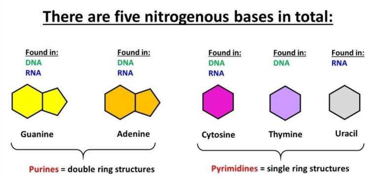
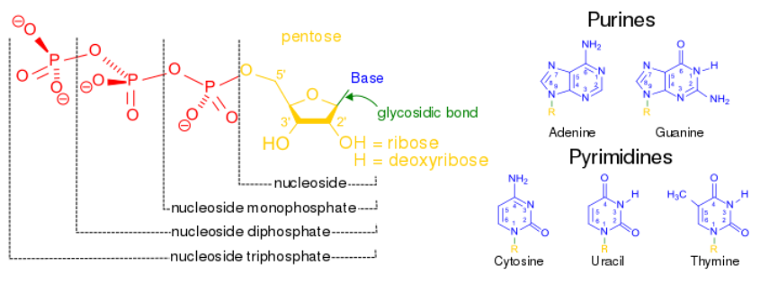
- The above figure shows nitrogenous base pairs of DNA. Adenine and Guanine are Purines (9 membered / double ring structures.) and the other two : Cytosine and Thymine are Pyrimidines (6 membered ring structures). See the image below for more clearance :
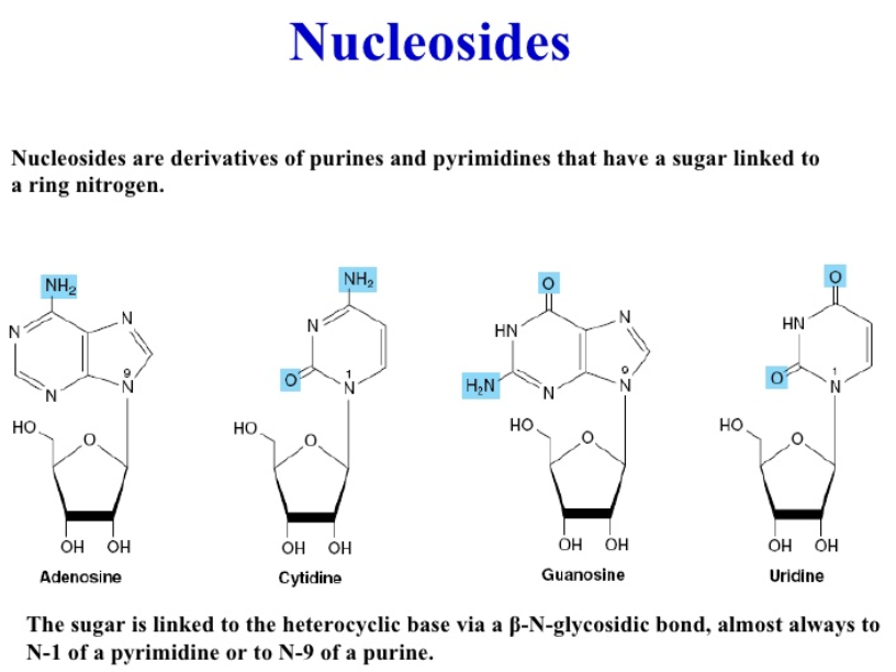
- The sugar pairs with nitrogenous base pairs via Glycosidic Linkage and the structure so formed is known as Nucleoside. If adenine pairs with deoxyribose sugar then the resulting structure is known as deoxyadenosine or if it pairs with ribose sugar then it is known as adenosine. Similarly other nucleosides are guanosine, deoxyguanosine, cytidine, deoxycytidine and uridine or deoxyuridine. Nitrogenous base pairs will always be linked at first carbon of the sugar molecules. Never at any other position.
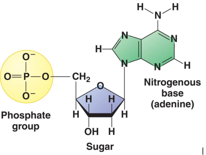
- When a phosphate group gets linked to 5C of the sugar via phospho ester linkage in a nucleoside it becomes a nucleotide or a deoxynucleotide (if deoxyribose sugar is present) as shown below.
- Overall structure of a nucleotide.
- A DNA molecule has many such nucleotides in the form of a chain. That is why we call DNA a polynucleotide chain.
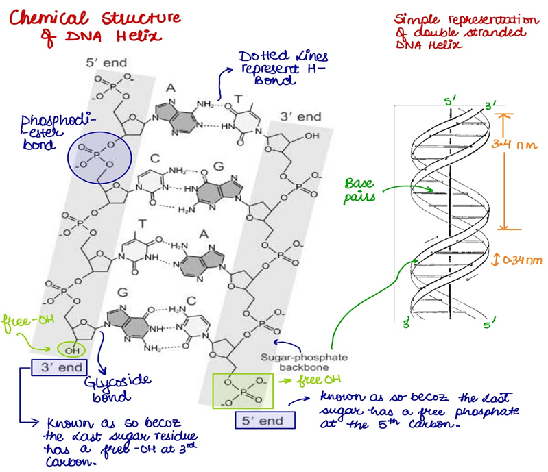
- One nucleotide is linked to another via a 3’-5’ phosphodiester linkage. These two nucleotides form a dinucleotide, soon another nucleotide gets linked to this dinucleotide to form a trinucleotide and then a tetranucleotide and so on. At last we get a polynucleotide chain whose one end has a free phosphate group attached to 5C of sugar so this end is known as 5’ end (pronounced as 5 prime end) while the other end of the polynucleotide chain has a free -OH group at 3C so this end is known as 3’ end (pronounced as 3 prime end).
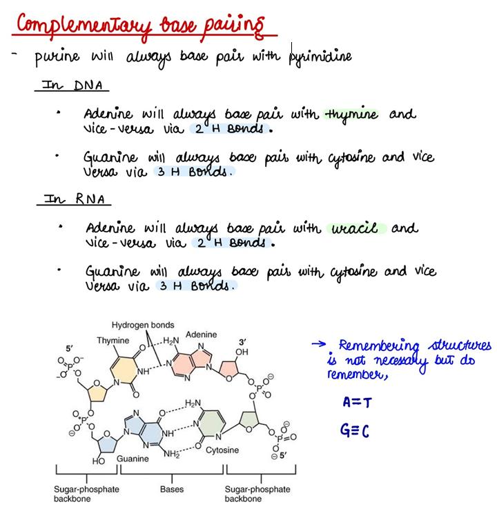
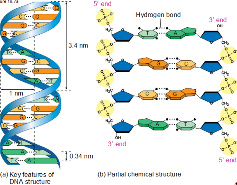
- DNA has two polynucleotide chains in opposite directions that is one strand has polarity 3’-5’ from down to top and another strand has 5’-3’ polarity from down to top and are linked to each other via hydrogen bond between the nitrogenous base pairs as shown below :
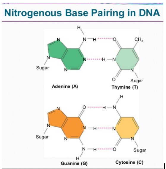
- As we can see in the above image, adenine is bonded to thymine by 2 hydrogen bonds and guanine is bonded to cytosine by 3 hydrogen bonds. These bondings show that a purine always base pairs with a pyrimidine and a pyrimidine will always base pair with a purine.
- This one bond between a purine and a pyrimidine is known as one base pair. The haploid human DNA has 3.3 X 10⁹ bp (base pairs). In a similar way the genome of E.coli bacteria has 4.6 X 10⁶ bp and Bacteriophage Lambda has 48502 bp.
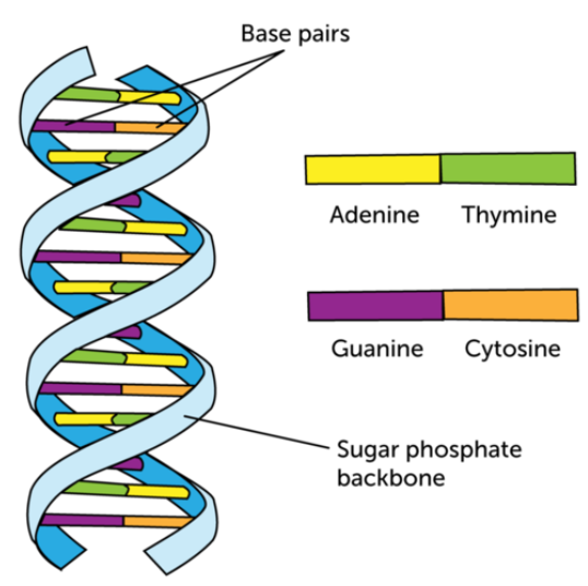
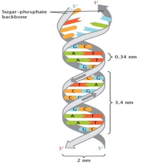
- The DNA has a double helical structure shown below :

DISCOVERY OF DOUBLE HELICAL STRUCTURE OF DNA
- DNA was first identified as a acidic substance in the nucleus by Friedrich Meischer in 1869. He named it as Nuclein but he couldn’t deduce the structure due to limitations of the equipment present at that time.
- In the year 1950s Maurice Wilkins and Rosalind Franklin, produced the first clear image of DNA by using X-ray diffraction. This picture came to be known as the X-ray diffraction data of crystallized DNA.
- In the year 1953 James Watson and Francis Crick, based on the X-ray diffraction picture and the observation of Erwin Chargaff proposed the double helical structure of DNA.
- Chargaff’s Rule : Ratio of Adenine:Thymine and Guanine:Cytosine are constant and equals to one. Which translates into : amount of purine always equals amount of pyrimidine in a double helical DNA. For eg : if a particular DNA sample has 20% Adenine then it will have 20% thymine to exactly base pair with the 20% adenine. So this 20% adenine and 20% thymine covers 40% out of the total 100% DNA content so the leftover 60% will be shared between Guanine (half of the remaining 60% which equals 30%) and Cytosine (half of the remaining 60% which equals 30%).
- It is made up of two polynucleotide chains having backbone of sugar-phosphate and the nitrogenous bases project inwards.
- The two polynucleotide chains are antiparallel (polarity wise). It means if one chain has the polarity 3’→5’ then the other has 5’ → 3’.
- The nitrogenous bases are linked via hydrogen bond. A purine will always pair with a pyrimidine and vice versa. Adenine is bonded with thymine via 2 H-Bonds and Guanine is bonded to Cytosine via 3 H-bonds.
- The two polynucleotide chains are coiled in a right handed fashion.
- The distance between two turns (pitch of the helix) is 3.4nm. Each turn of the right handed coiled DNA (B form of DNA) / B-DNA has 10 nucleotides which means that the distance between two adjacent base pairs is 3.4/10 = 0.34nm.
- The plane of one base pair stacks over the other. This provides additional stability in addition to the stability provided by H-bonding between base pairs.
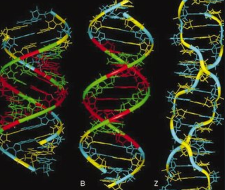
- Different forms of DNA are shown below:
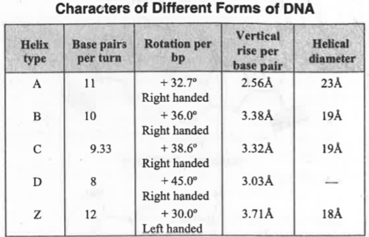
- Some different forms of DNA are shown in figure below. B -DNA is the most common form and the one studied by Crick & Watson.
SALIENT FEATURES OF DNA DOUBLE HELIX
Packaging of DNA Helix
- We know that human haploid DNA has 3.3 X 10⁹ bp and the distance between two base pairs is 0.34nm or 0.34 X 19−9m so the length of the DNA comes out to be approximately 2.2m because 6.6 X 109 bp X 0.34 X 10−9 m/bp = 2.2m, but the the size of nucleus is approximately 10−6m. So the question arises how such an amount of DNA can be packed in a teeny tiny nucleus of order 10−6m? Prokaryotes and Eukaryotes both have different ways of packing the DNA.
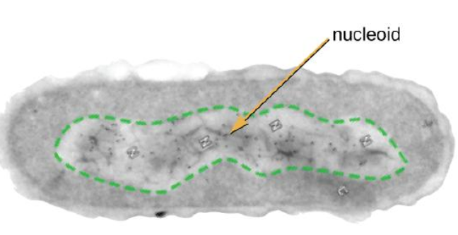
- Prokaryotes do not have a well defined nucleus but that does mean that their DNA is scattered throughout the cell. DNA is negatively charged because of the presence of phosphate groups so this negatively charged DNA is held by some positively charged proteins in the form of large loops in a region known as nucleoid.
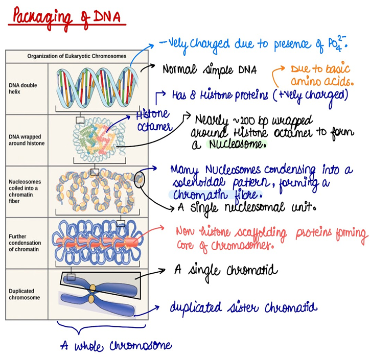
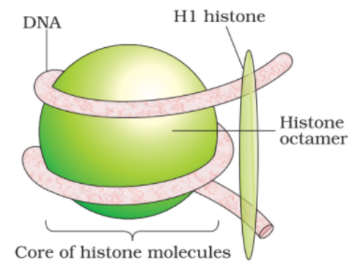
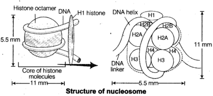
- Packaging of DNA in eukaryotes is much more complex than in prokaryotes. positively charged basic proteins known as histone proteins play a significant role in Packaging of DNA. These histone proteins are rich in basic amino acids such as lysine and arginine. 8 histone molecules are organized to a single unit known as histone octamer.
- The negatively charged DNA wraps itself around the positively charged histone octamer to form a structure known as nucleosome.
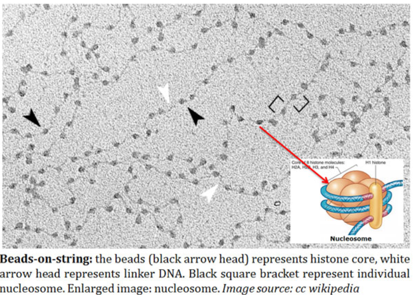
- A single nucleosome has around 200 base pairs wrapped around itself. Histone octamer consists of two copies of each of the four core histone proteins (H2A, H2B, H3 and H4). This form when seen under the electron microscope resembles beads on a string model where intensely colored bodies are nucleosomes.
- The DNA between two nucleosomes is known as Linker DNA.
- This ‘Beads on a string’ form of chromatin further condenses to chromatin fiber which further condenses and condenses into a typical chromosome shaped chromosome during the metaphase of cell cycle. Recall class 11 chapter : Cell Cycle & Cell Division. The packaging at this stage requires some additional non histone proteins also whose discussion is not necessary at this stage.
- At any normal point in a nucleus, some part of chromatin is loosely packed so that part could be transcribed. (The process of transcription will be discussed in the later sections.)That loosely packed part of chromatin is known as Euchromatin and this part stains lighter than the other parts.
- The part of chromatin which is densely packed and has no active transcription is going on (transcriptionally inactive) is known as Heterochromatin. This part stains darker than the other because it is more densely packed.
In Prokaryotes
In Eukaryotes
STRUCTURE & PACKAGING OF DNA
DEOXYRIBONUCLEIC ACID (DNA)
- DNA is a long chain of polynucleotides. A single nucleotide is made up of 3 components :
- Deoxyribose Sugar
- Nitrogenous Base Pair (either purine or pyrimidine) Recall class 11, chapter : Biomolecules.
- Phosphate Bond
- The above picture shows two types of pentose sugar (sugar having 5 carbon atoms) : Deoxyribose and Ribose. Deoxy in Deoxyribose means without oxygen (at second carbon atom highlighted in blue) and ribose is a 5C sugar represented by the green coloured structure.
- The above figure shows nitrogenous base pairs of DNA. Adenine and Guanine are Purines (9 membered / double ring structures.) and the other two : Cytosine and Thymine are Pyrimidines (6 membered ring structures). See the image below for more clearance :

- The sugar pairs with nitrogenous base pairs via Glycosidic Linkage and the structure so formed is known as Nucleoside. If adenine pairs with deoxyribose sugar then the resulting structure is known as deoxyadenosine or if it pairs with ribose sugar then it is known as adenosine. Similarly other nucleosides are guanosine, deoxyguanosine, cytidine, deoxycytidine and uridine or deoxyuridine. Nitrogenous base pairs will always be linked at first carbon of the sugar molecules. Never at any other position.
- When a phosphate group gets linked to 5C of the sugar via phospho ester linkage in a nucleoside it becomes a nucleotide or a deoxynucleotide (if deoxyribose sugar is present) as shown below.
- Overall structure of a nucleotide.
- A DNA molecule has many such nucleotides in the form of a chain. That is why we call DNA a polynucleotide chain.
- One nucleotide is linked to another via a 3’-5’ phosphodiester linkage. These two nucleotides form a dinucleotide, soon another nucleotide gets linked to this dinucleotide to form a trinucleotide and then a tetranucleotide and so on. At last we get a polynucleotide chain whose one end has a free phosphate group attached to 5C of sugar so this end is known as 5’ end (pronounced as 5 prime end) while the other end of the polynucleotide chain has a free -OH group at 3C so this end is known as 3’ end (pronounced as 3 prime end).
- DNA has two polynucleotide chains in opposite directions that is one strand has polarity 3’-5’ from down to top and another strand has 5’-3’ polarity from down to top and are linked to each other via hydrogen bond between the nitrogenous base pairs as shown below :

- As we can see in the above image, adenine is bonded to thymine by 2 hydrogen bonds and guanine is bonded to cytosine by 3 hydrogen bonds. These bondings show that a purine always base pairs with a pyrimidine and a pyrimidine will always base pair with a purine.
- This one bond between a purine and a pyrimidine is known as one base pair. The haploid human DNA has 3.3 X 10⁹ bp (base pairs). In a similar way the genome of E.coli bacteria has 4.6 X 10⁶ bp and Bacteriophage Lambda has 48502 bp.
- The DNA has a double helical structure shown below :


DISCOVERY OF DOUBLE HELICAL STRUCTURE OF DNA
- DNA was first identified as a acidic substance in the nucleus by Friedrich Meischer in 1869. He named it as Nuclein but he couldn’t deduce the structure due to limitations of the equipment present at that time.
- In the year 1950s Maurice Wilkins and Rosalind Franklin, produced the first clear image of DNA by using X-ray diffraction. This picture came to be known as the X-ray diffraction data of crystallized DNA.
- In the year 1953 James Watson and Francis Crick, based on the X-ray diffraction picture and the observation of Erwin Chargaff proposed the double helical structure of DNA.
- Chargaff’s Rule : Ratio of Adenine:Thymine and Guanine:Cytosine are constant and equals to one. Which translates into : amount of purine always equals amount of pyrimidine in a double helical DNA. For eg : if a particular DNA sample has 20% Adenine then it will have 20% thymine to exactly base pair with the 20% adenine. So this 20% adenine and 20% thymine covers 40% out of the total 100% DNA content so the leftover 60% will be shared between Guanine (half of the remaining 60% which equals 30%) and Cytosine (half of the remaining 60% which equals 30%).
- It is made up of two polynucleotide chains having backbone of sugar-phosphate and the nitrogenous bases project inwards.
- The two polynucleotide chains are antiparallel (polarity wise). It means if one chain has the polarity 3’→5’ then the other has 5’ → 3’.
- The nitrogenous bases are linked via hydrogen bond. A purine will always pair with a pyrimidine and vice versa. Adenine is bonded with thymine via 2 H-Bonds and Guanine is bonded to Cytosine via 3 H-bonds.
- The two polynucleotide chains are coiled in a right handed fashion.
- The distance between two turns (pitch of the helix) is 3.4nm. Each turn of the right handed coiled DNA (B form of DNA) / B-DNA has 10 nucleotides which means that the distance between two adjacent base pairs is 3.4/10 = 0.34nm.
- The plane of one base pair stacks over the other. This provides additional stability in addition to the stability provided by H-bonding between base pairs.
- Different forms of DNA are shown below:

- Some different forms of DNA are shown in figure below. B -DNA is the most common form and the one studied by Crick & Watson.
SALIENT FEATURES OF DNA DOUBLE HELIX
This topic often comes as a 5 mark question along with a diagram.


Packaging of DNA Helix
- We know that human haploid DNA has 3.3 X 10⁹ bp and the distance between two base pairs is 0.34nm or 0.34 X 19−9m so the length of the DNA comes out to be approximately 2.2m because 6.6 X 109 bp X 0.34 X 10−9 m/bp = 2.2m, but the the size of nucleus is approximately 10−6m. So the question arises how such an amount of DNA can be packed in a teeny tiny nucleus of order 10−6m? Prokaryotes and Eukaryotes both have different ways of packing the DNA.
- Prokaryotes do not have a well defined nucleus but that does mean that their DNA is scattered throughout the cell. DNA is negatively charged because of the presence of phosphate groups so this negatively charged DNA is held by some positively charged proteins in the form of large loops in a region known as nucleoid.
- Packaging of DNA in eukaryotes is much more complex than in prokaryotes. positively charged basic proteins known as histone proteins play a significant role in Packaging of DNA. These histone proteins are rich in basic amino acids such as lysine and arginine. 8 histone molecules are organized to a single unit known as histone octamer.
- The negatively charged DNA wraps itself around the positively charged histone octamer to form a structure known as nucleosome.
- A single nucleosome has around 200 base pairs wrapped around itself. Histone octamer consists of two copies of each of the four core histone proteins (H2A, H2B, H3 and H4). This form when seen under the electron microscope resembles beads on a string model where intensely colored bodies are nucleosomes.
- The DNA between two nucleosomes is known as Linker DNA.
- This ‘Beads on a string’ form of chromatin further condenses to chromatin fiber which further condenses and condenses into a typical chromosome shaped chromosome during the metaphase of cell cycle. Recall class 11 chapter : Cell Cycle & Cell Division. The packaging at this stage requires some additional non histone proteins also whose discussion is not necessary at this stage.
- At any normal point in a nucleus, some part of chromatin is loosely packed so that part could be transcribed. (The process of transcription will be discussed in the later sections.)That loosely packed part of chromatin is known as Euchromatin and this part stains lighter than the other parts.
- The part of chromatin which is densely packed and has no active transcription is going on (transcriptionally inactive) is known as Heterochromatin. This part stains darker than the other because it is more densely packed.
In Prokaryotes

In Eukaryotes


THE SEARCH FOR THE GENETIC MATERIAL
Grifith’s Transforming Principle
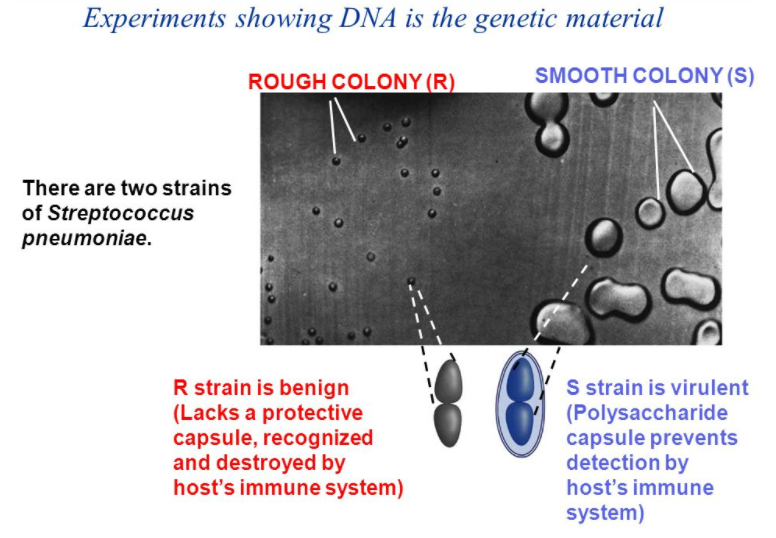
- Disease causing strain (virulent strain) : it forms smooth colonies so known as S-strain
- Non-Virulent (does not cause disease pneumonia) : it forms rough colonies because it does not have the mucous coat. It forms rough looking colonies so known as R-strain.
- Experiment done by Griffith :

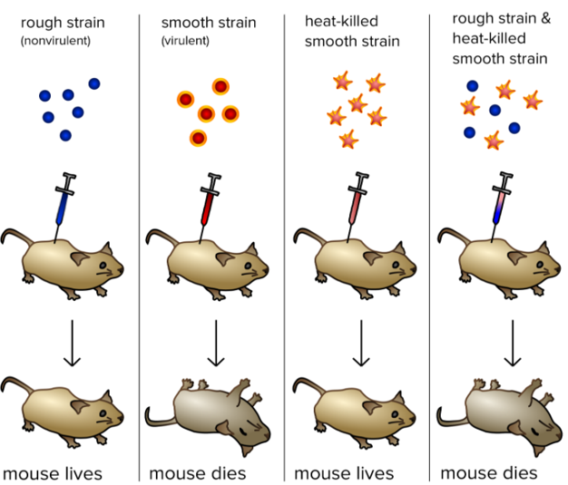
Explanation of above picture : Griffith injected S strain to healthy mice and after sometime the mice died due to disease pneumonia whereas when he injected another healthy mice with the R-strain of bacteria, the mice survived because the R-strain does not cause any disease. - Second phase of the experiment :
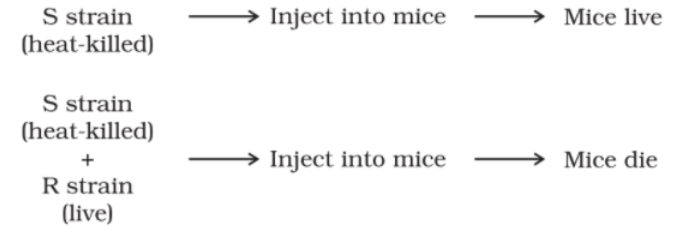
Explanation : Next Griffith injected the heat killed S-strain bacteria into the mice and the mice lived because the disease causing bacteria had died during the heat treatment so there were no live bacterias to cause any disease. But something unexpected happened when he injected the heat killed S-strain bacteria mixed with the live R-strain bacteria, the mice died. - He concluded that the R strain bacteria had somehow been transformed by the heat-killed S strain bacteria. Some ‘transforming principle’, transferred from the heat-killed S strain, had enabled the R strain to synthesise a smooth polysaccharide coat and become virulent. This must be due to the transfer of the genetic material.
- Griffith could not determine the biochemical nature of the genetic material which transformed the non virulent R strain into the virulent S-strain.
In the year 1928, when Griffith was experimenting using the bacteria Streptococcus pneumoniae which causes pneumonia. This bacteria has two strains:
Conclusion
Drawback
Biochemical Characterisation of Transforming Principle
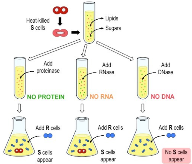
- The first experiment which showed that the genetic material is indeed DNA was performed by Oswald Avery, Colin MacLeod and Maclyn McCarty (1933-44) . Before their experiment, protein was thought to be the genetic material.
- They purified the biochemical components from the heat killed S-strain in three separate test tubes. They added live R-strain bacteria to each along with DNase in one test tube, Protease in second and RNase in third test tube. When the R-strain was cultured from the above test tubes, the R-strain from the test tube containing DNase did not transform into virulent S-strain while the R-strain transformed in the test tube containing protease and RNase suggesting protease and RNase do no affect the transformation.
- Observation : when the R strain from test tube 1 was injected in live mice, the mice survived.
- When the R-strain from test tube 2 and 3 was injected into separate healthy mices, they died of pneumonia.
- Explanation
- The first observation suggested that addition of DNase digested the DNA and when this mixture was injected to healthy mice, the mic did not die because the ‘Griffith’s transforming factor’ was the DNA from S-strain which was destroyed by addition of DNase.
- Second observation suggested that R strain from test tube 2 containing protease (protein digesting enzyme) has transformed into virulent strain which means that protein is not the genetic material since it was destroyed by the protease.
→ Similarly in test tube 3, RNase destroyed the RNA content but R strain had still transformed into virulent strain because the DNA from the S-strain was still intact.
But many scientists were not convinced by this experiment and as a result it was not highly accepted until the final set of experiments by Hershey and Chase in 1952 finally proved and convinced everyone that DNA is indeed the genetic material.
Alfred Hershey and Martha Chase Experiment (1952).
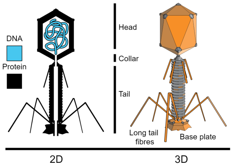
- They worked on T2 Bacteriophage which is a type of virus that attacks the bacteria E.coli. Now before proceeding into this experiment you need to look at the structure of this virus which is similar to viruses which you studied in class 11 in the diversity unit.
- Above figure shows 2 D and 3D structure of bacteriophage in which the head is made of protein and inside is DNA. other structures are proteinaceous in nature. The main debate at that time was whether the genetic material is DNA or Protein.
- This virus multiplies by injecting its genetic material in the bacteria E.coli.
- So Hershey and Chase labelled both the protein coat (shown in black colour in above diagram) and the DNA (shown in sky blue colour in above diagram) by radioactive molecules. DNA has phosphorus (in phosphodiester bond) so we used radioactive phosphorus (P32) to label it. Protein has some sulphur containing amino acids like Methionine and Cysteine so we used radioactive sulphur (S35) to label the protein coat.
- Some bacteriophages were grown in the culture medium containing radioactive sulphur and other bacteriophages were cultured in culture medium containing radioactive phosphorus. So we had 2 types of bacteriophages :
- Bacteriophages with radioactive sulphur labelled protein coat but normal DNA and
- Bacteriophages with radioactive phosphorus labelled DNA but normal protein coat.
- Stages of the experiment :
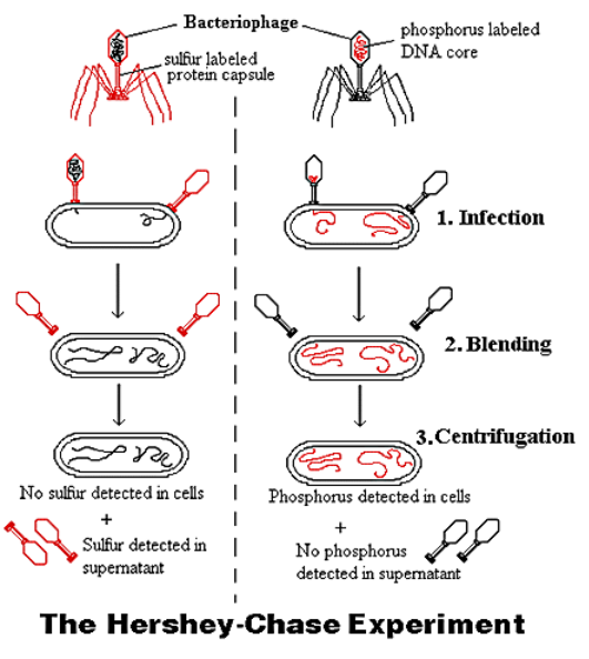
Explanation - The first stage is infection : virus attaches itself to the bacteria and injects its genetic material into the bacteria.
- The next stage is blending so as to remove the viral coat attached to the E.coli bacteria using a blender.
- Now to separate bacteria from viral coat, the third stage was centrifugation.
- Radioactive phosphorus was detected in the E.coli cells which were infected by Bacteriophage containing radioactive labelled DNA while no radioactivity was observed in the E.coli cells infected with bacteriophage labelled by radioactive sulphur.
- Radioactively labelled DNA of bacteriophage imparted radioactivity to the E.coli cells so DNA is the genetic material.
Observation
Conclusion
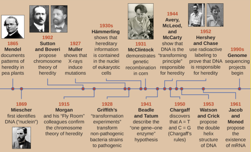
A Brief Timeline Of Experiments Leading To Identification Of DNA As Genetic Material :
THE SEARCH FOR THE GENETIC MATERIAL
- Genetic material ki search ke liye kafi alag alag experiment hue jinme se kuch jaruri hum abhi discuss karenge.
- Griffith ne Staphylococcus bacteria ke 2 strain : R-strain (jo disease cause nhi krta) and S-strain (jo disease cause krta hai).
- Jab unhone bacteria ki R strain mice me inject kiya toh mice me koi disease nhi hui and mice alive tha but jab unhone S-strain ko dusre healthy mice me inject kiya toh mice ko pneumonia hua and he died.
- Iske baad unhone S strain ko heat kill krke inject kiya toh mice alive tha and jan R-strain ko bhi heat kill krke inject kiya toh mice firse alive hi tha.
- Lekin jab unhone heat killed S strain bacteria ko live R-Strain bacteria ke sath mila kr inject kiya toh mice ki death ho gyi.
- Issi last step ke basis pe unhone apna Transforming Principle diya ki kisi cheez ke karan jo live R strain bacteria tha wo S-strain me transform ho gya jisse mice ko pneumonia hua and uski death hogyi.
- Limitation : Griffith yeah nai bta paye ki jis cheez ke karan R strain, S me transform hua uska biochemical nature kaisa hai, yani ki wo cheez DNA hai ya RNA hai ya Protein hai? But iss cheez ki discovery Macleod, McCarty ke experiment se mili.
- Ab in teno ne kiya yeah heat killed S strain se DNA,RNA, & Protein ko purify karke 3 test tube me dala.
- Ek test tube me unhone DNase daal diya jisne uss test tube me jitna bhi DNA tha usko digest kr dala, dusri test tube me unhone RNase daal diya jissey uss test tube ka RNA content digest ho gya and third test tube me unhone protease daal diye jinhone uss test tube ke protein content ko digest kar diyaa.
- Next unhone un test tube me live R-cells ko add kiya so that jo bhi cheez transformation ke liye responsible thi usko R-strain pickup krlega.
- Fir unhone har test tube se ke solution ko ek ek karke live mice me dala toh jiss test tube me DNase dala tha keval uss test tube ke solution ko jis mice me dala tha, uski death nahi hui baki dono mice dead thy.
- Toh iska matlab yeah ki genetic material DNA tha kyunki keval uske degradation par mice alive raha because the R-strain could not transform into the S-strain in absence of DNA from S-strain.
- Inhone bacteriophage ke sath kaam kiya jo ki ek aisa virus hai jo apna genetic material E.coli bacteria me inject krke, bacterial machinery ko use krke apny genetic material ko replicate karta hai.
- Toh inhone bacteriophage ke DNA ko radioactive phosphorus se label krdia and uske Protein coat ko radioactive sulphur se coat taki.
- Ab hog yeah ki jaise hi bacteriophage bacteria me apna genetic material inject karege, bacteria me radioactivity show hogi and agar bacteria me radioactive phosphorus mile toh genetic material DNA hai and agar radioactive sulphur mila toh genetic material Protein hoga.
- Toh unhone 2 plates me E.coli bacteria liye, ek plate me radioactive sulfur wale bacteriophage dale and ek plate me radioactive phosphorus waley bacteria daley. Iss stage ko infection stage kaha gya.
- Infection ke waqt bacteriophage bacteria ki membrane se attach ho jate hai toh unko remove karne ke humne infected bacteria ko blend and centrifuge kiya taki humare pass keval infected bacteria hi rahe.
- Iske baad jab unhone infected bacteris ko radioactivity ke liye test kiya toh keval unhe radioactive phosphorus mila jiska matlab tha ki DNA hi genetic material hai.
Griffith’s Experiment
Oswald Avery, Colin MacLeod and Maclyn McCarty (1933-44)
→ But abhi kuch scientists convinced ni thy ki DNA hi genetic material hai and kuch samay baad Hershey & Chase ke experiment ne yeah prove kr dia ki DNA hi genetic material hai.
Hershey & Chase Experiment
RNA WORLD
- Some viruses like tobacco mosaic virus (TMV), QB bacteriophage etc have RNA as their genetic material but the predominant genetic material of most of the living organisms is DNA due to its properties which were already discussed in the first section of the chapter. There are some differences between DNA and RNA which make the RNA a poor candidate for being the genetic material in complex living organisms.
- The genetic material should be stable enough not to change with different stages of life cycle, age or with change in physiology of the organism. Stability as one of the properties of genetic material was very evident in Griffith’s ‘transforming principle’ itself that heat, which killed the bacteria, at least did not destroy some of the properties of genetic Material. 2'-OH group present at every nucleotide in RNA is a reactive group and makes RNA labile (less stable) and easily degradable.
- RNA also displays catalytic properties which means its highly reactive but DNA does not show any such properties so DNA is not highly reactive and structurally more stable than RNA.
- RNA has uracil while DNA has thymine and thymine provides more stability to DNA than uracil could provide to RNA.
- RNA was the first genetic material. During the evolution of life, RNA acted as a catalyst and a genetic material both. But as we know, that being catalytic means RNA was highly reactive thus very unstable and more prone to mutations. So, DNA evolved from RNA with a few chemical properties. RNA is generally single stranded while DNA is normally double stranded. Double stranded DNA has additional stability, can resist harmful mutations and can repair itself.
- At this point this is enough and going beyond what we have already discussed here about the structure of RNA and DNA is really beyond the scope of class 12th biology.
Differences Between DNA and RNA
So among the two, DNA is a better genetic material.
RNA World
THE SEARCH FOR THE GENETIC MATERIAL
Grifith’s Transforming Principle
Generally comes as 3 mark short note question in CBSE exams.
- Disease causing strain (virulent strain) : it forms smooth colonies so known as S-strain
- Non-Virulent (does not cause disease pneumonia) : it forms rough colonies because it does not have the mucous coat. It forms rough looking colonies so known as R-strain.
- Experiment done by Griffith :

Explanation of above picture : Griffith injected S strain to healthy mice and after sometime the mice died due to disease pneumonia whereas when he injected another healthy mice with the R-strain of bacteria, the mice survived because the R-strain does not cause any disease. - Second phase of the experiment :

Explanation : Next Griffith injected the heat killed S-strain bacteria into the mice and the mice lived because the disease causing bacteria had died during the heat treatment so there were no live bacterias to cause any disease. But something unexpected happened when he injected the heat killed S-strain bacteria mixed with the live R-strain bacteria, the mice died. - He concluded that the R strain bacteria had somehow been transformed by the heat-killed S strain bacteria. Some ‘transforming principle’, transferred from the heat-killed S strain, had enabled the R strain to synthesise a smooth polysaccharide coat and become virulent. This must be due to the transfer of the genetic material.
- Griffith could not determine the biochemical nature of the genetic material which transformed the non virulent R strain into the virulent S-strain.
In the year 1928, when Griffith was experimenting using the bacteria Streptococcus pneumoniae which causes pneumonia. This bacteria has two strains:

Conclusion
Drawback

Biochemical Characterisation of Transforming Principle
- The first experiment which showed that the genetic material is indeed DNA was performed by Oswald Avery, Colin MacLeod and Maclyn McCarty (1933-44) . Before their experiment, protein was thought to be the genetic material.
- They purified the biochemical components from the heat killed S-strain in three separate test tubes. They added live R-strain bacteria to each along with DNase in one test tube, Protease in second and RNase in third test tube. When the R-strain was cultured from the above test tubes, the R-strain from the test tube containing DNase did not transform into virulent S-strain while the R-strain transformed in the test tube containing protease and RNase suggesting protease and RNase do no affect the transformation.
- Observation : when the R strain from test tube 1 was injected in live mice, the mice survived.
- When the R-strain from test tube 2 and 3 was injected into separate healthy mices, they died of pneumonia.
- Explanation
- The first observation suggested that addition of DNase digested the DNA and when this mixture was injected to healthy mice, the mic did not die because the ‘Griffith’s transforming factor’ was the DNA from S-strain which was destroyed by addition of DNase.
- Second observation suggested that R strain from test tube 2 containing protease (protein digesting enzyme) has transformed into virulent strain which means that protein is not the genetic material since it was destroyed by the protease.
→ Similarly in test tube 3, RNase destroyed the RNA content but R strain had still transformed into virulent strain because the DNA from the S-strain was still intact.
But many scientists were not convinced by this experiment and as a result it was not highly accepted until the final set of experiments by Hershey and Chase in 1952 finally proved and convinced everyone that DNA is indeed the genetic material.

Alfred Hershey and Martha Chase Experiment (1952).
- They worked on T2 Bacteriophage which is a type of virus that attacks the bacteria E.coli. Now before proceeding into this experiment you need to look at the structure of this virus which is similar to viruses which you studied in class 11 in the diversity unit.
- Above figure shows 2 D and 3D structure of bacteriophage in which the head is made of protein and inside is DNA. other structures are proteinaceous in nature. The main debate at that time was whether the genetic material is DNA or Protein.
- This virus multiplies by injecting its genetic material in the bacteria E.coli.
- So Hershey and Chase labelled both the protein coat (shown in black colour in above diagram) and the DNA (shown in sky blue colour in above diagram) by radioactive molecules. DNA has phosphorus (in phosphodiester bond) so we used radioactive phosphorus (P32) to label it. Protein has some sulphur containing amino acids like Methionine and Cysteine so we used radioactive sulphur (S35) to label the protein coat.
- Some bacteriophages were grown in the culture medium containing radioactive sulphur and other bacteriophages were cultured in culture medium containing radioactive phosphorus. So we had 2 types of bacteriophages :
- Bacteriophages with radioactive sulphur labelled protein coat but normal DNA and
- Bacteriophages with radioactive phosphorus labelled DNA but normal protein coat.
- Stages of the experiment :

Explanation - The first stage is infection : virus attaches itself to the bacteria and injects its genetic material into the bacteria.
- The next stage is blending so as to remove the viral coat attached to the E.coli bacteria using a blender.
- Now to separate bacteria from viral coat, the third stage was centrifugation.
- Radioactive phosphorus was detected in the E.coli cells which were infected by Bacteriophage containing radioactive labelled DNA while no radioactivity was observed in the E.coli cells infected with bacteriophage labelled by radioactive sulphur.
- Radioactively labelled DNA of bacteriophage imparted radioactivity to the E.coli cells so DNA is the genetic material.

Observation
Conclusion
A Brief Timeline Of Experiments Leading To Identification Of DNA As Genetic Material :

DNA REPLICATION
- Watson and Crick suggested the semiconservative replication of DNA in 1953 but no experimental proof was provided but nonetheless as we know today that they were indeed correct. The experiment proof is shown by Messelson and Stahl’s experiment in 1958.
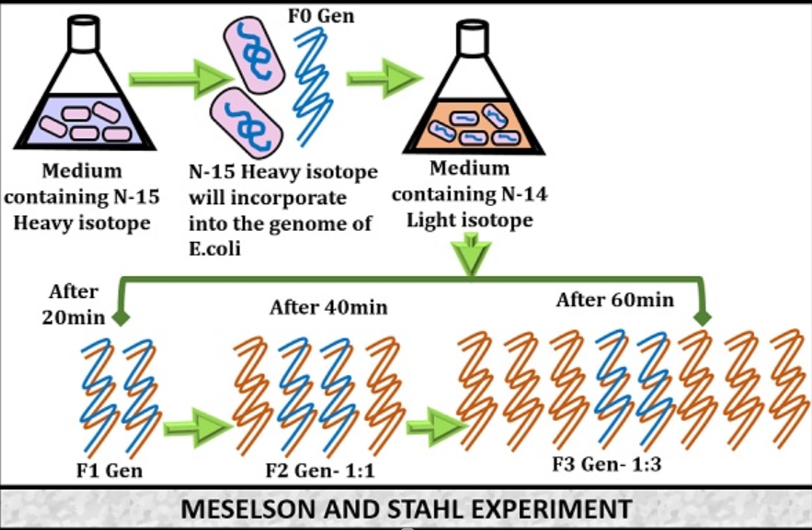
- They grew E.coli bacteria on a nutritional culture medium containing .15NH4Cl (.15N is the heavy isotope of nitrogen while N14 is the normal isotope of nitrogen) as the only nitrogen source for many generations so that this heavy nitrogen isotope N-15 was incorporated into newly synthesized DNA with each generation. Normal DNA has N14 while these E.coli cells had the heavy DNA as they had N15 so there was a difference in their density and the normal DNA and heavy DNA could be separated from a mixture by centrifugation in a CsCl (Caesium Chloride) density gradient where heavy DNA would sit at the base of test tube while normal DNA was found found above it.
- After growing E.coli in a heavy nitrogen medium for several generations they transferred cells to a medium containing normal nitrogen and took samples at definitive intervals. E.coli cells divide every 20 minutes.
- So the sample extracted just after 20 minutes had intermediate density value. Which means that this DNA was heavier than N14 DNA but lighter than the DNA with N15.
- Taylor and colleagues in 1958 conducted similar experiments on Vicia faba (faba beans) to show semiconservative mode of DNA replication, but instead of using heavy nitrogen isotope he used radioactive thymidine.
- The above image represents meselson and stahl experiment in a diagrammatic way. The first picture on the left hand side represents a double stranded red coloured DNA which has heavy nitrogen incorporated in it. Now when this DNA strand was centrifuged, this DNA sat at the bottom of the centrifuge tube. In the next stage when the E.coli were transferred to the normal nitrogen containing culture media their DNA replicated and the first sample was taken within the 20 minutes. The green colour in DNA represents normal N-14 nitrogen. This particular DNA sample has two types of DNA strand out of which one is of red colour and the other one is of green colour. so this is intermediate or we can say a hybrid DNA whose density will also be of intermediate value. Now what happened during the process of replication was that the original red colour DNA got separated into two strands which acted like a template using which new green coloured DNA strands were synthesized.This mode of replication is known as semi conservative mode of replication which means that every time a DNA replicates the new DNA will have one original parent stand while the daughter strand will be synthesized each time conserving 50% of the DNA so that the semi conservative nature of DNA replication.
- The experiment that we studied just now might not have made sense to a few of us because we really don’t know what exactly happened in there. So in this section we will study how DNA replicates itself and what does the term Semiconservative mode exactly means.
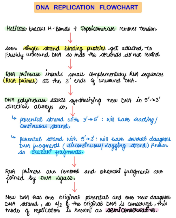
- There are several points in DNA from which DNA replication will start because eukaryotes have a very long DNA which means that the cell cannot really afford to replicate its DNA from one end to the other due to the energy constraints and other factors. During this process the cell will utilise a different set of enzymes each with its own role and function. Some of these enzymes will find the exact point in the DNA from where DNA replication will take place; and the point from where DNA replication takes place is known as origin of replication or we call it Ori point in short.
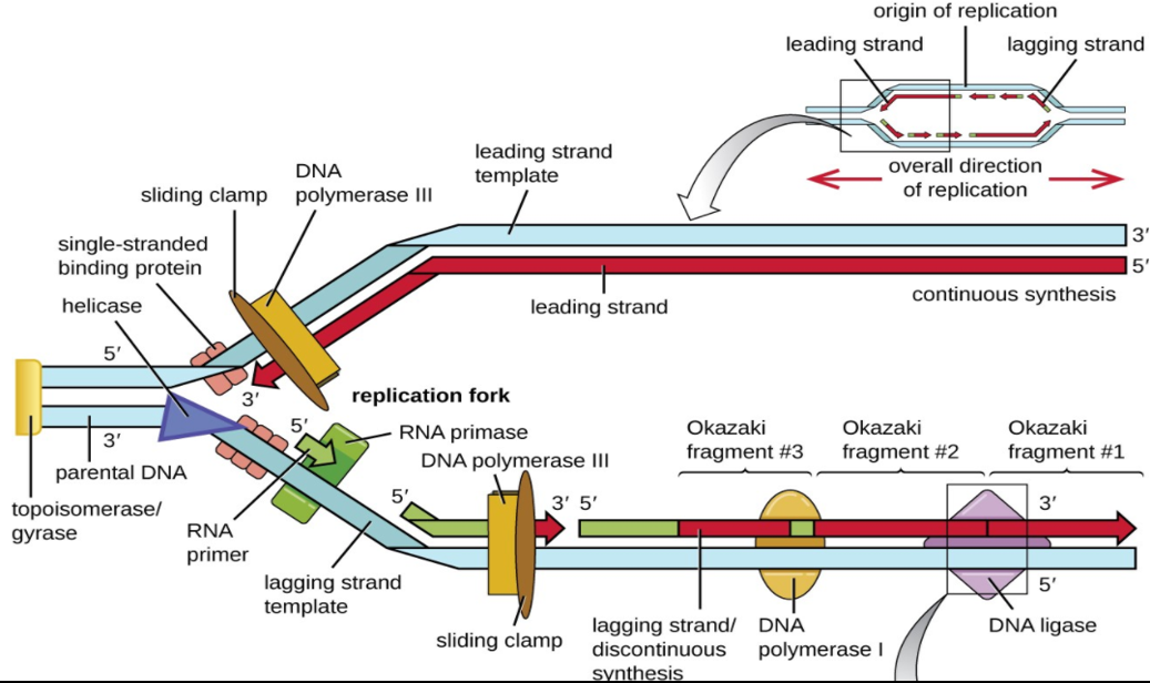
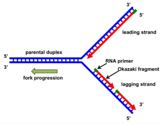
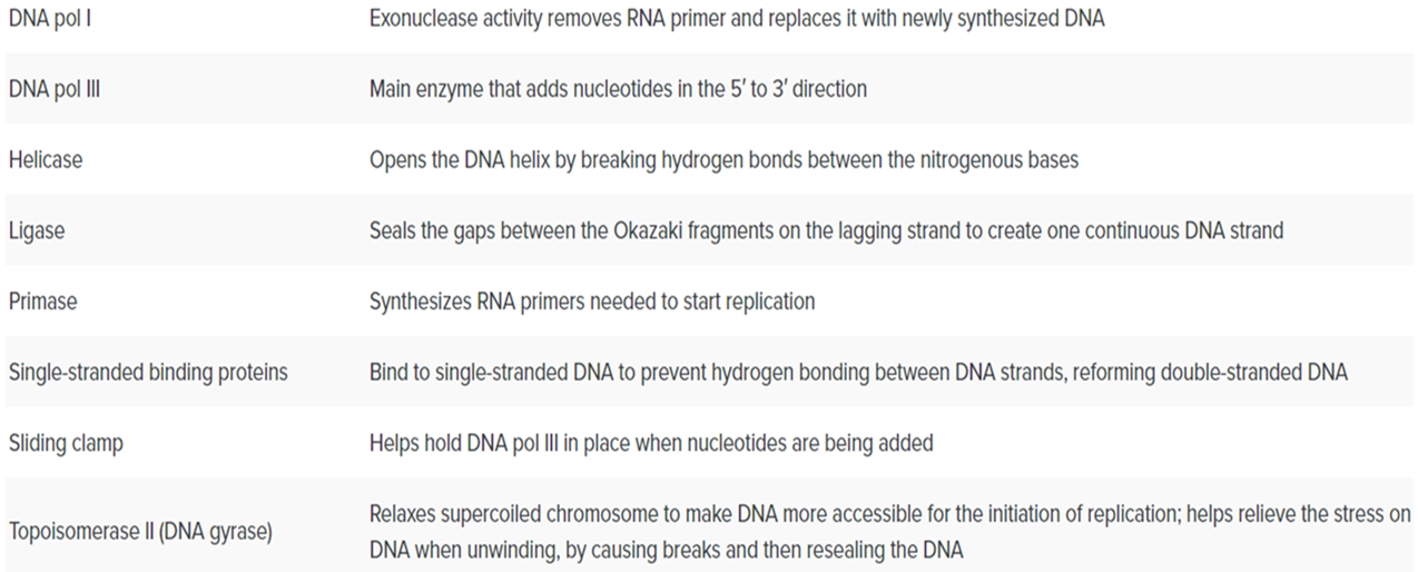
- The Above figure represents a structure which is known as a replication fork ( which is a Y shaped structure formed during the DNA replication). Enzyme DNA helicase unwinds the DNA at the ori point. Now remember that the DNA was coiled in a right-handed fashion. So unwinding by the enzyme DNA helicase creates a tension in the DNA and this tension is released by the enzyme DNA topoisomerase.
- In the next step single-stranded binding proteins get attached to the freshly separated strands so that these strands do not wind again. The single stranded binding proteins keep the replication fork open for the time during which the process of replication takes place.
- In the next step enzyme RNA primase attaches a short RNA primer to the replication fork which are represented by the small green fragments before red lines in the above diagram. The RNA primer is a short sequence of 5-8 nucleotides.
- RNA primers are attached at the 3 Prime end of the replication fork because the new DNA is always synthesized in 5’→3’ direction
This is so because the enzyme DNA dependent DNA polymerase (DNA Polymerase III) can add new nucleotides only in one direction, that is 5’→3’. - Also, the DNA dependent DNA polymerase cannot add new nucleotides from scratch. This enzyme needs a base from which it can synthesise a new DNA strand. That base is provided by an RNA primer which will be removed during the final stage of DNA replication so that the new DNA does not have any RNA fragment in it.
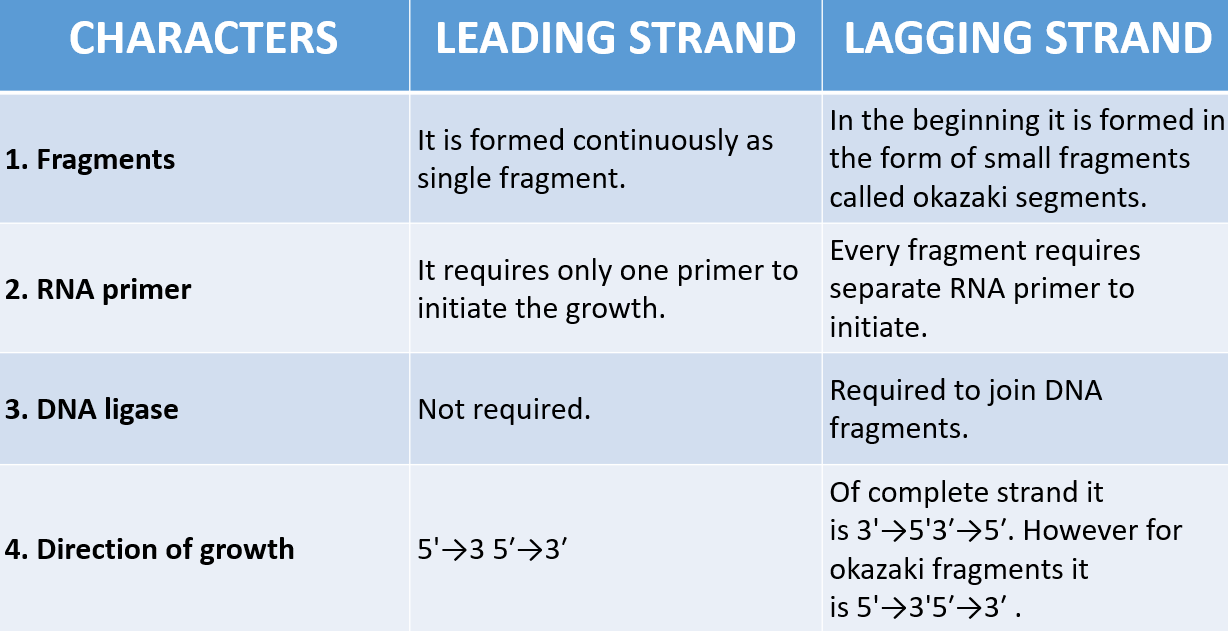
- As DNA polymerase can add new nucleotides only in one direction 5’→3’, the process of replication on template strand with the polarity 3’→5’ is continuous while the process of DNA replication on the another template strand will be discontinuous (discontinuous strand) in nature.
- Discontinuous replication occurs because only a small stretch of template strand is exposed at a time.
- These fragments on the discontinuous strand are known as okazaki fragments which will be joined by enzyme DNA ligase.
Meselson and Stahl’s Experiment
Explanation Of Image
The Machinery & The Enzymes Of DNA Replication
RNA WORLD
- Some viruses like tobacco mosaic virus (TMV), QB bacteriophage etc have RNA as their genetic material but the predominant genetic material of most of the living organisms is DNA due to its properties which were already discussed in the first section of the chapter. There are some differences between DNA and RNA which make the RNA a poor candidate for being the genetic material in complex living organisms.
- The genetic material should be stable enough not to change with different stages of life cycle, age or with change in physiology of the organism. Stability as one of the properties of genetic material was very evident in Griffith’s ‘transforming principle’ itself that heat, which killed the bacteria, at least did not destroy some of the properties of genetic Material. 2'-OH group present at every nucleotide in RNA is a reactive group and makes RNA labile (less stable) and easily degradable.
- RNA also displays catalytic properties which means its highly reactive but DNA does not show any such properties so DNA is not highly reactive and structurally more stable than RNA.
- RNA has uracil while DNA has thymine and thymine provides more stability to DNA than uracil could provide to RNA.
- RNA was the first genetic material. During the evolution of life, RNA acted as a catalyst and a genetic material both. But as we know, that being catalytic means RNA was highly reactive thus very unstable and more prone to mutations. So, DNA evolved from RNA with a few chemical properties. RNA is generally single stranded while DNA is normally double stranded. Double stranded DNA has additional stability, can resist harmful mutations and can repair itself.
- At this point this is enough and going beyond what we have already discussed here about the structure of RNA and DNA is really beyond the scope of class 12th biology.
Differences Between DNA and RNA
So among the two, DNA is a better genetic material.
RNA World
IMPORTANT POINTS
DNA REPLICATION
TRANSCRIPTION
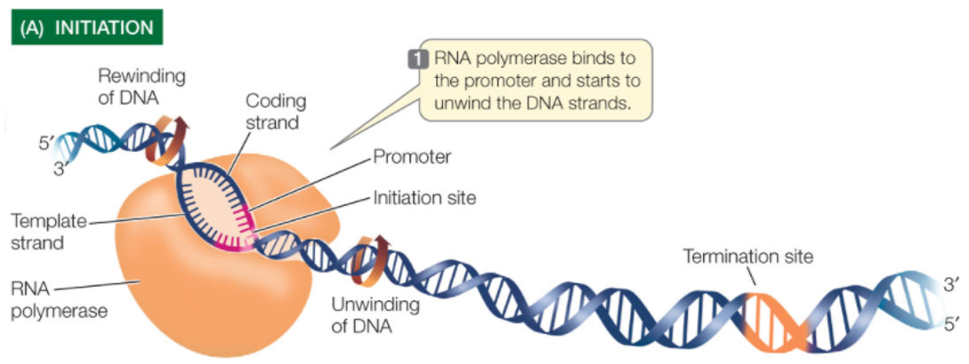
- The process of formation of RNA from DNA is known as transcription. One main point of difference in this process is remember that when we talked about nucleotides in the first section we discussed that thymine is absent in RNA and instead Uracil is present so the point where we have adenine in the DNA, it will base pair with Uracil during the process of transcription.
- The part of DNA segment that will be transcribed is known as transcription unit which consists of three regions :
- A Promoter : the part where enzymes bind to DNA to start transcription. Promoter is located towards the 5' end (upstream) of the structural gene in reference to the polarity of coding strand. This is the site where RNA polymerase will bind.
- The Structural Gene : the segment of NA that will be transcribed.
- A Terminator : The terminator is located towards 3' -end (downstream) of the coding strand and it usually defines the end of the process of transcription
- The main enzyme in replication was DNA dependent DNA polymerase. It was named so because this enzyme has the role of polymerising nucleotides into a polynucleotide chain so the name is DNA polymerase and this enzyme works with the help of DNA strand so the complete name for the enzyme became : DNA dependent DNA polymerase.
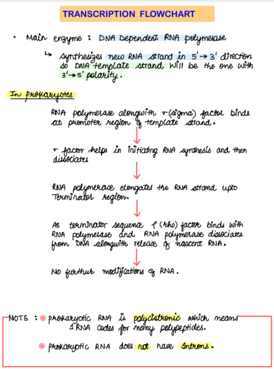
- This process of transcription is also somewhat similar to DNA replication that here only one strand will be copied and the end product is RNA. So the enzyme helping in this process is a RNA Polymerase but as this RNA Polymerase works on the DNA strand we call it DNA dependent RNA polymerase which is the main enzyme of transcription.
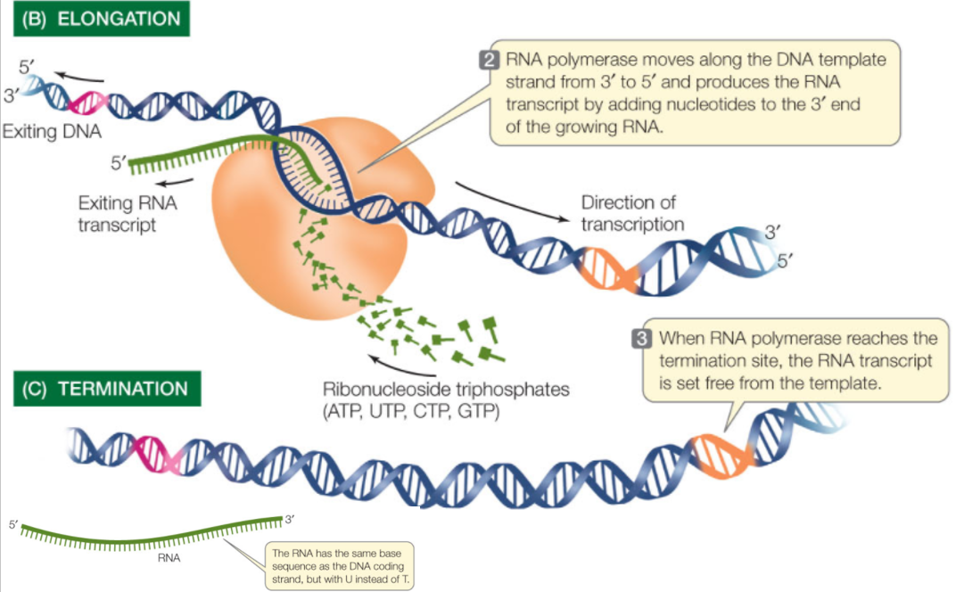
- Out of the two DNA strands only one will be transcribed. DNA dependent RNA polymerase can catalyse the polymerisation only in 5’→3’ so the strand with 3’→5’ polarity will act as template (the strand that will be transcribed).
NOTE : During transcription we will always have a single continuous nascent RNA which means that unlike in DNA replication, in transcription we do not have a discontinuous strand. - The other strand with 5’→3’ polarity does not code for anything and is still named as the Coding stand.
- In eukaryotes, a single structural gene codes for a single peptide chain or roughly (although not fully correct) we can say that in eukaryotes a single structural gene only codes for a single protein molecule. This means that structural genes in eukaryotes in monocistronic.
- On the other hand prokaryotes have a single structural gene for many different polypeptides which means that in prokaryotes a single structural gene will code for many different polypeptides so prokaryotes are Polycistronic.
- The transcription process is simple in bacteria as compared to the process in eukaryotes.
- The structural genes do not have any non coding intervening sequences (introns) in the transcriptional unit as they have in case of eukaryotes.
- They have a single DNA dependent RNA polymerase that catalyses transcription of all three types of RNA in prokaryotes.
- The whole process has just 3 steps
- Initiation
- Elongation
- Termination
- The process is initiated when RNA polymerase alongwith sigma factor (initiation factor) binds to the promoter region. Sigma factor initiates the process and RNA polymerase can only elongate the RNA strand.
- During the elongation step, sigma factor dissociates from RNA polymerase.
- At the termination site, RNA polymerase associates with Rho factor, the nascent RNA falls of and RNA polymerase gets dissociated from the DNA.
- Since transcription and translation (formation of protein from mRNA) take place in the same compartment (there is no separation of cytosol and nucleus in bacteria), many times the translation can begin much before the mRNA is fully transcribed.
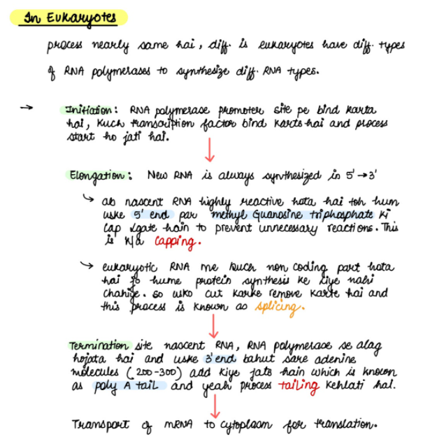
- In eukaryotes we have different polymerase enzymes for synthesis of different types of RNAs such as :
- RNA polymerase I transcribes rRNAs (28S, 18S, and 5.8S)
- The RNA polymerase II transcribes precursor of mRNA, the heterogeneous nuclear RNA (hnRNA).
- The RNA polymerase III is responsible for transcription of tRNA, 5srRNA, and snRNAs (small nuclear RNAs).
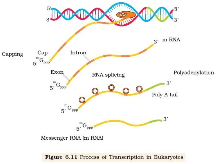
- Also, the nascent RNA in eukaryotes contain both functional / coding segments exons and the introns non coding / non functional segments. So after transcription these non functional segments have to be removed from the nascent RNA. This process of removing introns is known as splicing.
- hnRNA is highly reactive at the ends so it undergoes additional processing known as capping and tailing to make it stable enough so as not to react with other molecules unnecessarily.
- In capping methyl guanosine triphosphate is added at the 5’ end of hnRNA and in tailing, nearly 200-300 adenylate residues (Poly A tail) are added to the 3’ end of hnRNA.
- The fully processed hnRNA after all the splicing, capping and tailing is now known as mRNA and is stable enough to transport it to cytoplasm for translation.
Transcription Unit
Transcription In Bacteria
Transcription In Eukaryotes
NOTE:
First of all capping is done, then tailing to stabilize the nascent RNA and after that splicing is done.
DNA REPLICATION
- Watson and Crick suggested the semiconservative replication of DNA in 1953 but no experimental proof was provided but nonetheless as we know today that they were indeed correct. The experiment proof is shown by Messelson and Stahl’s experiment in 1958.
- They grew E.coli bacteria on a nutritional culture medium containing .15NH4Cl (.15N is the heavy isotope of nitrogen while N14 is the normal isotope of nitrogen) as the only nitrogen source for many generations so that this heavy nitrogen isotope N-15 was incorporated into newly synthesized DNA with each generation. Normal DNA has N14 while these E.coli cells had the heavy DNA as they had N15 so there was a difference in their density and the normal DNA and heavy DNA could be separated from a mixture by centrifugation in a CsCl (Caesium Chloride) density gradient where heavy DNA would sit at the base of test tube while normal DNA was found found above it.
- After growing E.coli in a heavy nitrogen medium for several generations they transferred cells to a medium containing normal nitrogen and took samples at definitive intervals. E.coli cells divide every 20 minutes.
- So the sample extracted just after 20 minutes had intermediate density value. Which means that this DNA was heavier than N14 DNA but lighter than the DNA with N15.
- Taylor and colleagues in 1958 conducted similar experiments on Vicia faba (faba beans) to show semiconservative mode of DNA replication, but instead of using heavy nitrogen isotope he used radioactive thymidine.
- The above image represents meselson and stahl experiment in a diagrammatic way. The first picture on the left hand side represents a double stranded red coloured DNA which has heavy nitrogen incorporated in it. Now when this DNA strand was centrifuged, this DNA sat at the bottom of the centrifuge tube. In the next stage when the E.coli were transferred to the normal nitrogen containing culture media their DNA replicated and the first sample was taken within the 20 minutes. The green colour in DNA represents normal N-14 nitrogen. This particular DNA sample has two types of DNA strand out of which one is of red colour and the other one is of green colour. so this is intermediate or we can say a hybrid DNA whose density will also be of intermediate value. Now what happened during the process of replication was that the original red colour DNA got separated into two strands which acted like a template using which new green coloured DNA strands were synthesized.This mode of replication is known as semi conservative mode of replication which means that every time a DNA replicates the new DNA will have one original parent stand while the daughter strand will be synthesized each time conserving 50% of the DNA so that the semi conservative nature of DNA replication.
- The experiment that we studied just now might not have made sense to a few of us because we really don’t know what exactly happened in there. So in this section we will study how DNA replicates itself and what does the term Semiconservative mode exactly means.
- There are several points in DNA from which DNA replication will start because eukaryotes have a very long DNA which means that the cell cannot really afford to replicate its DNA from one end to the other due to the energy constraints and other factors. During this process the cell will utilise a different set of enzymes each with its own role and function. Some of these enzymes will find the exact point in the DNA from where DNA replication will take place; and the point from where DNA replication takes place is known as origin of replication or we call it Ori point in short.
- The Above figure represents a structure which is known as a replication fork ( which is a Y shaped structure formed during the DNA replication). Enzyme DNA helicase unwinds the DNA at the ori point. Now remember that the DNA was coiled in a right-handed fashion. So unwinding by the enzyme DNA helicase creates a tension in the DNA and this tension is released by the enzyme DNA topoisomerase.
- In the next step single-stranded binding proteins get attached to the freshly separated strands so that these strands do not wind again. The single stranded binding proteins keep the replication fork open for the time during which the process of replication takes place.
- In the next step enzyme RNA primase attaches a short RNA primer to the replication fork which are represented by the small green fragments before red lines in the above diagram. The RNA primer is a short sequence of 5-8 nucleotides.
- RNA primers are attached at the 3 Prime end of the replication fork because the new DNA is always synthesized in 5’→3’ direction
This is so because the enzyme DNA dependent DNA polymerase (DNA Polymerase III) can add new nucleotides only in one direction, that is 5’→3’. - Also, the DNA dependent DNA polymerase cannot add new nucleotides from scratch. This enzyme needs a base from which it can synthesise a new DNA strand. That base is provided by an RNA primer which will be removed during the final stage of DNA replication so that the new DNA does not have any RNA fragment in it.
- As DNA polymerase can add new nucleotides only in one direction 5’→3’, the process of replication on template strand with the polarity 3’→5’ is continuous while the process of DNA replication on the another template strand will be discontinuous (discontinuous strand) in nature.
- Discontinuous replication occurs because only a small stretch of template strand is exposed at a time.
- These fragments on the discontinuous strand are known as okazaki fragments which will be joined by enzyme DNA ligase.

Meselson and Stahl’s Experiment

Explanation Of Image
The Machinery & The Enzymes Of DNA Replication

GENETIC CODE
- George Gamow argued that we have only 4 bases (adenine, guanine, thymine and cytosine) but the discovered amino acids for protein synthesis were 20, so it means that these bases form a combination of bases where each combination would code for a single amino acid. This was accepted but the next question was : How many bases would be there in a single combination set for a single amino acid?
- To answer this question, he proposed that these bases combine in a group of three base pairs known as a genetic code
- This proposition was very bold because if the genetic code was a triplet of bases then it would generate 43 = 64 genetic codons for only 20 amino acids. 64 genetic codes were even more than thrice the codons of what we needed because at that time it was thought that each amino acid had only one genetic codon. He failed to provide the proof that the genetic code was a triplet but nonetheless he was right.
- Finally experiments from contributions of Har Gobind Khorana and Marshall’s Nirenberg using Severo Ochoa enzyme helped in deciphering the genetic codons for the amino acids and from all the data, following table was prepared
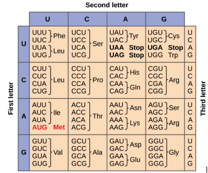
- In the above table table you can see that three codons (UAA, UGA & UAG) do not code for anything and they are stop codons leaving us with 61 codons.
- The salient features of genetic code are as follows:
- The codon is triplet. 61 codons code for amino acids and 3 codons do not code for any amino acids, hence they function as stop codons.
- Some amino acids are coded by more than one codon, hence the code is degenerate.
- The codon is read in mRNA in a contiguous fashion. There are no punctuations.
- The code is nearly universal: for example, from bacteria to human UUU would code for Phenylalanine (phe). Some exceptions to this rule have been found in mitochondrial codons, and in some protozoans.
- AUG has dual functions. It codes for Methionine (met) , and it also act as initiator codon.
- UAA, UAG, UGA are stop terminator codons.
- mRNA me codons hote ek series me hain bina kisi punctuation mark ke but when we read them, we read them as triplets. For eg: neeche wali image ko consider karo :

Ab iss image me AUGACGGAG…..UAG ek series me hain. The first codon will always be AUG and the last codon will always be any one of the three stop codons. - Addition or deletion of even a single base base will change the sequence after that mutation site. This could be explained by the following image from the NCERT.
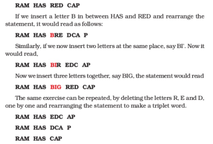
- The addition so B in the above image lead to a change in reading frame from RAM HAS RED CAP to RAM HAS BRE DCA P. That is the entire frame of reading has been shifted to a new phrase which doesn’t even make sense. In the same way addition of a new nucleotide in a sequence changes the reading frame and such mutations are known as frameshift mutations. Same goes with deletion of a single base pair.
- So in short, change in even a single nucleotide could prove fatal because that change might render the respective polypeptide non functional.
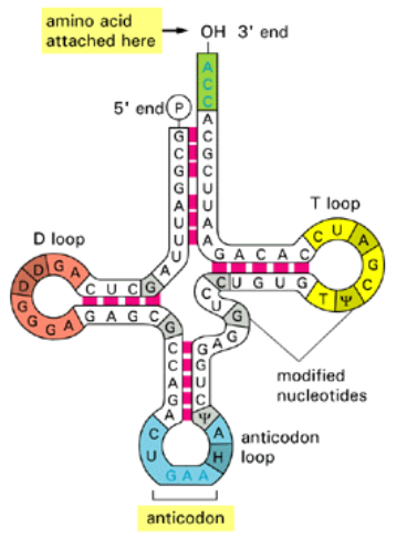
- The above image shows the clover leaf structure of a tRNA molecule. As you can see there are 3 different loops (where pink coloured complementary base pairing is absent)
1.)D-loop2.)Anticodon loop
3.)T-loop
out of which anticodon loop is our loop of interest at this time. - The anticodon loop has anticodons for the codons found on the mRNA. The amino acid corresponding to the anticodon on the anticodon loop is attached to the 3’ end of the tRNA molecule.
- The last three base pairs at the 5’ end are always CCA. This site accepts amino acids so sometime we call it an acceptor site
- The 5’ end has a phosphate group attached to it.
- There are no tRNAs for stop codons.
Mutations & Genetic Code
tRNA - The Adapter Molecule
IMPORTANT POINTS


TRANSLATION
- Translation refers to the process of polymerisation of amino acids to form a polypeptide or simply we can call it the process of formation of polypeptide (protein) from mRNA.
- A polypeptide is formed when a peptide bond is formed between amino acids as shown below :
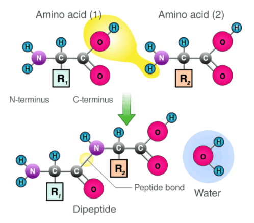
- Formation of this peptide bond requires energy so first we have to charge the amino acids in presence of ATP and then they are linked to their respective tRNAs. This step is commonly known as charging of tRNA or Aminoacylation of tRNA. This process is necessary because formation of peptide bond between two charged amino acids is energetically favoured.
- Ribosomes are protein factories which means that they are responsible for the process of translation. Ribosomes have 2 subunits : a larger and a smaller. The smaller subunit joins to the mRNA at the start codon (AUG), then from above the larger subunit joins and together they sandwich the mrNA.
- The region present before the start codon and after the stop codon is not translated, so those regions are known as untranslated regions (UTRs).
- Activation of Amino Acids : In the first step amino acid reacts with ATP to form amino acid-AMP Complex (activated amino acid) in presence of enzyme aminoacyl-tRNA synthetase and Mg2+. The enzyme aminoacyl-tRNA synthetase is different for each amino acid.
- Charging of tRNA / Aminoacylation of tRNA : The activate amino acid joins to the amino acid binding site of the respective tRNA and amino acid gets attached to the CCA site and results in formation of tRNA-amino acid complex known as charged tRNA.
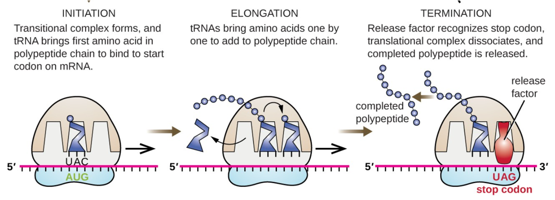
- Activation of Ribosomes : First the small subunit of ribosome joins the start codon and forms a initiation complex. Then larger subunit joins the smaller ribosomal unit in presence of specific concentration of Mg2+ ions forming a active ribosome
- Polypeptide Formation : This step involves 3 stages : initiation, elongation and termination of amino acid chain.
- Initiation of Polypeptide Chain : The initiator charged tRNA base pairs with the initiator codon at the A site of the large subunit of ribosome. This step requires some initiator factors and there are a few more steps but details of those are not required.
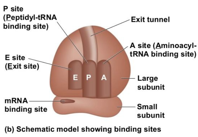
- Elongation of Polypeptide Chain : The ribosome moves forward while the mRNA is still so that the tRNA which was earlier at the A site is now at the P site and a new charged tRNA comes at the A site. Addition of each new amino acid requires certain proteins known as elongation factors.
- The enzyme peptidyl transferase is responsible for forming a peptide bond between the amino acid on P site and the amino acid on A site. The ribosome moves forwards, reads another codon so that now the first tRNA molecule is at E site, second tRNA molecule at P site and third tRNA at A site. The tRNA at E site, leaves the ribosome and ribosome keeps moving forward, reading every codon by codon and forming peptide bonds between amino acids until it reaches a stop codon for which there isn’t any tRNA molecule.
- Termination of Polypeptide Chain : We have 3 stop codons:
- UAG (also known as amber)
- UGA (also known as opal)
- UAA (also known as ochre)
- When ribosomes reaches stop codon and no tRNA associated with ribosome at A site then a release factor joins the stop codon at A site to add a water molecule to the completed polypeptide chain to hydrolyse the polypeptide chain tRNA that is at P site.
- THen ribosome splits again into larger and smaller subunits in presence of a dissociation factor.
Mechanism of Protein Synthesis
TRANSCRIPTION
REGULATION OF GENE EXPRESSION
- Each and every gene is regulated so that it is transcriptionally active only when its product (polypeptide) is needed. Some genes are expressed continuously until they are told not to while other genes express themselves only when their coded protein is needed by the cell. Also, some genes are expressed only in some organs and other genes are expressed in other organs while genes that are necessary for cell’s survival are expressed continuously throughout the body but mainly there are two types of gene regulations:
- Positive Regulation
- Negative Regulation
- In negative gene regulation the genes continue to express themselves until their activity is suppressed. This type of gene regulation is also known as repressible regulation.
- In positive gene regulation, the genes only express themselves when they are induced to do so. This type of regulation is also known as inducible regulation.
- Gene regulation is exerted at 4 levels :
- At transcriptional level when primary transcript is formed
- at processing level when splicing and terminal additions are made.
- transport of mRNA out of the nucleus into the cytoplasm.
- at translational level.
- Jacob and monod Khoob the functional unit of genetic material are Operons. Operon is a set of at least for 4 different genes :
- Regulator Gene : it forms a polypeptide for suppressing the activity of operator gene<
- Operator Gene : It allows functioning of the Operon when it is not inhibited by the polypeptide produced by regulator gene.
- Promoter Gene : this is the site where RNA polymerase will get attached for transcription of structural genes.
- Structural Genes : These are the genes which will be transcribed by RNA polymerase for synthesis of polypeptides.
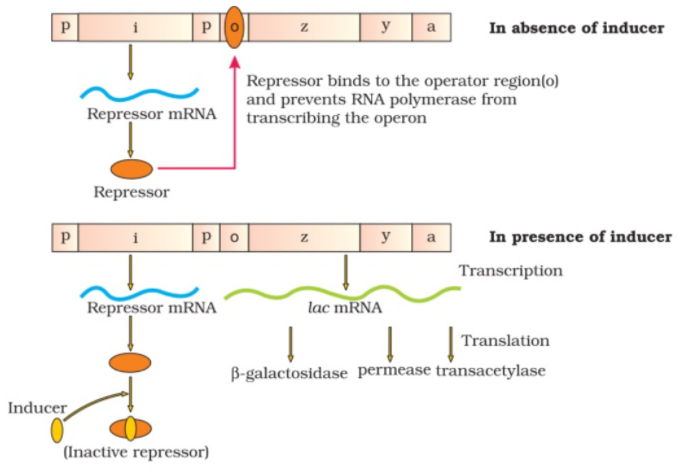
Lac Operon of E. coli
- Elucidated by Jacob & Monod
- Lac here refers to lactose sugar.
- Lac Operon is an inducible operon system in which the structural genes transcribed up Polycistronic mRNA molecule which produces three enzymes namely: beta galactosidase (from z-gene), permease (from y-gene) and transacetylase (from a-gene).
- beta galactosidase hydrolysis the lactose sugar into glucose and galactose molecules.
- Enzyme permease is required for entry of lactose into the E coli bacteria. It makes the bacterial membrane permeable for lactose sugar.
- Transacetylase can transfer acetyl group from acetyl coA to beta galactoside.
- Now when glucose sugar is present in the media and bacteria has access to glucose, the Lac Operon well stop However when bacteria has no access to glucose but lactose is present in the media at that time lactose sugar will act as inducer ( show in yellow colour oval shape) and it will bind to repressor so that repressor can no longer prevent from transcribing the Operon. Once the repressor is rendered inactive by the inducer then RNA polymerase transcribes the structural gene so that those three enzyme molecules which are necessary for lactose metabolism can be formed.
- When glucose is present for metabolism then the repressor will be synthesized so as to block the unnecessary transcription of genes which are required for lactose metabolism.
TRANSCRIPTION
- The process of formation of RNA from DNA is known as transcription. One main point of difference in this process is remember that when we talked about nucleotides in the first section we discussed that thymine is absent in RNA and instead Uracil is present so the point where we have adenine in the DNA, it will base pair with Uracil during the process of transcription.
- The part of DNA segment that will be transcribed is known as transcription unit which consists of three regions :
- A Promoter : the part where enzymes bind to DNA to start transcription. Promoter is located towards the 5' end (upstream) of the structural gene in reference to the polarity of coding strand. This is the site where RNA polymerase will bind.
- The Structural Gene : the segment of NA that will be transcribed.
- A Terminator : The terminator is located towards 3' -end (downstream) of the coding strand and it usually defines the end of the process of transcription
- The main enzyme in replication was DNA dependent DNA polymerase. It was named so because this enzyme has the role of polymerising nucleotides into a polynucleotide chain so the name is DNA polymerase and this enzyme works with the help of DNA strand so the complete name for the enzyme became : DNA dependent DNA polymerase.
- This process of transcription is also somewhat similar to DNA replication that here only one strand will be copied and the end product is RNA. So the enzyme helping in this process is a RNA Polymerase but as this RNA Polymerase works on the DNA strand we call it DNA dependent RNA polymerase which is the main enzyme of transcription.
- Out of the two DNA strands only one will be transcribed. DNA dependent RNA polymerase can catalyse the polymerisation only in 5’→3’ so the strand with 3’→5’ polarity will act as template (the strand that will be transcribed).
NOTE : During transcription we will always have a single continuous nascent RNA which means that unlike in DNA replication, in transcription we do not have a discontinuous strand. - The other strand with 5’→3’ polarity does not code for anything and is still named as the Coding stand.
- In eukaryotes, a single structural gene codes for a single peptide chain or roughly (although not fully correct) we can say that in eukaryotes a single structural gene only codes for a single protein molecule. This means that structural genes in eukaryotes in monocistronic.
- On the other hand prokaryotes have a single structural gene for many different polypeptides which means that in prokaryotes a single structural gene will code for many different polypeptides so prokaryotes are Polycistronic.
- The transcription process is simple in bacteria as compared to the process in eukaryotes.
- The structural genes do not have any non coding intervening sequences (introns) in the transcriptional unit as they have in case of eukaryotes.
- They have a single DNA dependent RNA polymerase that catalyses transcription of all three types of RNA in prokaryotes.
- The whole process has just 3 steps
- Initiation
- Elongation
- Termination
- The process is initiated when RNA polymerase alongwith sigma factor (initiation factor) binds to the promoter region. Sigma factor initiates the process and RNA polymerase can only elongate the RNA strand.
- During the elongation step, sigma factor dissociates from RNA polymerase.
- At the termination site, RNA polymerase associates with Rho factor, the nascent RNA falls of and RNA polymerase gets dissociated from the DNA.
- Since transcription and translation (formation of protein from mRNA) take place in the same compartment (there is no separation of cytosol and nucleus in bacteria), many times the translation can begin much before the mRNA is fully transcribed.
- In eukaryotes we have different polymerase enzymes for synthesis of different types of RNAs such as :
- RNA polymerase I transcribes rRNAs (28S, 18S, and 5.8S)
- The RNA polymerase II transcribes precursor of mRNA, the heterogeneous nuclear RNA (hnRNA).
- The RNA polymerase III is responsible for transcription of tRNA, 5srRNA, and snRNAs (small nuclear RNAs).
- Also, the nascent RNA in eukaryotes contain both functional / coding segments exons and the introns non coding / non functional segments. So after transcription these non functional segments have to be removed from the nascent RNA. This process of removing introns is known as splicing.
- hnRNA is highly reactive at the ends so it undergoes additional processing known as capping and tailing to make it stable enough so as not to react with other molecules unnecessarily.
- In capping methyl guanosine triphosphate is added at the 5’ end of hnRNA and in tailing, nearly 200-300 adenylate residues (Poly A tail) are added to the 3’ end of hnRNA.
- The fully processed hnRNA after all the splicing, capping and tailing is now known as mRNA and is stable enough to transport it to cytoplasm for translation.
Transcription Unit
Transcription In Bacteria


Transcription In Eukaryotes

NOTE:
First of all capping is done, then tailing to stabilize the nascent RNA and after that splicing is done.
HUMAN GENOME PROJECT
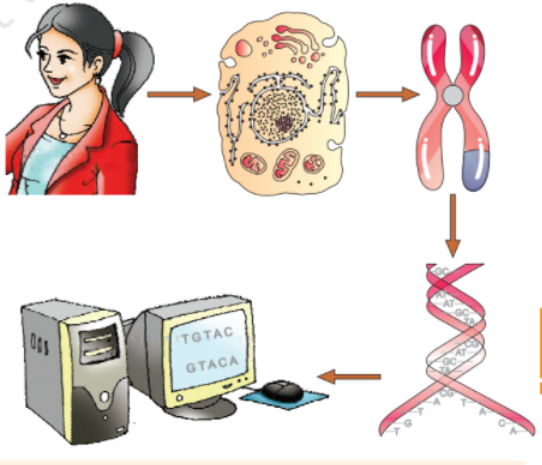
- The Human Genome Project was launched in the year 1990 so as to sequence the whole genome of humans. haploid human DNA has nearly 3 X 10⁹ bp and the cost of sequencing per base pair is 3 US dollars. so the whole project would take approximately 9 billion US dollars. HGP was closely associated with the rapid development of a new area in biology known as bioinformatics.
- The project was completed in 2003. so it took 13 years to complete
- To identify all the approximately 20000 to 25000 genes in human DNA.
- To determine the sequences of all the base pairs.
- Store the information in databases
- Improved tools for data analysis.
- Transfer related technology to other sectors such as industries
- Address the ethical, legal and social issues (ELSI) that may arise from the project.
- The methods involved two major approaches :
- Expressed Sequence Tags : this approach focused on identifying all the genes that are expressed as RNA.
- Sequence Annotation : This was the blind approach of simply sequencing the whole set of genome including all the coding and non coding sequences and then later on functions were assigned to the different regions of the sequence.
- For the purpose of Sequencing total DNA from a cell is isolated and converted in two random fragments of smaller sizes and these fragments were cloned into suitable hosts using specialised vectors. The common hosts were bacteria and yeast, And the vectors were called as a bacterial artificial chromosome (BAC) and yeast artificial chromosome (YAC).
- The Human Genome contains 3164.7 million base pairs.
- The average gene consists of 3000 bases but size varies greatly with the largest known human gene being dystrophin at 2.4 million bases.
- The number of genes is estimated at 30,000 which is much lower than the previous estimates of 80,000 to 140000 genes.
- Almost 99.9% nucleotides bases are exactly the same in all people.
- The functions are unknown or over 50% of the discovered genes.
- Less than 2% of the genome codes for protein.
- Repeated sequences make up very large portion of the human genre these are the DNA sequences that are repeated many times, sometimes hundred to thousand times.
- Chromosome 1 has most genes : 2968, while chromosome Y has the lowest number of chromosomes, 231.
- There are nearly 1.4 million locations where single base DNA differences single nucleotide polymorphism (SNPs) occur in humans.
Goals of HGP
Methodologies
Salient Features of Human Genome
GENETIC CODE
- Humare DNA me jitni bhi informational hai wo nitrogenous base pair me stored hai. Lekin ek akele base pair jaisey ki sirfA ya keval G ka koi matlab nahi hai. Lekin ab DNA me toh yhi sab hai and inhi base pairs ke tranlation se ek protein banta hai jiska koi apna khud ka function hoga.
- Toh jo yeah protein formation ki information hai wo inhi base pair me coded hai toh base pair ka ek aisa group jiska khud ka vajud ho ki bhyii base pair ke iss group se let’s say methionine amino acid banega.
- Scientists ko yeah baat pasand aagyi and unhone maan liya ki chalo theek hai, nitrogenous base pair ka ek group hai jisme ek amino acid ki information coded hai or aisey hai sabhi 20 amino acids ke liye ek particular coded group hoga base pairs jisko hum aaj se codon kahenge. Sab scientists ne maan liya ki okk. Theek hai bhyii. Yeah sensible baat lag rhi hai or humne maan liya.
- Par fir kisi ne puch liya ki ab sawal yeah hai ki ek codon me nucleotide kitne hain? Yani ki jo aapne har ek amino acid ke liye ek codon btaya hai usme 2 base pairs hain ya 3 base pair hai ya 4 base pair hai wo btaiyeah?
Toh iska jawab diya George Gamow ne. - Unhone kha ki bhyii humare pass keval 4 base pair hain or amino acid acid 20. Toh jo bhi codon hai unme inhi 4 base pair me kuch hoga. Fir unhone guess karna shuru kiya let’s say ek codon me agar 2 hi base pair huy toh iss hisab se 42 humare pass keval 16 codon bne but amino acid toh 20 han and ek amino acid ke liye atleast ek codon toh hoga.
- Next unhone socha ki maybe ek codon me 3 base pair ho toh iss hypothesis ke according we have 43 = 64 codons jo bahut jyada hai as compared to 20 amino acids. Ab iske baad agar 44 ko consider karte toh or jyada codon bn jate toh George ji 3 par set hogye ki ek codon me 3 base pairs hote hain. Ab yeah baat unhone oron ko bataie toh unhone hath hila diya ki bhyii proof do aise biology me math ka hum kya karenge. Ab proof unpe tha ni toh kisine unko seriously nahi liya.
- Kuch samay baad Har gobind khorana and Nireneberg & Leder ke experiment ne yeah prove kar diya ki genetic codon me 3 hi base pair hote hain. Experiment apne syllabus me nahi hai toh we won’t discuss it here now.
- codon hamesha triplet hoga. Jaisey ki AUG codes for methionine amino acid.
- Ek code ek hi amino acid ko code karega. (degeneracy of genetic code).
- Ek amino acid ke liye ek se jyada codon possible hai but reverse is not true.
- Codon nearly universal hai.
- UAA, UAG, UGA - yeah 3 stop codon hain, yani ki yeah teeno kisi nhi amino acid ke liye code nahi karte.
- codon ko padhte waqt koi coma, semicolon vgarah kuch ni aata, and codon is always read in a continuous manner.
- Mutation ka matlab hai - badalav. Genetic codon me agar ek bhi base pair ki jagah humne koi dusra base pair aagya and uski vajah se amino acid badal gya toh this is mutation.
- Ek example consider karo ki humare pass koi protein hai jisme CUU koi ek codon hai which codes of leucine amino acid and maanlo agar is C ki jagah G lga diya toh codon ab badal kar GUU ho gya which codes for valine amino acid ya fir G ki jagah U aagya toh now codon is UUU which codes for amino acid pehnylalanine.
- Toh ek point par bhi kuch change kiya toh uski vajah se amino acid change hojata hai which in turn might change the structure of functional protein jiske bahut harmful effects ho sakte hain.
- Ab yaad karo Sickle cell anemia ko jisme haemoglobin ki beta chain me 6th position par glutamic acid ki jagah valine amino acid aa jata hai bas issi ke karan Hb ki oxygen transport krne ki ability bahut jyada decrease kar jati hai.
- Dusre tarike ki mutation me there could be addition or deletion of base pairs. Mannlo koi chotu sa sequence hai UUUUUUUUU which is read as UUU UUU UUU and codes for Phe-Phe-Phe but isme . isme addition mutation hui jiske karan 7th position par ab ek A add hogya and now the codon reads UUU UUU AUU U which codes for Phe-Phe-Ile and last wala U akela reh gya toh dekho is addition ke karan ab hum codon ko 3rd code par padh hi AUU rahe hain instead of the original UUU so such mutations are known as Frameshift Mutations jo humare reading frame ko hi shift krde.
- Same goes for deletion. Try to read the sequence AUGAUGAUG after deleting U from the 5th position and then compare it with the original amino acid sequence. You should get same result with a shift of reading frame.
- tRNA is transfer-RNA. Yani ki yeah amino acid ko vahan transport karega jha translation ho rhi hai.
- Iska accepted structure clover leaf model hai jo niche diya gya hai.
- Yeah actual me ek polynucleotide ki lambi chain hai jisme ek hi chain me bases me complementary base pairing hony ke karan iska clover leaf life structure milta hai and jha jha base pairing nahi ho pati vha Loops ki formation ho jati hai.
- So we have 3 loops jinme se apne kaam ki keval anti codon loop hai. Iss loop me jo sabse neeche 3 base pair hote hain wo mRNA par jo codon hai uske sath complementary base pairing karte hain.
- tRNA ke 3’ end par hamesha humy CCA-3’ sequence milegi and iss sequence ke baad tRNA se respective amino acid attach hoga during translation. Amino acid tRNA se hamesha attached nahi rehta.
- 5’ end par as usual ek phosphate moiety milti hai.
- We have separate tRNA for each of the codon but we have no tRNA for the stop codons.
Inn sab discoveries ke baad genetic codon ko jab or ache se scientist ne samjha toh unhone kuch features btaye genetic codon ke jaisey ki
Genetic Code & Mutation
tRNA - The Adapter Molecule
DNA FINGERPRINTING
- As we already know that almost 99.9% of DNA is same in all humans so DNA printing was developed by Alec Jeffreys which is a technique to identify repeated sequences in the human genome the produces a pattern of band in electrophoresis which is unique for every individual.
→ Satellite DNA contains highly repetitive sequences of DNA and these sequences show a high degree of polymorphism (different forms). - Imagine a hypothetical small sequence, lets say AAGGTTCC (a simple sequence) which is repeated anywhere from 30-40 times on chromosome 15 (again hypothetical). So this sequence has been repeated 30-40 times which is Variable number of tandem repeats (=how many times a sequence is repeated).
- We have many such repeat sequences which are unique for a person
- Steps Of DNA Fingerprinting
- Isolation of DNA,
- Digestion of DNA by restriction endonucleases,
- Separation of DNA fragments by electrophoresis,
- Transferring (blotting) of separated DNA fragments to synthetic membranes, such as nitrocellulose or nylon,
- Hybridization using labelled VNTR probe, and
- Detection of hybridized DNA fragments by autoradiography.
- The above image shows DNA fingerprinting results from a crime scene compared with the result of two suspects.
- The DNA pattern in column C is from a crime scene and we have let’s say two suspects : A and B. Match the pattern in column C to the pattern in column B. We can clearly see that they are exactly the same which means that DNA sample C from the crime scene is of suspect B and not of suspect A.
- Forensic Science: Biological materials used for DNA fingerprinting are: Blood, Hair, Saliva, Semen, Body tissue cells etc. DNA isolated from the evidence sample can be compared through the VNTR (Variable number of tandem repeats) prototype. It is useful in solving crimes like murder and rape.
- Paternity and Maternity Determination: A Person accedes to his or her VNTRs from his or her parents. Parent-child VNTR prototype analysis has been used to solve disputed cases. This information can also be used in inheritance cases, immigration cases.
- Personal Identification: It utilizes the idea of using DNA fingerprints as a sort of genetic barcode to identify individuals.
- Diagnosis of Inherited Disorders: It is also useful in diagnosing inherited disorders in both prenatal and newborn babies. These disorders may include cystic fibrosis, hemophilia, Huntington’s disease, familial Alzheimer’s, sickle cell anemia, thalassemia, and many others.
- Development of Cures for Inherited Disorders:By studying the DNA fingerprints of relatives who have a history of some particular disorder, DNA prototypes associated with the disease can be ascertained.
- Detection of AIDS: By comparing the band of HIV “RNA” (converted to DNA using RT PCR) with the bands formed by the man’s blood, a person suffering with AIDS can be identified.
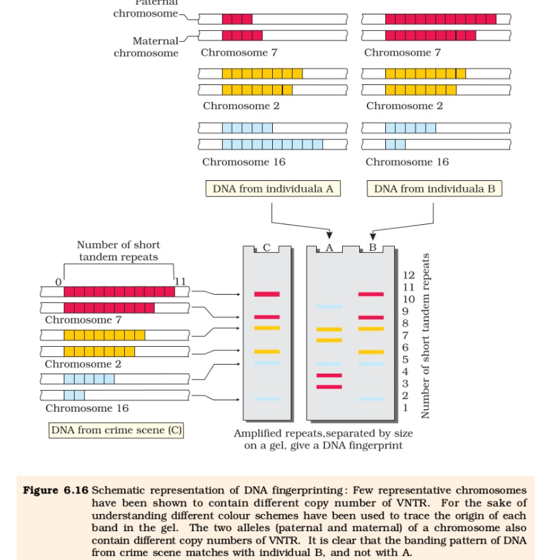
Explanation of Image
Applications Of DNA Fingerprinting
GENETIC CODE
- George Gamow argued that we have only 4 bases (adenine, guanine, thymine and cytosine) but the discovered amino acids for protein synthesis were 20, so it means that these bases form a combination of bases where each combination would code for a single amino acid. This was accepted but the next question was : How many bases would be there in a single combination set for a single amino acid?
- To answer this question, he proposed that these bases combine in a group of three base pairs known as a genetic code
- This proposition was very bold because if the genetic code was a triplet of bases then it would generate 43 = 64 genetic codons for only 20 amino acids. 64 genetic codes were even more than thrice the codons of what we needed because at that time it was thought that each amino acid had only one genetic codon. He failed to provide the proof that the genetic code was a triplet but nonetheless he was right.
- Finally experiments from contributions of Har Gobind Khorana and Marshall’s Nirenberg using Severo Ochoa enzyme helped in deciphering the genetic codons for the amino acids and from all the data, following table was prepared

- In the above table table you can see that three codons (UAA, UGA & UAG) do not code for anything and they are stop codons leaving us with 61 codons.
- The salient features of genetic code are as follows:
- The codon is triplet. 61 codons code for amino acids and 3 codons do not code for any amino acids, hence they function as stop codons.
- Some amino acids are coded by more than one codon, hence the code is degenerate.
- The codon is read in mRNA in a contiguous fashion. There are no punctuations.
- The code is nearly universal: for example, from bacteria to human UUU would code for Phenylalanine (phe). Some exceptions to this rule have been found in mitochondrial codons, and in some protozoans.
- AUG has dual functions. It codes for Methionine (met) , and it also act as initiator codon.
- UAA, UAG, UGA are stop terminator codons.
- mRNA me codons hote ek series me hain bina kisi punctuation mark ke but when we read them, we read them as triplets. For eg: neeche wali image ko consider karo :

Ab iss image me AUGACGGAG…..UAG ek series me hain. The first codon will always be AUG and the last codon will always be any one of the three stop codons. - Addition or deletion of even a single base base will change the sequence after that mutation site. This could be explained by the following image from the NCERT.

- The addition so B in the above image lead to a change in reading frame from RAM HAS RED CAP to RAM HAS BRE DCA P. That is the entire frame of reading has been shifted to a new phrase which doesn’t even make sense. In the same way addition of a new nucleotide in a sequence changes the reading frame and such mutations are known as frameshift mutations. Same goes with deletion of a single base pair.
- So in short, change in even a single nucleotide could prove fatal because that change might render the respective polypeptide non functional.
- The above image shows the clover leaf structure of a tRNA molecule. As you can see there are 3 different loops (where pink coloured complementary base pairing is absent)
1.)D-loop2.)Anticodon loop
3.)T-loop
out of which anticodon loop is our loop of interest at this time. - The anticodon loop has anticodons for the codons found on the mRNA. The amino acid corresponding to the anticodon on the anticodon loop is attached to the 3’ end of the tRNA molecule.
- The last three base pairs at the 5’ end are always CCA. This site accepts amino acids so sometime we call it an acceptor site
- The 5’ end has a phosphate group attached to it.
- There are no tRNAs for stop codons.
Mutations & Genetic Code
tRNA - The Adapter Molecule

SUMMARY
- Nucleic acids are long polymers of nucleotides. While DNA stores genetic information, RNA mostly helps in transfer and expression of information.
- Though DNA and RNA both function as genetic material, but the DNA being chemically and structurally more stable is a better genetic material.
- However, RNA is the first to evolve and DNA was derived from RNA. The hallmark of the double stranded helical structure of DNA is the hydrogen bonding between the bases from opposite strands.
- The rule is that Adenine pairs with Thymine through two H-bonds, and Guanine with Cytosine through three H-bonds.
- This makes one strand complementary to the other. The DNA replicates semiconservatively, the process is guided by the complementary H-bonding. A segment of DNA that codes for RNA may in a simplistic term can be referred to as gene.
- During transcription also, one of the strands of DNA acts as a template to direct the synthesis of complementary RNA.
- In bacteria, the transcribed mRNA is functional, hence can directly be translated.
- In eukaryotes, the gene is split. The coding sequences, exons, are interrupted by non-coding sequences, introns. Introns are removed and exons are joined to produce functional RNA by splicing.
- The messenger RNA contains the base sequences that are read in a combination of three (to make triplet genetic code) to code for an amino acid. The genetic code is read again on the principle of complementarity by tRNA that acts as an adapter molecule. There are specific tRNAs for every amino acid. The tRNA binds to specific amino acid at one end and pairs through H-bonding with codes on mRNA through its anticodons.
- The site of translation (protein synthesis) is ribosomes, which bind to mRNA and provide a platform for joining amino acids.
- One of the rRNA acts as a catalyst for peptide bond formation, which is an example of RNA enzyme (ribozyme).
- Translation is a process that has evolved around RNA, indicating that life began around RNA. Since, transcription and translation are energetically very expensive processes, these have to be tightly regulated.
- Regulation of transcription is the primary step for regulation of gene expression. In bacteria, more than one gene is arranged together and regulated in units called operons. Lac operon is the prototype operon in bacteria, which codes for genes responsible for metabolism of lactose. The operon is regulated by the amount of lactose in the medium where the bacteria are grown. Therefore, this regulation can also be viewed as regulation of enzyme synthesis by its substrate.
- Human genome project was a mega project that aimed to sequence every base in the human genome. This project has yielded much new information. Many new areas and avenues have opened up as a consequence of the project. DNA Fingerprinting is a technique to find out variations in individuals of a population at DNA level. It works on the principle of polymorphism in DNA sequences. It has immense applications in the field of forensic science, genetic biodiversity and evolutionary biology.
TRANSLATION
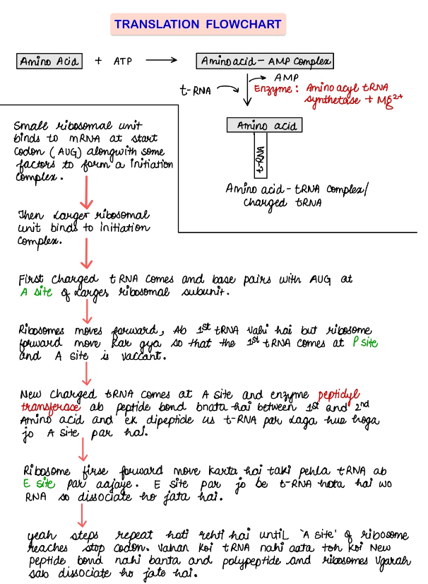
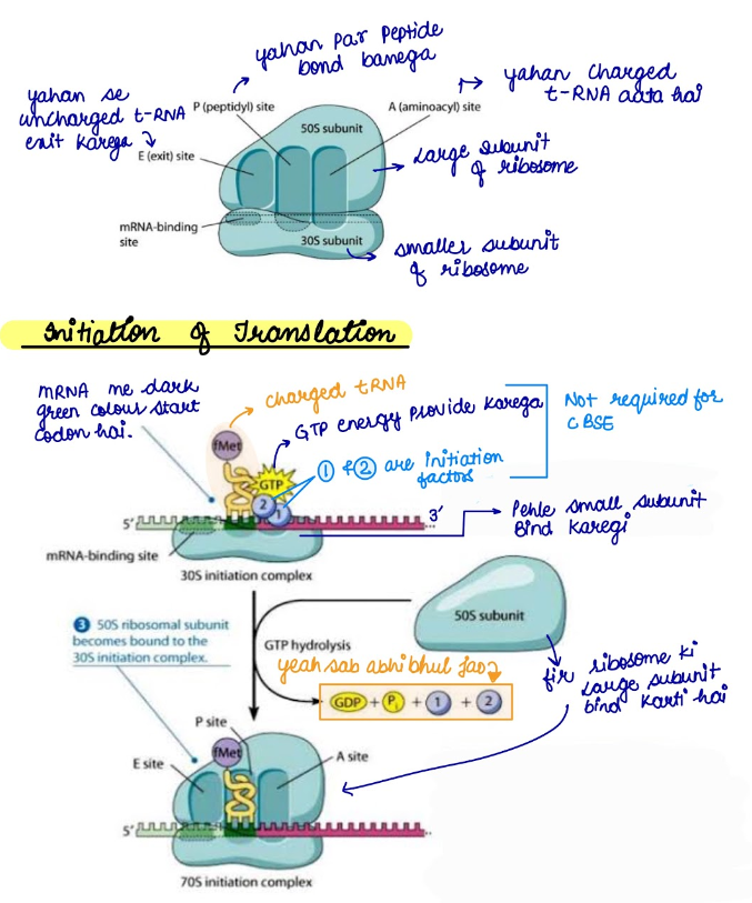
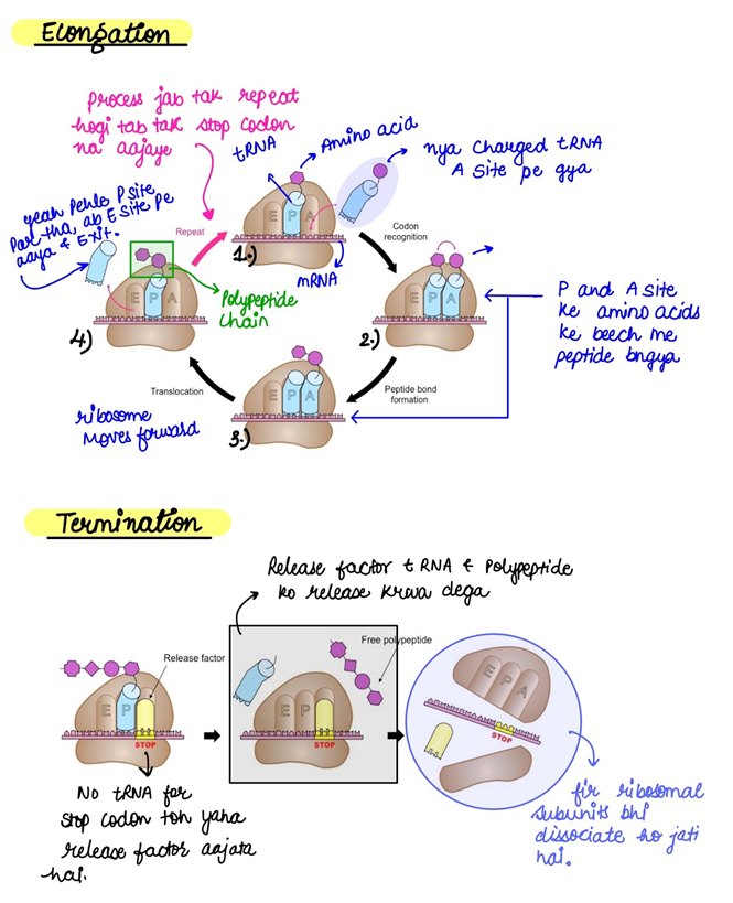
TRANSLATION
- Translation refers to the process of polymerisation of amino acids to form a polypeptide or simply we can call it the process of formation of polypeptide (protein) from mRNA.
- A polypeptide is formed when a peptide bond is formed between amino acids as shown below :

- Formation of this peptide bond requires energy so first we have to charge the amino acids in presence of ATP and then they are linked to their respective tRNAs. This step is commonly known as charging of tRNA or Aminoacylation of tRNA. This process is necessary because formation of peptide bond between two charged amino acids is energetically favoured.
- Ribosomes are protein factories which means that they are responsible for the process of translation. Ribosomes have 2 subunits : a larger and a smaller. The smaller subunit joins to the mRNA at the start codon (AUG), then from above the larger subunit joins and together they sandwich the mrNA.
- The region present before the start codon and after the stop codon is not translated, so those regions are known as untranslated regions (UTRs).
- Activation of Amino Acids : In the first step amino acid reacts with ATP to form amino acid-AMP Complex (activated amino acid) in presence of enzyme aminoacyl-tRNA synthetase and Mg2+. The enzyme aminoacyl-tRNA synthetase is different for each amino acid.
- Charging of tRNA / Aminoacylation of tRNA : The activate amino acid joins to the amino acid binding site of the respective tRNA and amino acid gets attached to the CCA site and results in formation of tRNA-amino acid complex known as charged tRNA.
- Activation of Ribosomes : First the small subunit of ribosome joins the start codon and forms a initiation complex. Then larger subunit joins the smaller ribosomal unit in presence of specific concentration of Mg2+ ions forming a active ribosome
- Polypeptide Formation : This step involves 3 stages : initiation, elongation and termination of amino acid chain.
- Initiation of Polypeptide Chain : The initiator charged tRNA base pairs with the initiator codon at the A site of the large subunit of ribosome. This step requires some initiator factors and there are a few more steps but details of those are not required.
- Elongation of Polypeptide Chain : The ribosome moves forward while the mRNA is still so that the tRNA which was earlier at the A site is now at the P site and a new charged tRNA comes at the A site. Addition of each new amino acid requires certain proteins known as elongation factors.
- The enzyme peptidyl transferase is responsible for forming a peptide bond between the amino acid on P site and the amino acid on A site. The ribosome moves forwards, reads another codon so that now the first tRNA molecule is at E site, second tRNA molecule at P site and third tRNA at A site. The tRNA at E site, leaves the ribosome and ribosome keeps moving forward, reading every codon by codon and forming peptide bonds between amino acids until it reaches a stop codon for which there isn’t any tRNA molecule.
- Termination of Polypeptide Chain : We have 3 stop codons:
- UAG (also known as amber)
- UGA (also known as opal)
- UAA (also known as ochre)
- When ribosomes reaches stop codon and no tRNA associated with ribosome at A site then a release factor joins the stop codon at A site to add a water molecule to the completed polypeptide chain to hydrolyse the polypeptide chain tRNA that is at P site.
- THen ribosome splits again into larger and smaller subunits in presence of a dissociation factor.

Mechanism of Protein Synthesis

REGULATION OF GENE EXPRESSION
- Hume pta hai ki har protein ke liye we have encode information in our genes. But kuch genes ke product hume kabhi kabhi chaiyeah hoty hain and kuch genes aisey hoty hain jinke product ki hume hamesha jarurat hoti hai. For example aapne sabhi ne class 11 me glycolysis padhan hai. Usme har step pe different enzymes use hote hain. Har enzyme ke liye ek different gene hota hai jiska regularly transcription hota rehta hai but kuch gene aisey hote hain jinke product ki requirement hume kisi ek particular time par hoti hai and baad me nahi. Toh humy genes to regulate karna padta hai jiske 2 tareeke hain :
- Positive / inducible Regulation : genes ki transcription keval tab hogi jab uski jarurat hogi.
- Negative /Repressible Regulation : gene khud ko tab tak express karta rahega jab tak usko koi rok na de. Yani ki repressible regulation.
- Har gene ko regulate karne ke liye we have different genes associated with jo ek unit ki tarah function karte hain and issi unit ko hum Operon kehte hain jisme generally 4 different genes hote hain :
unit of genetic material are Operons. Operon is a set of at least for 4 different genes :- Regulator Gene : it forms a polypeptide for suppressing the activity of operator gene
- Operator Gene : It allows functioning of the Operon when it is not inhibited by the polypeptide produced by regulator gene.
- Promoter Gene : this is the site where RNA polymerase will get attached for transcription of structural genes.
- Structural Genes : These are the genes which will be transcribed by RNA polymerase for synthesis of polypeptides.
- Yaha par lac ka matlab hai lactose sugar. Lac operon basically lactose sugar ke metabolism ke liye responsible hota hai - in bacteria.
- Elucidated by Jacob & Monad.
- Generally bacteria glucose use krte hain toh unko lactose ke metabolism wale genes k product ki requirement nahi hoti isiliye normally lac operon off hota hai. But jab bacteria ko glucose nahi milta energy production ke liye but lactose available toh bacteria lac open ko switch on karta hai (inducible operon).
- lac operon me following genes included hote hain:
p = promoter gene
i = inhibitor gene
O = operator gene
3 structural genes which are : Z gene, Y gene and A gene. - Normal condition me jab glucose available hota hai toh lac operon ka inhibitor gene ek repressor mRNA ki synthesis karta hai jissey Repressor protein ka synthesis hoga which in turn binds with operator gene jiske karan structural gene ki transcription ruk jati hai.
- Ab jab glucose nahi hai toh cell me lactose aaya, lactose repressor protein ke sath bind kar gya jiski vajah se ab repressor protein operator gene se bind karke structural gene ki transcription ko inhibit na kar paye.
- Ab hogi teeno structural genes ki transcription. Z gene ke transcription se jo mRNA milta hai uss se enzyme beta galactosidase ka synthesis hoga which breaks down lactose sugar into glucose and galactose and yhan se ab cell ko indirectly glucose mil gya.
- Y gene ke transcription se jo mRNA milta hai uss se permease ki formation hoti hai jo membrane ki lactose sugar ke liye permeability increase krta hai taki cell jyada se jyda lactose ka uptake kr paye.
- A gene ki transcription se jo mRNA form hua, ussey transacetylase enzyme ki formation hoti hai which helps enzyme beta galactosidase in breaking down the lactose sugar.
- Yeah tha lac operon.
LAC OPERON
REGULATION OF GENE EXPRESSION
- Each and every gene is regulated so that it is transcriptionally active only when its product (polypeptide) is needed. Some genes are expressed continuously until they are told not to while other genes express themselves only when their coded protein is needed by the cell. Also, some genes are expressed only in some organs and other genes are expressed in other organs while genes that are necessary for cell’s survival are expressed continuously throughout the body but mainly there are two types of gene regulations:
- Positive Regulation
- Negative Regulation
- In negative gene regulation the genes continue to express themselves until their activity is suppressed. This type of gene regulation is also known as repressible regulation.
- In positive gene regulation, the genes only express themselves when they are induced to do so. This type of regulation is also known as inducible regulation.
- Gene regulation is exerted at 4 levels :
- At transcriptional level when primary transcript is formed
- at processing level when splicing and terminal additions are made.
- transport of mRNA out of the nucleus into the cytoplasm.
- at translational level.
- Jacob and monod Khoob the functional unit of genetic material are Operons. Operon is a set of at least for 4 different genes :
- Regulator Gene : it forms a polypeptide for suppressing the activity of operator gene<
- Operator Gene : It allows functioning of the Operon when it is not inhibited by the polypeptide produced by regulator gene.
- Promoter Gene : this is the site where RNA polymerase will get attached for transcription of structural genes.
- Structural Genes : These are the genes which will be transcribed by RNA polymerase for synthesis of polypeptides.
Lac Operon of E. coli
- Elucidated by Jacob & Monod
- Lac here refers to lactose sugar.
- Lac Operon is an inducible operon system in which the structural genes transcribed up Polycistronic mRNA molecule which produces three enzymes namely: beta galactosidase (from z-gene), permease (from y-gene) and transacetylase (from a-gene).
- beta galactosidase hydrolysis the lactose sugar into glucose and galactose molecules.
- Enzyme permease is required for entry of lactose into the E coli bacteria. It makes the bacterial membrane permeable for lactose sugar.
- Transacetylase can transfer acetyl group from acetyl coA to beta galactoside.
- Now when glucose sugar is present in the media and bacteria has access to glucose, the Lac Operon well stop However when bacteria has no access to glucose but lactose is present in the media at that time lactose sugar will act as inducer ( show in yellow colour oval shape) and it will bind to repressor so that repressor can no longer prevent from transcribing the Operon. Once the repressor is rendered inactive by the inducer then RNA polymerase transcribes the structural gene so that those three enzyme molecules which are necessary for lactose metabolism can be formed.
- When glucose is present for metabolism then the repressor will be synthesized so as to block the unnecessary transcription of genes which are required for lactose metabolism.

HUMAN GENOME PROJECT
- Human genome project 1990 me shuru hua tha and it ended in 2003. Iska main goal tha ki human ke genome me jitne genes hai unko identity karo and unki sequence ka hai wo pta lagan and basically ek digital database tyaar karna taki information sabke sath share ki ja sake.
- 2 approaches thi HGP ke liye :
- EST : expressed sequence tag basically focused thi for identification of all the genes that are expressed as RNA. yani ki isme keval DNA ka coding part hi sequence kiya gya tha.
- Sequence Annotation : iss process me pura ka pura DNA hi sequence kiya gya jisme coding or non coding dono sequences include ki gyi thi.
- Toh HGP me human cell se DNA ko isolate kiya gya, restriction enzymes ki help se uski bahut sari choti choti fragments ki gyi jinko gel electrophoresis se separate kiya gya and the vectors ko use karke unko suitable host me clone kiya gya. Common hosts were bacteria and yeast and common vectors thy - YAC & BAC.
- Iss part se generally question yeah aata hai ki : what are the salient features of Human Genome jo niche waley section me pointwise diye gye hain. Be sure to memorise those points.
HUMAN GENOME PROJECT
- The Human Genome Project was launched in the year 1990 so as to sequence the whole genome of humans. haploid human DNA has nearly 3 X 10⁹ bp and the cost of sequencing per base pair is 3 US dollars. so the whole project would take approximately 9 billion US dollars. HGP was closely associated with the rapid development of a new area in biology known as bioinformatics.
- The project was completed in 2003. so it took 13 years to complete
- To identify all the approximately 20000 to 25000 genes in human DNA.
- To determine the sequences of all the base pairs.
- Store the information in databases
- Improved tools for data analysis.
- Transfer related technology to other sectors such as industries
- Address the ethical, legal and social issues (ELSI) that may arise from the project.
- The methods involved two major approaches :
- Expressed Sequence Tags : this approach focused on identifying all the genes that are expressed as RNA.
- Sequence Annotation : This was the blind approach of simply sequencing the whole set of genome including all the coding and non coding sequences and then later on functions were assigned to the different regions of the sequence.
- For the purpose of Sequencing total DNA from a cell is isolated and converted in two random fragments of smaller sizes and these fragments were cloned into suitable hosts using specialised vectors. The common hosts were bacteria and yeast, And the vectors were called as a bacterial artificial chromosome (BAC) and yeast artificial chromosome (YAC).
- The Human Genome contains 3164.7 million base pairs.
- The average gene consists of 3000 bases but size varies greatly with the largest known human gene being dystrophin at 2.4 million bases.
- The number of genes is estimated at 30,000 which is much lower than the previous estimates of 80,000 to 140000 genes.
- Almost 99.9% nucleotides bases are exactly the same in all people.
- The functions are unknown or over 50% of the discovered genes.
- Less than 2% of the genome codes for protein.
- Repeated sequences make up very large portion of the human genre these are the DNA sequences that are repeated many times, sometimes hundred to thousand times.
- Chromosome 1 has most genes : 2968, while chromosome Y has the lowest number of chromosomes, 231.
- There are nearly 1.4 million locations where single base DNA differences single nucleotide polymorphism (SNPs) occur in humans.
Goals of HGP
Methodologies

Salient Features of Human Genome
DNA FINGERPRINTING
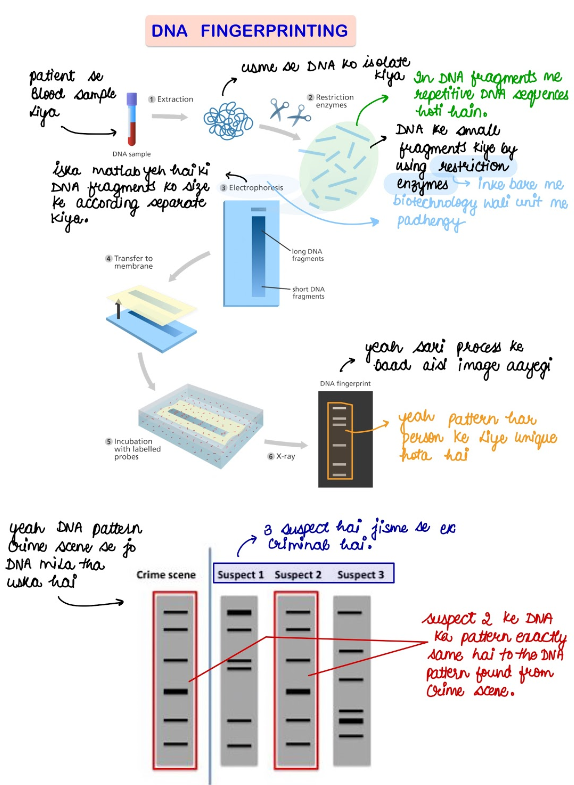
DNA FINGERPRINTING
- As we already know that almost 99.9% of DNA is same in all humans so DNA printing was developed by Alec Jeffreys which is a technique to identify repeated sequences in the human genome the produces a pattern of band in electrophoresis which is unique for every individual.
→ Satellite DNA contains highly repetitive sequences of DNA and these sequences show a high degree of polymorphism (different forms). - Imagine a hypothetical small sequence, lets say AAGGTTCC (a simple sequence) which is repeated anywhere from 30-40 times on chromosome 15 (again hypothetical). So this sequence has been repeated 30-40 times which is Variable number of tandem repeats (=how many times a sequence is repeated).
- We have many such repeat sequences which are unique for a person
- Steps Of DNA Fingerprinting
- Isolation of DNA,
- Digestion of DNA by restriction endonucleases,
- Separation of DNA fragments by electrophoresis,
- Transferring (blotting) of separated DNA fragments to synthetic membranes, such as nitrocellulose or nylon,
- Hybridization using labelled VNTR probe, and
- Detection of hybridized DNA fragments by autoradiography.
- The above image shows DNA fingerprinting results from a crime scene compared with the result of two suspects.
- The DNA pattern in column C is from a crime scene and we have let’s say two suspects : A and B. Match the pattern in column C to the pattern in column B. We can clearly see that they are exactly the same which means that DNA sample C from the crime scene is of suspect B and not of suspect A.
- Forensic Science: Biological materials used for DNA fingerprinting are: Blood, Hair, Saliva, Semen, Body tissue cells etc. DNA isolated from the evidence sample can be compared through the VNTR (Variable number of tandem repeats) prototype. It is useful in solving crimes like murder and rape.
- Paternity and Maternity Determination: A Person accedes to his or her VNTRs from his or her parents. Parent-child VNTR prototype analysis has been used to solve disputed cases. This information can also be used in inheritance cases, immigration cases.
- Personal Identification: It utilizes the idea of using DNA fingerprints as a sort of genetic barcode to identify individuals.
- Diagnosis of Inherited Disorders: It is also useful in diagnosing inherited disorders in both prenatal and newborn babies. These disorders may include cystic fibrosis, hemophilia, Huntington’s disease, familial Alzheimer’s, sickle cell anemia, thalassemia, and many others.
- Development of Cures for Inherited Disorders:By studying the DNA fingerprints of relatives who have a history of some particular disorder, DNA prototypes associated with the disease can be ascertained.
- Detection of AIDS: By comparing the band of HIV “RNA” (converted to DNA using RT PCR) with the bands formed by the man’s blood, a person suffering with AIDS can be identified.

Explanation of Image
Applications Of DNA Fingerprinting
SUMMARY
- Nucleic acids are long polymers of nucleotides. While DNA stores genetic information, RNA mostly helps in transfer and expression of information.
- Though DNA and RNA both function as genetic material, but the DNA being chemically and structurally more stable is a better genetic material.
- However, RNA is the first to evolve and DNA was derived from RNA. The hallmark of the double stranded helical structure of DNA is the hydrogen bonding between the bases from opposite strands.
- The rule is that Adenine pairs with Thymine through two H-bonds, and Guanine with Cytosine through three H-bonds.
- This makes one strand complementary to the other. The DNA replicates semiconservatively, the process is guided by the complementary H-bonding. A segment of DNA that codes for RNA may in a simplistic term can be referred to as gene.
- During transcription also, one of the strands of DNA acts as a template to direct the synthesis of complementary RNA.
- In bacteria, the transcribed mRNA is functional, hence can directly be translated.
- In eukaryotes, the gene is split. The coding sequences, exons, are interrupted by non-coding sequences, introns. Introns are removed and exons are joined to produce functional RNA by splicing.
- The messenger RNA contains the base sequences that are read in a combination of three (to make triplet genetic code) to code for an amino acid. The genetic code is read again on the principle of complementarity by tRNA that acts as an adapter molecule. There are specific tRNAs for every amino acid. The tRNA binds to specific amino acid at one end and pairs through H-bonding with codes on mRNA through its anticodons.
- The site of translation (protein synthesis) is ribosomes, which bind to mRNA and provide a platform for joining amino acids.
- One of the rRNA acts as a catalyst for peptide bond formation, which is an example of RNA enzyme (ribozyme).
- Translation is a process that has evolved around RNA, indicating that life began around RNA. Since, transcription and translation are energetically very expensive processes, these have to be tightly regulated.
- Regulation of transcription is the primary step for regulation of gene expression. In bacteria, more than one gene is arranged together and regulated in units called operons. Lac operon is the prototype operon in bacteria, which codes for genes responsible for metabolism of lactose. The operon is regulated by the amount of lactose in the medium where the bacteria are grown. Therefore, this regulation can also be viewed as regulation of enzyme synthesis by its substrate.
- Human genome project was a mega project that aimed to sequence every base in the human genome. This project has yielded much new information. Many new areas and avenues have opened up as a consequence of the project. DNA Fingerprinting is a technique to find out variations in individuals of a population at DNA level. It works on the principle of polymorphism in DNA sequences. It has immense applications in the field of forensic science, genetic biodiversity and evolutionary biology.
