TISSUES for Class 9 BIOLOGY
INTRODUCTION
- All of us know that we organisms (either unicellular or multicellular) are made up of cells. In unicellular organisms a single cell performs all the activities like digestion, respiration, etc but in multicellular organisms there are different types as well as groups of cells that carry out different functions with higher efficiency.
So in multicellular, we can say that cells form groups, cells that perform a single task often group together. This grouping of cells together to perform a function efficiently is called a tissue. The tissue cells have the same structure and they perform the same function.
There are different levels of organisation like shown in the image below :
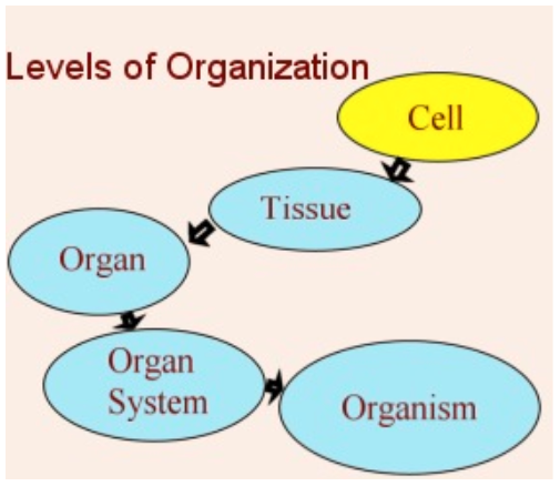
CHAPTER WEIGHTAGE
- This is an important chapter and many questions are asked from this. Isme almost sabhi topics are important like Plant tissue, Animal tissue, types of plant or animal tissue etc. etc.
BASICS OF PLANT AND ANIMAL TISSUE
- Tissue group of cells hote hain jinka similar structure hota hai or yeh ek saath ek specific kaam krte hain. Jaise animals mein, muscles cells movement karwati hai, nerve cells signal or message bhejti hain from one part of the body to the other or blood food or oxygen ko carry krta hai to different parts of the body. Ab qki har cell ka apna specific kaam hota hai islia hum keh skte hain ki cells mein division of labour hota hai. Tissues ki microscopic study ko Histology kehte hain.
BASICS OF PLANT AND ANIMAL TISSUE
- In animals, muscle cells are responsible for movement, nerve cells are responsible for carrying signals and messages from one part of the body to the other and blood is responsible for transportation of food and oxygen to different parts of the body.
In plants, there are vascular tissues that are responsible for carrying food and water to different parts of the plant. Thus there is a kind of division of labour that exists in the cells of multicellular organisms.
Division of Labour refers to the distribution of different functions among different parts of the organism's body which get specialized for the particular function. A particular function, inside an organ is performed by a group of specialized cells located at a definite place in the body.
A group or collection of similar or dissimilar cells that performs a common function and have a common origin is known as Tissues. The microscopic study of tissues is known as Histology.
PLANT TISSUE
- Har plant tissue ka apna specific kaam hota hai. Plant tissue do tarah ke hote hain: Meristematic tissue and Permanent tissue.
- Apical meristem: Yeh wo hote hain jo ki plant ke badhti hui stems ki or roots ki tips par hote hain. Islia inki wajah se hi stems or roots ki length badhti hai. Is tarah ki growth ko primary growth bhi kehte hain.
- Lateral meristem: Yeh plant mein sides mein hota hai. Jaise ki stemki side me. Inki wajah se plants ka diameter or girth yani ki circumference badhta hai jisse plant ki thickness badhti hai. Yeh plants ke stem or roots mein bark or vascular bundles mein hota hai.
- Intercalary meristem: Yeh plants mein leaves ke base pe, internodes or nodes me hota hai. Iski wajah se leaves or internodes ki growth hoti hai.
- Simple permanent tissue
- Complex permanent tissue
- Parenchyma: Yeh sabse simple or starting ki cells hoti hain plant ki. Inki cells thin walled hoti hain or inme dense cytoplasm hota hai jisme nucleus, vacuoles and baki cell organelles present hoti hai. parenchyma tissue me jo cells hote, unme apas me thoda bahut space reh jata hai jisko intercellular space kehte hai. The cells are isodiametric, i.e., equally expanded on all sides.
Iski cell wall cellulose ya pectin ki bani hoti hai. Parenchyma plants mein mostly jagah present hota hai jaise ki stems, roots, leaves, fruits, flowers, etc.
Agar inme chloroplast ho toh aise parenchyma ko chlorenchyma bola jata hai and chlorenchyma photosynthesis me help karta hai.
Yeh plant ko rigidity or support provide karta hai, food or nutrients store krta hai. Also yeh metabolic activities ke lia site provide krta hai. - Collenchyma: Yeh tissue bhi parenchyma ke jaise living hi hote hain. Yeh cells epidermis ke neeche hote hain in dicot stem, midrib and petioles. Is tissue ki cell walls to thin hoti hai lekin cells ke corners pe cellulose ka extra deposition hota hai. Yeh tissue leaves or young tissues ko flexibility provide krta hai jisse wo bend ho sake lekin break na ho. Inki cells mein usually intercellular space nai hota hai. Also yeh monocots ke roots, stem or leaves mein nahi hote.
- Sclerenchyma: Yeh dead cells hoti hain or inme protoplasm nai hota hai. In cells ki secondary walls extremely hard hoti hai kyuki unme lignin ka deposition hota hai jisse unhe lignified cells bhi kehte hain. Cell wall excessive thick hone ki wajah se unme cell cavity bhi nai hoti hai yani unme intercellular spaces bhi nai hote hai. Kai baar thick cells mein thin areas bhi hote hain jinhe pits kehte hain. Yeh cells aapas mein middle lamella ke through jude hote hain.
Sclerenchyma mein do tarah ke cells hote hain: Fibres and Sclereids. Yeh cells ya to definite layers ya fir patches mein hoti hai. Sclerenchyma stems, roots, veins of leaves, ya fir seeds or nuts ke covering mein hota hai. Example, coconut ka husk isi tissue se bana hota hai. Yeh tissue fruit ke mesocarp mei present hota hai or inse rope, mats ya fr brushes bnaya ja skta hai. - Xylem: Yeh tissue water or organic materials ko transport karne mein help karta hai. Sath hi sath yeh plant ko mechanical strength bhi provide krta hai. Xylem further four types ka hota hai:
- Tracheids
- Vessels
- Xylem parenchyma
- Xylem fibres
- Phloem: Yeh ek living conducting tissue hota hai jo ki organic solutes or food ke translocation mein help karta hai. Isme tubes to hoti hain lekin unka koi mechanical function nai hota hai. Phloem four types ke components se milkar bana hota hai:
- Sieve tubes
- Companion cells
- Phloem parenchyma
- Phloem fibres
Meristematic tissue:
Meristematic tissue mein undifferentiated cells hoti hain jo ki kisi bhi plant ka base yani building block hoti hain. Yeh hamesha divide hoti rehti hai isi wajah se yeh length or girth badhane mein help krti hain. Yeh cells kuch specific locations tak hi rehte hain. Inke types hain:
Kya aap jante hain ki intercalary meristems mein vacuoles kyu nahi hota? Jaise ki hame pta hai ki vacuoles mein khanaa or paani store hota hai or intercalary tissues inhe store nai krte balki bnate hain. Islia intercalary meristems mein vacuoles nahi hota.
Permanent Tissue:
Jo meristematic tissue hota hai wo divide hoke ek speecific role play krta hai and further divide hone ki ability ko loose kr deta hai. Or isi tissue ko ab permanent tissue kehte hain. Jis process se meristematic tissue khud ko convert krte hain permanent tissue mein ek specific size, shape, function se, uss process ko differentiation kehte hain. Permanent tissue mature cells se bana hota hai jinme already growth and differentiation ho chuka hai or further divide karne ki ability temporarily ya permanently ja chuki hai.
Permanent tissue ki cells living bhi ho skti hai or dead bhi. Jo living cells hoti hain wo large, thin walled hoti hain jiske cytoplasm mein vacuole present hota hai or agar cells dead hain to un cells ki wall thick hoti hai or usme koi cytoplasm bhi nai hota.
Permanent tissue do tarah ka hota hai:
Simple permanent tissue: Yeh wo tissue hai jo ek hi tarah ke cells se milakr bna hota hai or jin kacommon origin or function hota hai. Yeh further parenchyma, sclerenchyma or collenchyma mein classified hote hain.
Complex permanent tissue:
In tissues mein ek se jada cells hoti hai jinka common origin or function hota hai. Inka main kaam hota hai water, minerals ya organic solutes ka transportation krwana.
Yeh do tarah ke hote hain: Xylem and Phloem. Dono xylem or phloem conducting tissues hote hain ya jinhe vascular tissues bhi kehte hain jo milkar vascular bundles form krte hain.
PLANT TISSUE
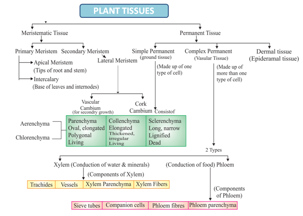
- Plant tissue is a collection of similar cells performing an organized function for the plant. Each plant tissue is specialized for a unique purpose. The plant tissue may be primarily classified into two groups:
- Meristematic Tissues
- Permanent Tissues
- The meristematic tissue has the quality of self-renewal.
- The cells of the meristematic tissues are similar in structure and have thin and elastic cellulosic cell walls.
- The meristematic cells can be spherical, rectangular, polygonal or oval in shape.
- These cells are compactly arranged and do not contain any intercellular space between them.
- The meristematic tissue has the quality of self-renewal.
- They have very small and few vacuoles.
- The protoplasm of these cells is very dense and has large and prominent nuclei.
- Each cell contains dense or abundant cytoplasm.
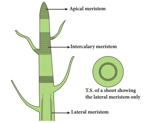
- Apical meristem: It is situated at the growing apex or tips of the stems and roots and also at the apices of the leaves. So, these cells are responsible for the increases in the length of the stems and roots and for linear growth of an organ i.e primary growth. Example, root apical meristem and shoot apical meristem.
- Lateral meristem: It is arranged at the parallel to the sides of an organ (stems or leaves) and consists of initials which divide mainly in one plane. It is responsible for the increase in diameter and girth(circumference) of the organ i.e secondary growth. They are found beneath the cork cambium(bark) and in vascular bundles of dicot roots and stems (called vascular cambium). Cambium is responsible for growth in thickness or secondary growth.
- Intercalary meristem: It is located in between the regions of permanent tissues i.e at the base of the leaves, internodes and nodes. So it is responsible for the growth of leaves and internodes eg, stems of grasses and other monocots.
- Simple permanent tissue
- Complex permanent tissue
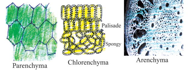
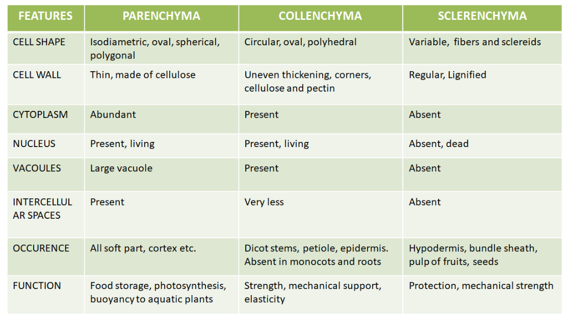
- Parenchyma: It forms the bulk of the plant body and is the most simple and unspecialized primitive tissue. It consists of thin walled cells and encloses a dense cytoplasm which contains a small nucleus, surrounds a large central vacuole and has intercellular spaces between them. We can also say that parenchyma cells also have living protoplasm and perform metabolic processes. The cell wall is made up of cellulose or calcium pectate. As these cells are living, they possess power of division.
The cells are rounded or isodiametric i.e equally expanded on all sides, oval, cylindrical, rectangular, stellate or long spindle like. The parenchyma is widely distributed in plant bodies such as stem, roots, leaves, flowers and fruits. The parenchyma tissue is found in epidermis, cortex, pericycle, mesophyll of cells, pulp of fruits and endosperm of seeds. It is also distributed in pith, medullary rays and packing tissue in xylem and phloem.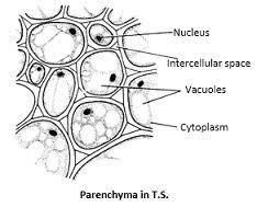
Functions of Parenchyma:- Stores food and nutrients.
- Provides support and foundation.
- Involved in growth and development.
- Provide mechanical rigidity to the plants.
- They are the site of all metabolic activities.
- Helps in regeneration, healing and repair of wounds.
- Involved in the movement of water and other nutrients.
- If chloroplast is present, the parenchyma tissue is called Chlorenchyma and it performs photosynthesis e.g, the mesophyll of leaves.
- In aquatic plants, large air cavities are present in the ground tissue. These cavities store gases and provide buoyancy to aquatic plants to help them float. Such parenchyma is called Aerenchyma
- Collenchyma: These tissues are also living like parenchyma. The cells of collenchyma are located below the epidermis of dicot stem, midrib and petiole. The cell walls are thin but have deposition of extra cellulose at the corners of the cells.
The tissue provides flexibility to the leaves and young stems of the plant so that they can bend without breaking. In this tissue, intercellular spaces are generally absent. These cells are elongated in shape and contain few chloroplasts. They are generally absent in monocot roots, stems and leaves.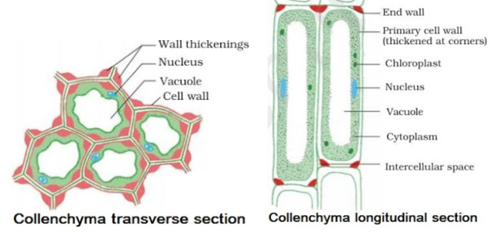
Functions of Collenchyma:- It provides mechanical support and elasticity in young dicotyledonous stems.
- It manufactures sugar and starch if their cells possess chloroplast.
- Present at the margins of some leaves and it resists the tearing effect of the wind.
- Sclerenchyma: These are dead cells and do not contain protoplasm. These cells have hard and extremely thick secondary walls due to uniform deposition of lignin. Such cell walls are called lignified.
Due to excessive thickening of the cell wall, its cell cavity (lumen) becomes nearly absent. The cells of sclerenchyma are closely packed without intercellular spaces. Sometimes, thin areas are found in thick cell walls which are called pits. These cells are joined with the help of conspicuous middle lamella (made up of pectin, lignin and protein).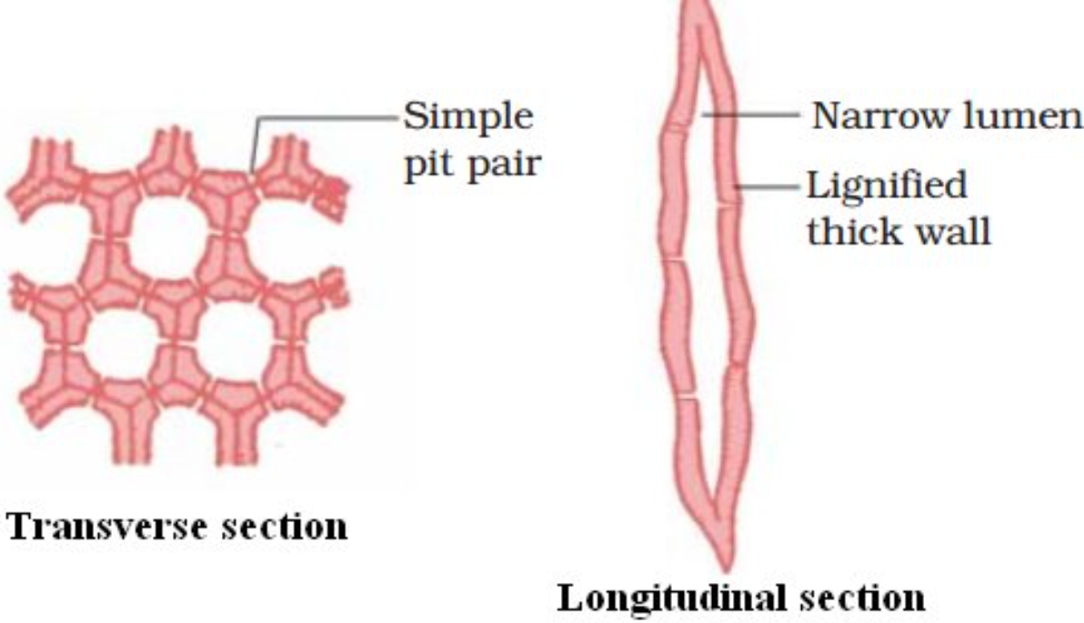
Cells of sclerenchyma are of two types: Fibres and Sclereids.- Fibres: It consists of very long, narrow, thick and lignified cells. These cells vary in length from 1mm to 550mm in different plants. These are usually pointed at both ends and are usually clustered into strands and look polygonal in the transverse section.
- Sclereids: These are short and possess extremely thick lamellated lignified walls with long tubular simple pits. They are short and vary greatly in shape (spherical, oval, cylindrical, T-shaped, dumbbell shaped and stellate).
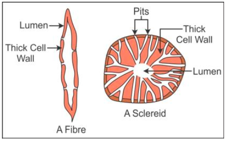
The sclerenchyma occurs either in patches or definite layers. They are found in stems, roots, veins of leaves, hard covering of seeds and nuts. They also occur in hard seed coats, hard endocarp of almond and coconut, grit of apple, pear and guava. Like, husk of coconut is made up of sclerenchymatous tissue. It is present in mesocarp of fruit of coconut and yield fibre used for mats, ropes, cords, brushes, etc.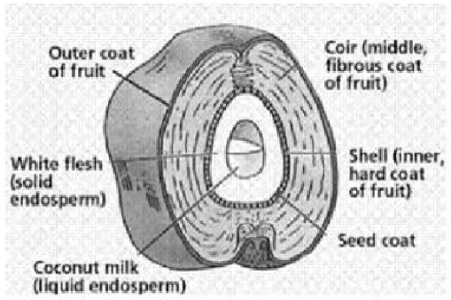
Functions of Sclerenchyma:- It provides mechanical strength, rigidity, flexibility, and elasticity to the plant parts.
- It provides a protective covering around seeds and nuts.
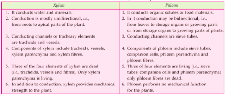
- Xylem
- Phloem
- Xylem: It is a chief vascular and mechanical tissue responsible for conduction of water and inorganic solutes.
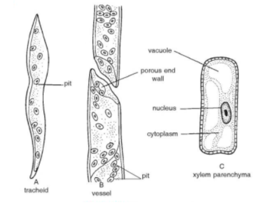
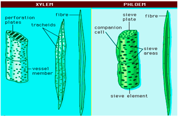
Xylem is composed of four different types of cells:- Tracheids: These are elongated tubes like dead cells with tapering ends. The end walls are oblique and remain intact and possess bordered pits. They also conduct water and the walls are hard and lignified.
- Vessels: These are shorter and wider than tracheids. They are very long tube-like structures(for conduction of water and minerals) formed by placing cells one upon the other and their end walls are either absent or have perforations. Vessels are found mainly in angiosperms.
- Xylem parenchyma: These are living parenchymatous cells which help in lateral conduction of organic solutes and storage of food.
- Xylem fibres: These lignified fibres provide mechanical strength to the plant body.
Functions of Xylem:- To carry water and mineral salts upward from the roots to the different aerial parts of the plant.
- It provides mechanical strength to the plant body.
- Phloem: It is the chief food conducting tissue of vascular plants. It contains tubes but performs no mechanical function. It is regarded as a living conducting tissue responsible for translocation of organic solutes.
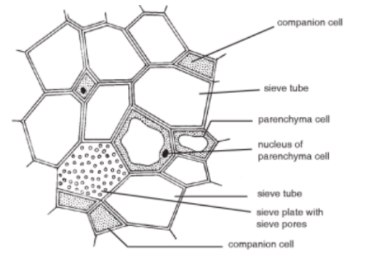
Phloem is composed of following four components:- Sieve tubes: These are slender tube-like structures composed of elongated thin walled cells placed one above the other forming a continuous tube. The end walls of sieve tube elements are called sieve plates which are perforated by numerous pores. At maturity, the nucleus of each sieve tube degenerates but cytoplasm persists. The plastids, mitochondria, endoplasmic reticulum and dictyosomes are absent.
- Companion cells: These are living cells generally associated with sieve tubes is a small thin walled cell containing dense and active cytoplasm and large elongated nucleus. The common wall between sieve tube and companion cells shows presence of fine pits which are traversed by plasmodesmata.
- Phloem parenchyma: These are thin walled, living cells of parenchyma of phloem. They have the function of storage and slow lateral conduction of food.
- Phloem fibres: They are thick walled, elongated spindle shaped dead cells which have narrow lumen. They also provide mechanical strength to the tissue.
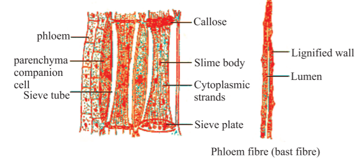
Functions of Phloem:- They transport food materials from leaves to the storage organ and later to growing regions of the plant body.
- The phloem is also a pathway to signaling molecules and has a structural function in the plant body.
Meristematic Tissue:
Meristematic tissues have undifferentiated cells which form the building blocks of the specialized plant structures. They include the group or cluster of cells which remain in continuous state of division or retain their power of division. These cells have the ability to divide continuously and help in increasing the length and girth of the plant.Characteristics of Meristematic tissues:
The meristematic cells are restricted to some specific regions and thus they can be classified on the basis of area of plants where they are located:
Do you know why there are no vacuoles in the intercalary meristem?
Vacuoles are responsible for the storage of food and water. The intercalary tissues do not store them rather are responsible for manufacturing them. Moreover, vacuoles contain sap which provides rigidity to a cell. This property of vacuoles may not allow the intercalary tissues to divide and manufacture new cells. Hence vacuoles are not present in intercalary meristem.
Permanent Tissue:
Permanent tissues originate from the meristematic tissue as cells derived from the division of the meristematic tissue take up the specific role and lose the ability to divide. The process by which cells of the meristematic tissues convert themselves into a permanent tissue by taking a fixed shape, size and function is called differentiation. They are made up of mature cells that have undergone growth and differentiation thus they lose their ability to divide and form more cells.The cells of these tissues may be living or dead. The living cells of these tissues are large, thin walled enclosing vacuolated cytoplasm and dead cells are usually thick walled without having cytoplasm. Thickening may be regular or irregular.
Permanent tissue is of two types:
Simple permanent tissue:
These tissues are composed of similar types of cells having common origin and function. These tissues are made up of one type of cells. They are further classified into parenchyma, collenchyma and sclerenchyma.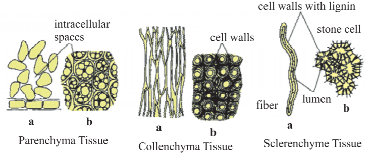
Epidermis:
The outermost layer of the cell is known as the Epidermis. It is the protective tissue and covers the entire plant. It is a thin layer of single cells but in places with less water, the epidermis of the plants can become thick in order to avoid frequent water loss.The cells are flat and they have no intercellular spaces between them. The outer walls of the epidermal cells are thick and the inner walls are thin. The epidermal cells often have long hair-like structures in roots which facilitate the absorption of water.
The main function of the epidermis is to protect the plants from fungi, water loss and any injuries by secreting a wax-like water-resistant substance (called cuticle) on the surface of the plants which protects them.
Stomata:
It is a pore-like structure that is present in the epidermis of the leaves. These pores are enclosed by two cells that have a similar shape as a kidney. These are called Guard cells of stomata. These are modified epidermal cells and are responsible for the exchange of gases and transpiration.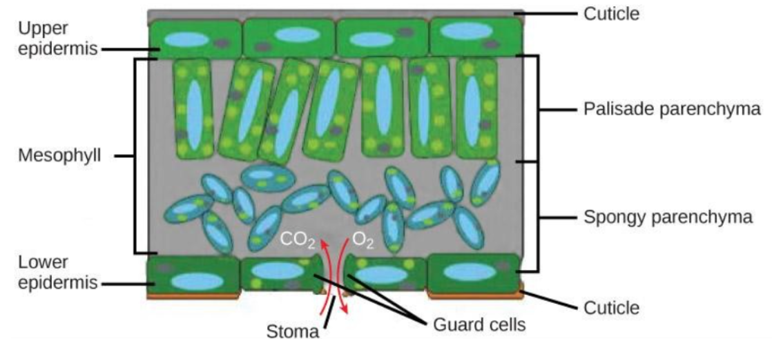
Complex permanent tissue:
These tissues consist of more than one type of cells with common origin and working together as a unit to perform a common function. These tissues are mainly concerned in transportation of water, mineral nutrients and organic solutes like food materials.They are of following two types:
Both xylem and phloem are conducting tissues and also known as vascular tissues which constitutes a vascular bundle.
ANIMAL TISSUE
- Animal cells ek sath milkar animal tissue banate hain. In tissues ka structure, function or origin vary karta hai. Jaise ki, muscles, blood, lymph, nerves, etc ye sabhi animal tissue hain. Yeh tissue chaar tarah ke hote hain.
- Epithelial tissue
- Muscular tissue
- Connective tissue
- Nervous tissue
- Squamous epithelium: Yeh thin, flat, irregular-shaped cell hoti hai jo ek compact tissue form krte hain or yeh tissue dikhne mein aisa lagta hai jaise ki floor ki tiles ek sath judi hoti hain. Islia ise Pavement epithelium bji kehte hain. Yeh epithelium blood vessels,nose, mouth, urinary tubules, alveoli of the lungs, tongue aur skin ki lining form krta hai.
- Cuboidal epithelium: Iss epithelium ki cells cubes jaisi hoti hai or yeh kidney tubules, thyroid vesicles, small salivary ducts, pancreatic ducts or glands jaise sweat glands, salivary glands, and exocrine glands wagerah ki lining mein hota hai.
- Columnar epithelium: Yeh cells pillar ke jaise hote hain. Yani ki lambe jada or chaude(wide) kam hote hai. In cells ka nuclei bhi elongated hota hai. Yeh epithelium stomach, intestine or gall bladder ki lining form krta hai.
- Ciliated epithelium: Is epithelium mein cuboidal ya columnar koi bhi cells ho skti hai lkin fark yeh hai ki inme cilia present hota hai. Cilia ek thread ke jaisa structure hota hai jo arise hota hai basal granule se or yeh movement mein help krta hai of substances like urine, eggs or sperms, etc.
- Glandular epithelium: Iss epithelium mein columnar cells hoti hain jo ki chemicals secrete krte hai. Yeh epithelium mainly glands mein hota hai like in gastric glands, intestinal glands, etc.
- Striated muscle: Inhe skeletal, striped or voluntary muscles bhi kehte hain. Skeletal inhe isliye kehte hain kyuki yeh bones se attached hote hain or movements krwate haain. Striped inhe islia kehte hain qki inme alternate light or dark stips hote hain or voluntary islia kehte hain qki inh hum apni will ke according move kar skte hain. Yeh muscle cells multi nucleated hoti hain or har cell ka apna plasma membrane hota hai jisko sarcolemma kehte hain. Yeh muscles tongue, imbs pharynx or beginning of oesophagus or body wall mein present hota hai.
- Unstriated muscles: Inhe smooth muscles bhi kehte hain or inme koi stripes bhi nai hoti hai. Inke cytoplasm ko sarcoplasm kehte hain or yeh cells long, narrow spindled with pointed ends hoti hain jinme ek hi nucleus present hota hai. Yeh loose connective tissue ki help se jude rehte hain. Inhe visceral or involuntary muscles bhi kehte hain. Yeh hamari alimentary canal ki wall mein ya internal organs, ducts of glands, blood vessels, stomach, intestines, etc mein present hote hain.
- Cardiac muscles: Inme striated or smooth dono muscles ke characteristics hote hain. Inki cells ke charo aur sarcolemma, sarcoplasm or fibres ke end ke beech mein intercalated disc bhi hoti hai. Yeh tissues heart, pulmonary veins or superior vena cava mein hote hain. Inka kaam hai throughout lifetime contract or relax krna bina thake or sath hi sath blood ko pump or distribute krna puri body mein.
- Areolar tissue
- Dense regular connective tissue
- Adipose tissue
- Skeletal tissue
- Vascular tissue
- Areolar tissue(Loose connective tissue):Yeh transparent, jelly-like sticky matrix hota hai jisme bhut sare fibres present hote hain. Yeh fibres do tarah ke hote hain: (a) White collagen fibre(jo protein se bana hota hai, jise collagen kehte hain), (b) Yellow elastic fibres(jo ek protein se milkar baana hota hai jise elastin kehte hain). Collagen flexibility or strength provide krta hai aur elastic fibre elasticity provide krta hai.
- Dense regular connective tissue:Yeh nature mein loose and elastic hote hain or inki cells densely packed hoti hai. Isme tendons or ligaments hote hain.
- Tendons: Yeh skeletal muscles ko bones se connect karte hain. Inme flexibility to limited hoti hai lekin strength bhut hoti hai.
- Ligaments: Yeh ek bone ko dusre bone se connect karte hain.Ligaments ki cells mein thoda sa matrix bhi hota hai. Yeh yellow elastic tissue se bane hote hain jisme bhut sare collagen fibres hote hain jo areolar tissue se jude hote hain. Sath hi sath inme elasticity bhi hoti hai.
- Adipose tissue: Yeh alag alag tissues ko organs se connect karte hain or unhe support bhi provide karte hain. Inka origin mesoderm se hota hai.
Connective tissue kuch cells se milkar bana hota hai jinhe elastin or collagen kehte hain. Connective tissues mein jo fibres hain wo unhe elasticity, flexibility or strength provide karta hai. Jo adipose tissue hai wo sabse jada skin ke neeche, internal organs mein or yellow bone marrow mein hota hai. - Skeletal tissue: Yeh tissue rigid skeleton form karte hain jo ki locomotion mein bhi help karte hain or hamare vital organs ko protection provide karte hain. Yeh do tarah ke hote hain:
- Cartilage: Yeh jada tar embryonic stage mein hota hai or ek tarah ke supporting skeleton ka kaam krta hai. Bhut sare cartilage to bache jb adult ho jate hain to bone mein convert ho jate hain. Cartilage ears mein, nose mein, between bones of vertebral columns mein bhi hote hain.
Cartilage mein chondrocytes cells present hote hain jo collagen fibre secrete karte hai jisse hame additional strength milti hai. Yeh chondrocyte cells ek cavity mein hoti hai jinhe lacunae kehte hain. Yeh 2-4 cells ke group mein hote hain. Cartilage elasticity provide karne ke sath sath firm bhi rhta hai. Inme ek solid matrix present hota hai jo ki protein or sugar se bana hota hai. Cartilage hamari body ke bhut sare parts ko support and flexibility provide krta hai. - Bones: Yeh hamari body ka hardest connective tissue hota hai jo hamari body ki shape or uske posture ko maintain krta hai. Bones mein collagen fibre or calcium bhut jada amount mein hota hai jo use strength provide krta hai.
Bones ki cells ko osteocytes kehte hain jo lacunae mein present hoti hai or matrix secrete karte hain. Jo osteocytes cells ka cytoplasm hai wo tiny channels banata hai jise canaliculi kehte hain jiski wajah se osteocytes or cappilaries mein communication ho pata hai.
Spongy bones ek core mein present hote hain matlab andar ki side spongy bone hoti hai or uske bahar charo aur compact bone hoti hai. Osteocytes matrix ke concentric layers mein hote hain jise lamellae kehte hain. Capillaries or nerves ek central channel ke through pass hoti hai jise Haversian canals kehte hain. In canal ke charo aur bhi canal present hote hain.
Bones ek endoskeleton ya jise dhacha kehte hai wo banate hain. Yeh hame movement provide krne ke sath sath hamare body ke delicate parts like tissues and organs ko support bhi provide karta hai.
- Cartilage: Yeh jada tar embryonic stage mein hota hai or ek tarah ke supporting skeleton ka kaam krta hai. Bhut sare cartilage to bache jb adult ho jate hain to bone mein convert ho jate hain. Cartilage ears mein, nose mein, between bones of vertebral columns mein bhi hote hain.
- Fluid tissue (Vascular tissue): Isme ek fluid matrix hota hai jisme cells freely float karti rehti hain. Is matrix mein fibres nai hota. Isme Blood or Lymph included hota hai.
- Blood: Plasma mein bhut sari cells hoti hain jisse blood banta hai. Hamare blood mein red blood cells, white blood cells or platelets hoti hain. WBC ek tarah ka defence system create krta hai jo ki koi bhi bacteria ya foreign material se fight krtaa hai or hamari body ko protect krta hai. RBC mein haemoglobin hota hai jo ki oxygen ko transport krta hai. Platelets clotting ka kaam krta hai yani agar kahi cut lagta hai to wha seblood nikalta hai to platelets us blood ke nikalne ko rokta hai. Plasma mein proteins, water, hormones, salts hote hain jo ise different parts tk transport karta hai.
- Lymph: Yeh ek colorless fluid hota hai jo ki WBC’s ko puri body mein leke leke jata hai in lymphatic vessels. Kuch organs hote hain hamari body mein jo lymph produce karte hain lymphoid organs kehte hain or lymphoid system form krte hain. Jisme se kuch hai lymph nodes and tonsils.
Lymph ka kaam hai blood ke volume ko maintain karne or bacteria ya virus ko body se remove krke immunity maintain karna. Yeh oxygen or minerals ko puri body mein leke jata hai or body se carbon dioxide leke body ke bahar release karwata hai. Yeh hamare intestine se fat ko absorb krta hai or puri body mein transport karta hai.
- Dendrites: Yeh short branches hote hain jo cell body se nikalti hai. Yeh neuron ke surface area ko badhate hai. Yeh dusre neurons se chemical signal lete hain or use electrical signal mein convert krke neuron ki cell body mein bhejte hain. Kisi bhi neuron mein ek dentrite bhi ho skta hai or bhut sare bhi.
- Cell Body: Inka main kaam hai cell ki functionality ko maintain karna. Inka signal ke transmission mein koi active role nai hota hai. Yeh kuch protein bhi secrete krte hai jo ki neurons ko thik se kaam krne mein helk karta hai. Yeh normal cell ki tarah hi hota hai jisme mitochondria, golgi apparatus ya baki cell organelles hote hai jisse cell ape kaam kr pati hai.
- Axon: Yeh ek long fibre hota hai jo cell body se nikalta hai. Yeh neuron ko baki neurons, ya cells ya organs se bhi connect karta hai through nerve terminals. Jitna jada axon ka diameter hota hai utni hi jaldi signal transfer hota hai. Ispe ek insulating substance ki covering hoti hai jise mylein kehte hain.
Epithelium tissue:
Yeh sabse simple tissue hota hai animals mein. Yeh ek tarah se protection ka kaam karata hai or iski cells tightly packed hote hain jiski wajah se yeh continuous sheets form krta hai jo ki body or internal organs ko cover karta hai. Like, skin, mouth, alimentary canal, lungs, etc. Tightly packed hone ki wajah se inme rare hi intercellular matrix hota hai. Yeh intercellular junctions ki wajah se ek sath jude rhte hai. Yeh tissue do tarah ke hote hain, Simple or Stratified.
Epithelium tissue ek barrier ka kaam karta hai jo ki different organs or systems ko separate rakhta hai. Yeh tissue andar ki cells ko dry hone se, injury, chemical effect, viral or bacterial infections se protect karta hai. In tissues ki wajah se hi water or minerals ka absorption hota hai or waste products ka elimination hota hai. Epithelial tissue 5 types ke hote hain:
Muscle tissue:
Yeh tissue muscle cells se bane hote hain jo ki contraction karte hain. Inme ek se jada nuclei bhi ho skte hain. Inke parallel arrangement ki wajah se hi hamari body mein movement hoi hai yani ki contractile proteins ka muscle cells mein contraction or relaxation hota hai. Muscle cells teen tarah ke hote hain:
Connective tissue:
Yeh ek tissue ko dusre tissue se bind or join karne ka kaam karte hain jisse ek protective sheath form ho jati hai organs ke charo auror organs ek dusre se seperate rhte hain or apna apna kaam krte haain jaise ki materials ko transport krna from one part to another. Yeh cells living hoti hai or inka number bhi kam hota hai. Yeh tissue supporting framework banate hain of cartilage and bones. Yeh cells loosely bound hoti hai or intercellular matrix mein present hoti hai. Yeh matrix chaar tarah ka hota hai: – Dense, Rigid, Fluid or Jelly-like.
Connective tissue panch tarah ka hota hai:
Nervous tissue:
Yeh tissue hamare nervous system ki main tissue hai. Yeh hamare body ke functions ko regulate krti hai. Brain, Spinal Cord, and nerves milkar nervous tissue banata hai. Inhi ki wajah se stimuli ody ke ek part se dusre part mein transport hota hai.
Neurons chemical neurotransmitters secrete karta hai jisse message hamare organs se brain mein jata hai or fr brain hame action krne ke lia btata hai. Do neurons ke beech mein gap hota hai jise synapse kehte hain jaha par chemical release hota hai. Har neuron kr teen main parts hote hain: Dendrites, Axon and Cell body.
ANIMAL TISSUE
- The animal cells are grouped together to form animal tissue. These tissues vary in their structure, function and origin. For eg, muscles, blood, lymph, nerves, etc.
- Epithelial tissue
- Muscular tissue
- Connective tissue
- Nervous tissue
- They act as a barrier and separate different organs and systems from each other.
- These cells protect the underlying cells from drying, injury and chemical effect. They also protect the body from viral and bacterial infections.
- They help in absorption of water and nutrients.
- They help in elimination of waste products.
- Some of these perform secretory function and secrete substances like saliva, sweat, enzymes, etc.
- Squamous epithelium
- Cuboidal epithelium
- Columnar epithelium
- Ciliated epithelium
- Glandular epithelium
- Squamous epithelium:
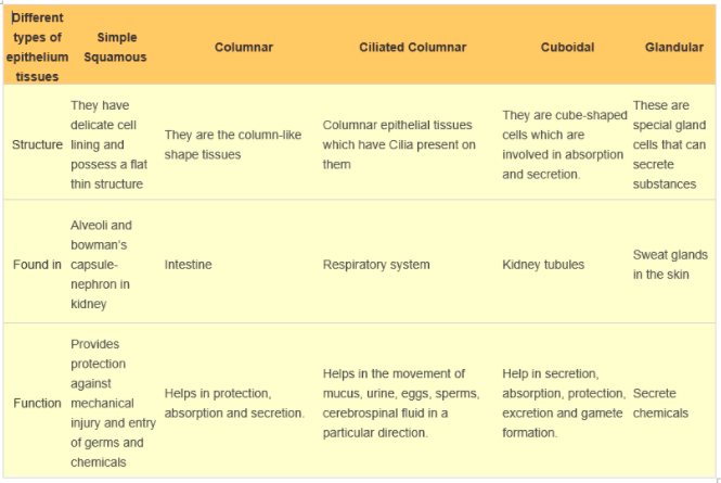
It is a thin, flat, irregular-shaped cell which fits together like floor tiles to form a compact tissue. Therefore, it is also known as Pavement epithelium. It forms the lining of the blood vessels,nose, mouth, the coelom, urinary tubules, alveoli of the lungs and covering of the tongue and skin.
Functions:- This epithelium protects the underlying parts against abrasion(mechanical injury), entry of germs, chemicals and drying.
- It also forms a selectively permeable surface through which filtration occurs.
- It also helps in excretion, gas exchange and secretion of coelomic fluid.
- Cuboidal epithelium:
This epithelium consists of cube-like cells. These cells are polygonal in surface view and square in section. This epithelium is found in kidney tubules, thyroid vesicles, lines of the small salivary ducts, pancreatic ducts and in glands(like, sweat glands, salivary glands, and exocrine glands). It also covers the ovaries and lines the sperm-producing tubules.
Functions:- It provides mechanical support.
- It helps in protection, secretion, absorption, excretion and gamete formation.
- Columnar epithelium:
It consists of cells which are taller than wider i.e pillar-like. The nuclei are towards the base of the cell. Sometimes the free end of the cells have a brush border containing microvilli. It forms the lining of the stomach, intestine and colon forming mucous membrane. It also forms the lining of the gall bladder, mammary glands ducts and parts of urethra.
Functions:- It helps in absorption(eg, stomach, intestine) and secretion(eg, mucus by goblet cells or mucus membrane).
- It also helps in protection.
- Epithelium of the intestine is specialized for absorption of water and digested food.
- Ciliated epithelium:
This epithelium consists of cuboidal or columnar cells which bear thread-like cytoplasmic outgrowths called cilia on their free surfaces. Such cells form the ciliated epithelium. Cuboidal ciliated epithelium lines the certain parts of sperm ducts. Columnar ciliated epithelium lines the nasal passage, trachea(wind-pipe), terminal bronchioles(lungs), kidney tubules, oviducts(fallopian tubes) and ventricles of the brain.
Function:- The rhythmic concerted beating of the cilia helps in the movement of mucus, urine, eggs, sperms and cerebrospinal fluid in a particular direction.
- Glandular epithelium:
The columnar epithelium is often modified to form glands to secrete chemicals. It lines the glands such as gastric glands, pancreatic lobules, intestinal glands, etc. - Striated muscle
- Smooth muscle
- Cardiac muscle
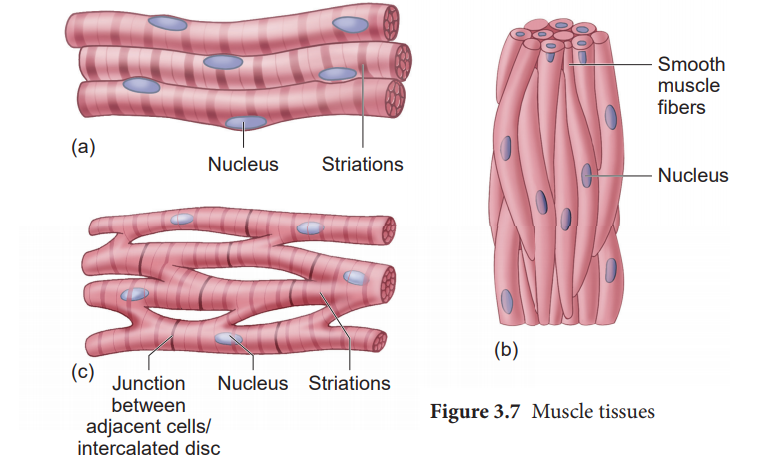
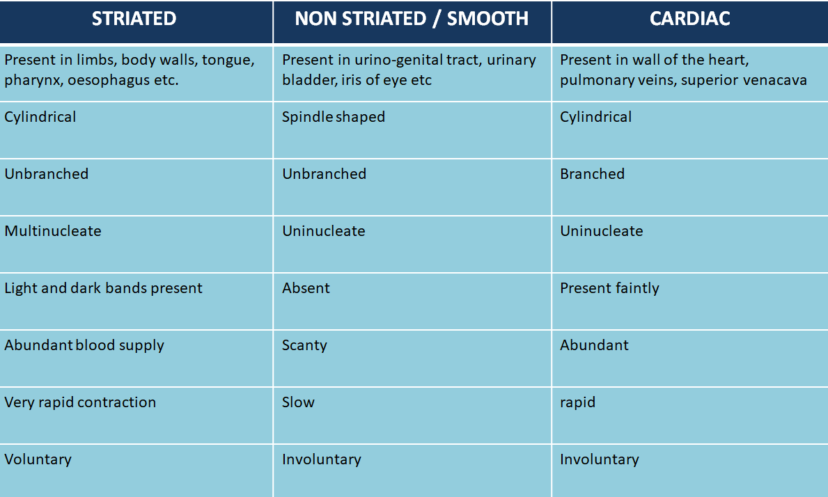
- Striated muscle: They are also known as skeletal(as they are attached to the bones and responsible for movements), striped(as the entire fibre shows alternate light or dard stripes) or voluntary muscles(as they work according to our will). These are long, non-tapering, cylindrical and unbranched muscle fibers. These cells are multinucleated. Each muscle cell is enclosed in a thin but distinct plasma membrane, called sarcolemma. The striated muscles are found in the body wall and the limbs. They also occur in the tongue, pharynx and beginning of oesophagus.
Functions:- These are powerful and undergo rapid contraction so these muscles may get tired and need rest.
- They help in movement of body parts and locomotion and also in several voluntary movements of the body.
- Unstriated muscles: They are also called smooth muscles because they do not show any stripes or striations across the muscle fibre. They are long, narrow spindles shaped with pointed ends and there is a single centrally located nucleus in the centre of cytoplasm called sarcoplasm. They are generally shorter than striated muscle fibre. They are held together by loose connective tissue. Each muscle cell is enclosed in a plasma membrane They are also called visceral or involuntary muscles.
The smooth muscle tissue occurs within the walls of hollow(tubular) visceral organs except the heart. They also occur in the wall of the alimentary canal and internal organs, ducts of glands, urogenital ducts, blood vessels, stomach, intestine, ureter, bronchi, iris of the eye, etc.
Functions:- They do not work according to our will so called involuntary muscles.
- Smooth muscles contractions are rhythmic. These muscles contract slowly but can remain contracted for a long period of time.
- Cardiac muscles: They show characteristics of both striated and smooth muscles. These muscles are short, cylindrical fibres which are branched and joined end to end to form a network. Each fibre or cell is surrounded by sarcolemma, cytoplasm(sarcoplasm) with longitudinal myofibrils and a centrally located nucleus(uninucleated).
Generally intercalated discs occur between the ends of fibres. The intercellular spaces of these muscles are filled with abundant loose connective tissue supplied with the blood capillaries. These muscles have stripes of light and dark bands. Cardiac muscles are confined to the wall of the heart, pulmonary veins and superior vena cava.
Functions:- Cardiac muscles contract and relax rapidly, rhythmically and tirelessly throughout the lifetime.
- These muscles help to pump and distribute the blood to the various parts of the body.
- They attach organs and tissues together.
- They store fat in the form of adipose tissues.
- They help in repairing tissues.
- They prevent the organs from mechanical shocks.
- The organs also help in defence.
- Areolar tissue
- Dense regular connective tissue
- Adipose tissue
- Skeletal tissue
- Vascular tissue
- Areolar tissue(Loose connective tissue): This tissue acts as a filter in between the spaces present inside the organs of the body. It consists of a transparent, jelly-like sticky matrix having numerous fibres. Its fibres are of two types: (a) White collagen fibre(made of protein called collagen), (b) Yellow elastic fibres(formed of protein called elastin). Collagen provides flexibility and strength whereas elastic fibre provides elasticity.
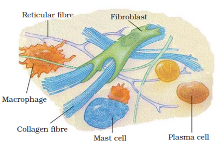
Function:- It acts as supporting and packing tissue between organs lying in the body cavity.
- It helps in repairing other tissues as well.
- It is found in the skin and bone marrow.
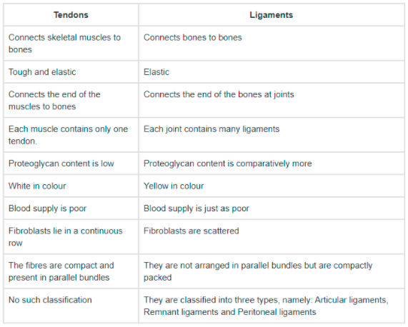
- Dense regular connective tissue: It consists of ordered and densely packed fibres and cells. These are loose and elastic in nature. They are secreted by the surrounding connective tissue cells. This tissue is the principal component of tendons and ligaments.
- Tendons: These are cord-like structures that transmit muscular force to a bone. They have limited flexibility but very great strength. They are tough, in-elastic bundles of white collagen fibres bound together by areolar tissue. They connect skeletal muscles to bones.
- Ligaments: They are cords formed by yellow elastic tissue in which many collagen fibres are bound together by areolar tissue. A ligament connects two bones together. It has an elasticity which facilitates the connection. The cells of ligaments have a little matrix.
- Adipose tissue: It supports and connects different tissues and organs of the body. They are widely distributed in every part of the body. They originate from the mesoderm (the middle germinal layer of the embryo).
Connective tissue is made up of a few cells present in the intercellular framework of protein fibres secreted by the cells, known as collagen or elastin. The cells also secrete a thin gel of polysaccharides, which together with fibres make matrix or ground substance.
The elasticity, flexibility and strength of the connective tissues are due to fibres. The function and types of connective tissues depend on the nature of the intercellular substance present. The adipose tissue is abundant below the skin, between the internal organs and in yellow bone marrow.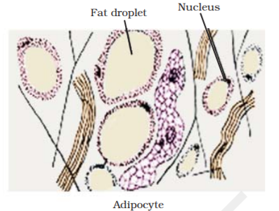
Functions:- It serves as a fat reservoir.
- It provides shape to the limbs and the body.
- It keeps visceral organs in position and forms shock absorbing cushions around the kidneys and eye balls.
- It acts as an insulator and thus regulates body temperature.
- Skeletal tissue: They form the rigid skeleton which supports the body, helps in locomotion and provides protection to many vital organs. They are of two types:
- Bone
- Cartilage
- Cartilage: Cartilage is mostly present in the embryonic stages and works as a supporting skeleton. Most of the cartilage is replaced by bones in adults, however, it supports some structures in adults too. The cartilage is present between the bones of the vertebral column, in the external ear, nose and hands.
The cartilage consists of chondrocytes cells, which are enclosed in a hard, rubbery matrix, secreted by them. They secrete collagen fibres also, which provide additional strength. Chondrocytes lie in the cavities known as lacunae, in a group of 2-4 cells or singly. Cartilage possesses elasticity, but is firm too. They lack nerves, blood and lymph vessels. They contain a solid matrix made of protein and sugar. They have a homogenous matrix. It provides support and flexibility to various parts of our body. - Bones: It is the hardest connective tissue and helps in maintaining the shape and posture of the body, it protects internal organs. They are rich in collagen fibres and calcium, which give strength.
The cells of the bone are known as osteocytes. They are present in lacunae and secrete the matrix. There is substantial blood supply in bony tissues. The cytoplasmic extension of osteocytes makes tiny channels known as canaliculi. These channels help in communication among osteocytes and capillaries.
Spongy bone is present in the core surrounded by the compact bone. Osteons is the spindle-shaped unit present in the compact bone. Osteocytes are present in the concentric layers of the matrix in each osteon, called lamellae. Capillaries and nerves pass through a central channel known as Haversian canals. Haversian canals are surrounded by lamellae.
There is a central marrow cavity made up of spongy tissues (marrow). The yellow marrow contains fat, whereas red marrow produces blood cells.
Bones form the endoskeleton of vertebrates. They provide levers for movements and support for soft parts of the body. They also protect delicate tissues and organs.
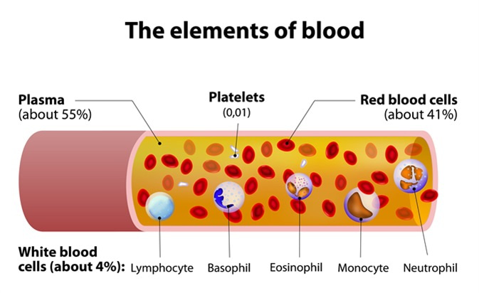
- Fluid tissue: It is also known as vascular tissue. It consists of a fluid matrix in which are suspended free floating cells. The matrix is devoid of fibres, flows freely and is not secreted by the cells it contains. It includes both
- Blood
- Lymph
- Blood: Blood is made up of various cells present in the plasma. The blood contains red blood cells (RBCs), white blood cells (WBCs) and platelets. RBCs have haemoglobin and transport oxygen. WBCs form a defence system and protect from foreign antigens. Platelets are important for blood clotting. Plasma contains proteins, water, hormones, salts, etc. to transport to different parts of the body. The main function of blood is to transport gases, food, waste materials and hormones in the body.
- Lymph: Lymph is a colourless fluid that carries white blood cells throughout the human body in lymphatic vessels. There are lymphoid organs present in the body that produce lymph and together form the lymphatic system. Some of them are lymph nodes and tonsils. It consists of two part Lymph Plasma and Lymph Corpuscles
Functions of Lymph:- It carries oxygen and minerals to the cells in the body and carries back carbon dioxide and waste materials back into the blood.
- It keeps the body cells moist.
- It maintains the volume of the blood.
- It helps in eliminating harmful bacteria and viruses from the body and hence is responsible for the immunity of human beings.
- It absorbs fats from the intestine and transports them throughout the body.
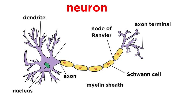
- Dendrites
- They are tree-like extensions (highly-branched) at the beginning of a neuron.
- They increase the surface area of the neuron.
- They receive chemical signals from different neurons of the body.
- They then convert these chemical signals into electrical signals and pass them to the neuron cell body.
- A neuron can have a single dendrite or multiple dendrites.
- Cell Body
- Also called soma.
- The main function of the cell body and nucleus of the neuron is to maintain the functionality of the cell.
- It does not play an active role in the transmission of the signal.
- It produces proteins that are required by different parts of the neuron to work properly.
- It contains different cell organelles such as mitochondria, Golgi apparatus etc that perform various functions of the cell.
- Axon
- Neurons have one axon in general.
- It is a long structure that connects the cell body to the terminals and it also connects with other neurons, cells and organs of the body through nerve terminals.
- It allows in fast transmission of signals. The larger the diameter of the axon the faster it will transmit signals.
- It is covered with a special insulating substance called myelin. It helps in rapid transmission of signals.
- Neurons generate and carry out nerve impulses. They produce electrical signals that are transmitted across distances, they do so by secreting chemical neurotransmitters.
- Responds to stimuli.
- Carries out communication and integration.
- Provides electrical insulations to nerve cells and removes debris.
- Carries messages from other neurons to the cell body.
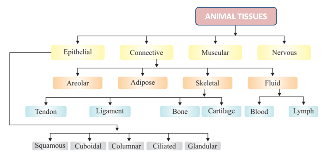
On the basis of function, the animal tissue can be broadly classified into following four types:
Epithelium tissue:
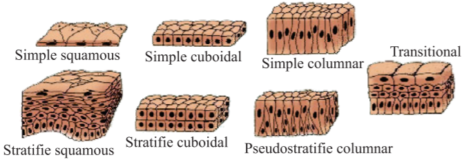
It is the simplest tissue. It is the protective tissue and these cells are tightly packed and they form a continuous sheet which covers the external surface of the body and viscera(internal organs) or lining the cavities. Eg, skin, mouth, alimentary canal, lungs, etc. These cells are tightly packed and contain little or no intercellular matrix. These are held together by intercellular junctions. Epithelial tissue may be simple (composed of a single layer of cells) or stratified (composed of several layers of cells).
Functions of epithelium cells:
Types of Epithelial tissue
Muscle tissue:
These tissues are made up of muscle cells and form the contractile tissue. These cells are elongated and large-sized so they are also called muscle fibres. It may contain one or more nuclei. The muscle cells are parallel arranged and contract in a definite direction which causes movements of the body parts. The movements are brought by contractions and relaxations of the contractile protein in muscle cells. The movements of the internal organs like the heart or alimentary canal are all caused by muscle tissue.On the basis of location, structure and function they are of following three types:
 <
<
Connective tissue:
It serves the function of binding and joining one tissue to another forming protective sheath and packing material around the various organs and separates them so that they don't interfere with each others activities, like transporting materials from one part to another, forming supporting framework of cartilage and bones for the body, etc. These cells are living, separated and low in number. They are loosely bound cells present in an intercellular Matrix. This matrix can be of different types – Dense, Rigid, Fluid or Jelly-like.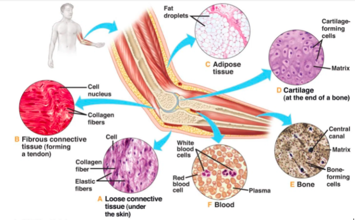
Functions:
Types of connective tissue:
Nervous tissue:
Nervous or the nerve tissue is the main tissue of our nervous system. It monitors and regulates the functions of the body. Nervous tissue consists of two cells: nerve cells or neurons and glial cells, which helps transmit nerve impulses and also provides nutrients to neurons. Brain, Spinal Cord, and nerves are composed of nervous tissue. They are specialized for being stimulated to transmit stimuli from one to another part of the body rapidly.Nervous tissue makes up for the CNS and PNS of the nervous system. Neurons secrete chemical neurotransmitters which are responsible for stimulating other neurons as a result of a stimuli. Presence of specialization at axonal terminals called synapsis Nerve cells live long, cannot be divided and replaced(except memory cells).
Each neuron is composed of three parts:
Functions:
SUMMARY
- Tissue is a group of cells similar in structure and function. Plant tissues are of two main types – Meristematic and Permanent.
- Meristematic tissue is the dividing tissue present in the growing regions of the plant.
- Permanent tissues are derived from meristematic tissue once they lose the ability to divide. They are classified as simple and complex tissues. Parenchyma, collenchyma and sclerenchyma are three types of simple tissues. Xylem and phloem are types of complex tissues.
- Animal tissues can be epithelial, connective, muscular and nervous tissue.
- Depending on shape and function, epithelial tissue is classified as squamous, cuboidal, columnar, ciliated and glandular.
- The different types of connective tissues in our body include areolar tissue, adipose tissue, bone, tendon, ligament, cartilage and blood.
- Striated, unstriated and cardiac are three types of muscle tissues.
- Nervous tissue is made of neurons that receive and conduct impulses.