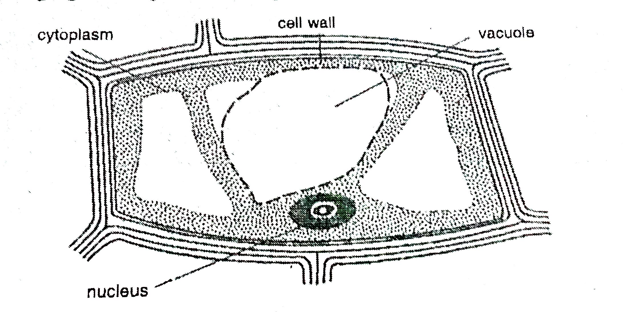
A student prepared the temporary mount of stained leaf peel. After observing the slide under microscope he drew the following sketch. Identify and name the parts labelled as A B C and D.
A Chloroplast B Stomatal pore or stoma C Guard cell D Nucleus
Related Solutions
Identify the observed various parts of temporary mount of well stained leaf peel, when focused under the high power of a microscope.
View SolutionIdentify the observed various parts of temporary mount of well stained leaf peel, when focused under the high power of a microscope.
View SolutionA student is observing the temporary mount of a leaf peel under a microscope. Draw
labelled diageram of the structure (of stomata) as seen under the microscope.
View SolutionA student is observing temporary mount of leaf peel under the microscope. Draw labelled diagram of the structure of the stomata as seen under the microscope.
View SolutionA student is observing the temporary mount of a leaf peel under a microscope. Draw labelled diagram of the structure (of stomata) as seen under the microscope.
View SolutionTo prepare stained temporary mounts of (a) onion peel, and (b) human cheek cells and to record observations and draw their labelled diagram. The cels are first focussed in microscope under
View SolutionTo prepare stained temporary mounts of (a) onion peel, and (b) human cheek cells and to record observations and draw their labelled diagram. We generally mount the material in the slide
View SolutionWhich stain is used while preparing a temporary mount of a leaf peel ?
View SolutionTo prepare stained temporary mounts of (a) onion peel, and (b) human cheek cells and to record observations and draw their labelled diagram. Temporary mount of a tissur is made in
View SolutionWhy was the temporary mount of the leaf epidermal peel pinkish-red under the microscope?
View SolutionWhy was the temporary mount of the leaf epidermal peel pinkish-red under the microscope?
View SolutionAmit is observing temporary mount of an onion peel stained with safranin under a microscope. The colour of the cell wall appears...
View SolutionAmit is observing temporary mount of an onion peel stained with safranin under a microscope. The colour of the cell wall appears...
View SolutionTo prepare stained temporary mounts of (a) onion peel, and (b) human cheek cells and to record observations and draw their labelled diagram.


Safranin is a reagent that is used to stain
View SolutionTo prepare stained temporary mounts of (a) onion peel, and (b) human cheek cells and to record observations and draw their labelled diagram. Human cheek cells are commonly stained with
View SolutionList the steps of preparation of temporary mount of a leaf peel to observe stomata.
View SolutionList the steps of preparation of temporary mount of a leaf peel to observe stomata.
View SolutionRecord your observations when a stained a mounted leaf peel is viewed by you under high power (45 xx ) microscope.
View SolutionA well stained leaf peel mount when observed under the high power of a microscope shows nuclei in:
View SolutionWhile preparing a temporary stained mount of a leaf epidermal peel, how is the extra stain removed?
View Solution