With the help of a neat and labelled diagram describe the anatomy of 'human eye'. Explain the mechanism of vision.
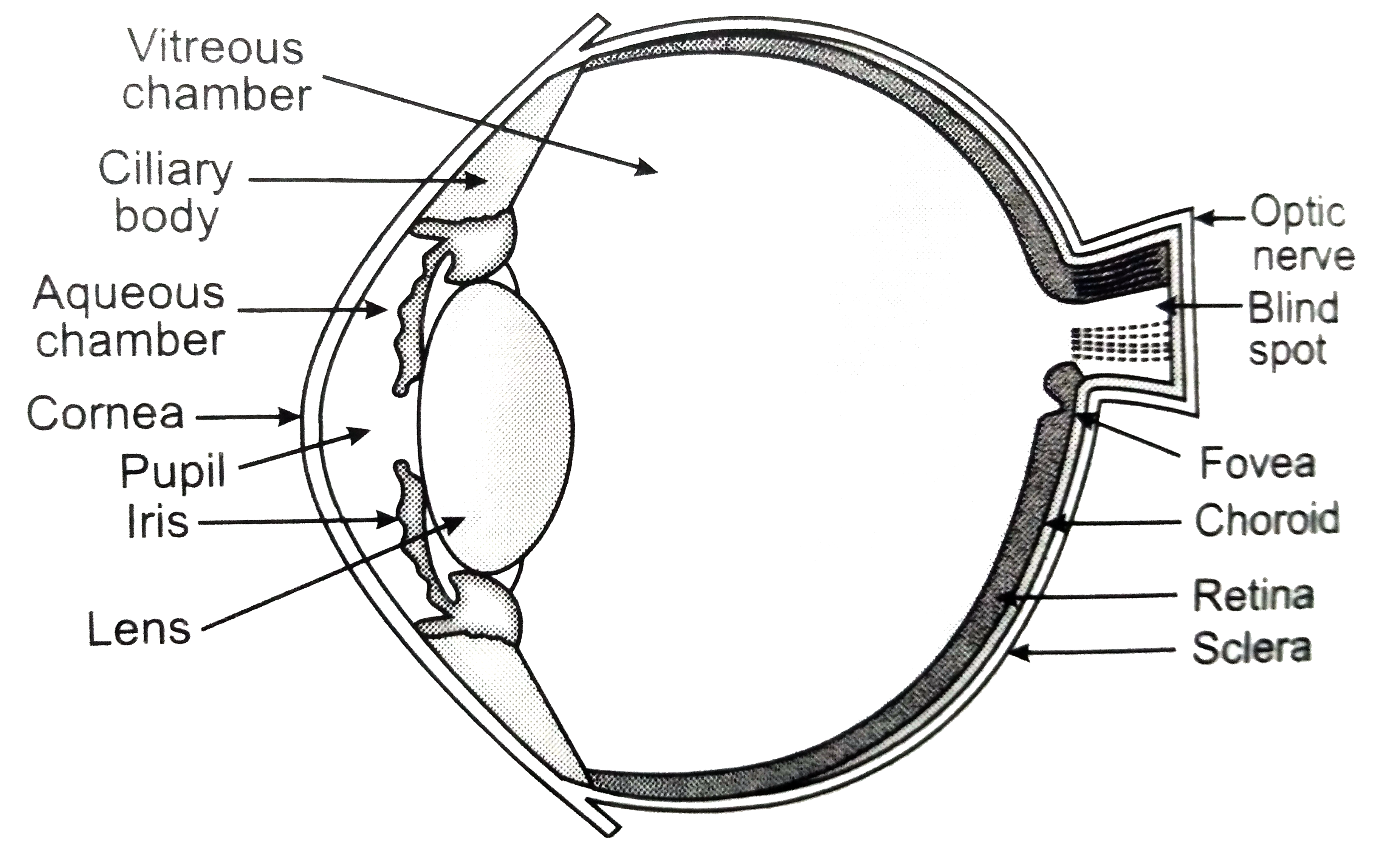
Anatomy of human eye :
(i) Human eye ball is spherical in structure.
(ii) It consist of three layers :
(a) Outer layer of dense connective tissue forms the sclera.
(b) Middle layer choroid.
(c) Innermost layer is retina.
(iii) Cornea : The transparent anterior portion of sclera is cornea.
(iv) Conjunctiva : Thin transparent membrane in front of cornea.
(v) Anterior part of choroid is thick and forms ciliary body.
(vi) Iris : Forward segment of the ciliary body pigmented and opaque known as iris.
(vii) Lens : Transparent crystalline lens held in position by ligaments of ciliary body.
(viii) Pupil : The aperture surrounded by iris in front of lens is known as pupil.
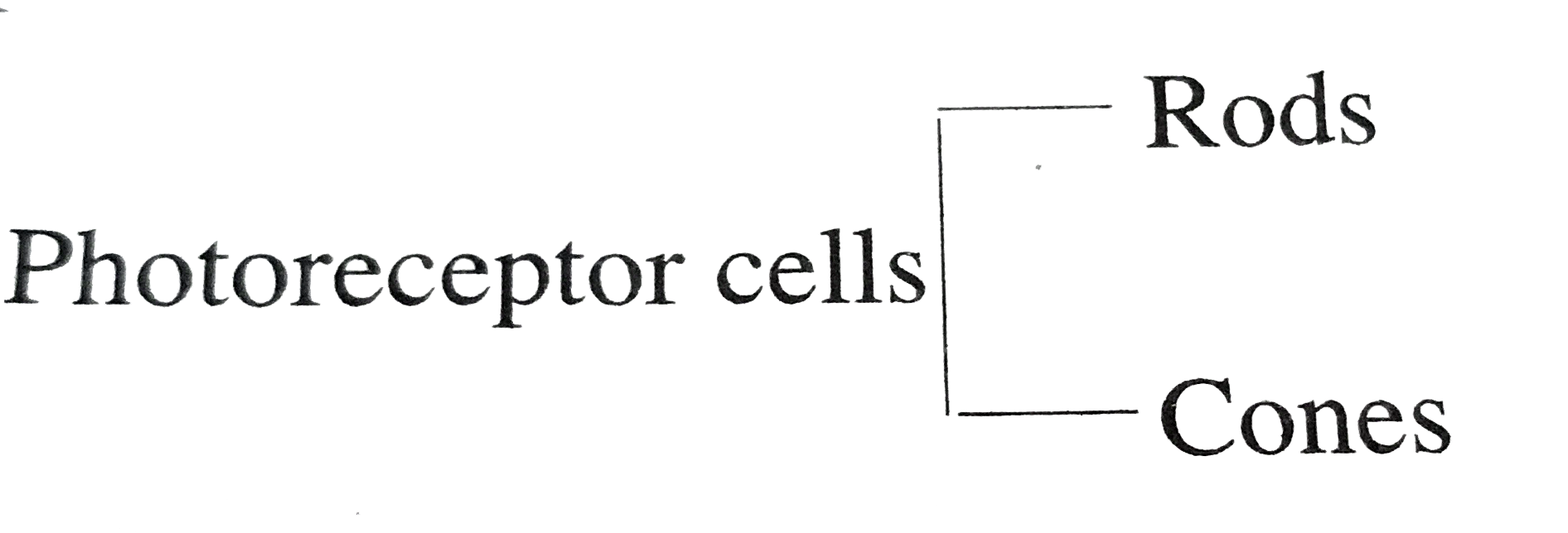
(ix) Retina : Consists of three sub-layers-ganglion cells, bipolar cells and photoreceptor cells.

(x) Optic nerve leaves the eye at a point slightly away from median posterior pole of the eyeball. Rods and Cones are absent in this region hence known as blind spot.
(xi) A yellowish pigmented spot called Mocula lutea is present lateral to blind spot with a central pit termed as fovea.
(xii) The aqueous chamber is a pace between cornea and lens. It contains thin watery fluid known as aqueous humour.
(xiii) The large space between the lens and retina is vitreous chamber.
(xiv) It is filled with transparent gelatinous fluid known as vitreous humour. The aqueous humour and vitreous humour maintain the shape of the eyeball. Mechanism of Vision : The light rays (visible wavelength) from the objects pass through the cornea and lens, focused on the retina of the eye. Rods and cone cells contains photopigments which are conjugated proteins. They are composed of opsin (a protein) and retinol (Vitamin A derivative). When light induces dissociation of retinol from opsin, the structure of opsin changes, and the permeability of retinal cells also changes. An action potential is generated which is carried via bipolar neurons and ganglion, conducted by optic nerves to visual cortex of brain. Through this the neural impulses are analyzed and image formed on the retina is recognised. Neural impulses are analyzed and image is formed on retina.

Topper's Solved these Questions
- MARCH 2018BOOK - GURUKUL PUBLICATION - MAHARASHTRA PREVIOUS YEAR PAPERSCHAPTER - MARCH 2018EXERCISE - SECTION-II
24 Videos
- MARCH 2017BOOK - GURUKUL PUBLICATION - MAHARASHTRA PREVIOUS YEAR PAPERSCHAPTER - MARCH 2017EXERCISE - SECTION-II
24 Videos
- MARCH 2019BOOK - GURUKUL PUBLICATION - MAHARASHTRA PREVIOUS YEAR PAPERSCHAPTER - MARCH 2019EXERCISE - SECTION C
23 Videos
Similar Questions
With the help of a neat and labelled diagram, describe anatomy of human eye. Explain the mechanism of vision .
View SolutionWith the help of a neat and labelled diagram, describe reflex arc.
View SolutionWith the help of a neat and labelled diagram, describe reflex arc.
View SolutionWith the help of a neat and labelled diagram, describe the human male reproductive system.
View SolutionWith the help of a neat labelled diagram, describe the Geiger-Marsden experiment.
View SolutionWith the help of a neat and labelled diagram describe steps in recombinant DNA technology.
View SolutionWith the help of a neat and labeled diagram describe Watson and Crick's model of DNA.
View SolutionDraw a labeled diagram of male reproductive system.
View SolutionWith the help of a neat and labelled diagram, describe Wastson and Crick's model of DNA.
View SolutionWith the help of a neat and labelled diagram describe the development of female gametophyte of angiosperms.
View Solution
