Joints
Joints
Joints are very crucial parts of human body they help in beding and movement of different parts of body.. Here, in this chapter, we will study joints that would make you get the right knowledge. We will further study the types of joints that would help you in learning it more accurately.
What are Joints?
A joint is a structure where two or more adjacent elements of the skeletal system are separated. It all depends on the joint types as to whether the separated elements may or may not move one another.

What are the types of Joints?
Joints are classified into two types – temporally and structurally. It depends on the joint function by which each classification is associated.

In refard to the temporally, joints prove to be either transient or permanent. The bones of a transient joint are seen to be fused together sooner or later. But it should be noted that it always after birth. Except the middle ear and those between the lower jaw and the braincase, all the joints of the skull prove to be transient. Such fusion is called arthrodesis. Permit movement is joint by all permanent and some transient joints.
There happens to be two basic structural types of joint which are diarthrosis and synarthrosis. In diarthrosis fluid is present whereas in synarthrosis there is no fluid.
Synarthrosis
There are three classes of Synarthrosis which are fibrous, symphysis, and cartilaginous.
- Fibrous joints: When it comes to the fibrous joints the white connective tissue (collagen) fibres separates the articulating parts. It passes from one part to the other. It should be noted that there are two types of fibrous joints: suture and gomphosis. Here a suture is formed by the fibrous covering, or periosteum. It is only yin the roof and sides of the braincase that sutures are found in the adult. It is also found in the upper part of the face as well. Sutures prove to be transient. The direct conversion of the sutures into bone affects the fusion. On the other hand, a gomphosis happens to be a fibrous mobile peg-and-socket joint. These are permanent joints which long lasts like the roots of the teeth until they are damaged by disease.
- Symphysis: The joint in which the body (physis) of one bone meets the body of another is a symphysis (fibrocartilaginous joint). All but two of the symphyses lie in the vertebral (spinal) column. On the other hand and all but one contain fibrocartilage as a constituent tissue.
- Cartilaginous: It is also known as synchondroses. These are parts of bones where prior to ossification, it passes through a cartilaginous stage. The ossifying parts of a bone are separated by juxta-epiphyseal plates. At these plates, the growth of the whole bone takes place. Here, it should be noted that all synchondroses are transient.
Related Videos:
- Synovial Joints
- Mechanism of Muscle Contraction
- Actin is present in thin filament
- Human skeleton has 206 bones
- There are 11 pairs of ribs in man
Diarthroses
Structure and elements of synovial joints
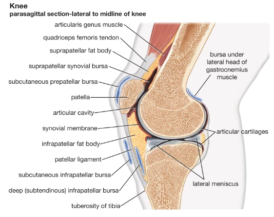
The synovial bursas are found to be lined with synovial membrane which appears to be closed, thin-walled sacs. In between the structures, bursas are found that glide upon each other. Synovia is the bursal fluid which is exuded by the synovial membrane.
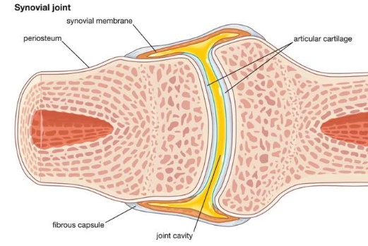
There is a wall enclosing a joint cavity that is wholly filled with synovial fluid in a synovial joint. The wall is composed of two layers: an outer complete fibrous layer and an inner incomplete synovial layer. The parts of the outer layer are either chondrified as articular cartilages or partly ossified as sesamoid. There is a presence of collagen in the fibrous layer.

Articular cartilage
Articular cartilage is the one that covers the articulating part of a bone. It can beeasily cut by a sharp knife unlike bone. It also proves to be deformable and elastic. When it comes to the part of the cartilage nearest to the bone, it is found to be impregnated with calcium salts.
Coming to the classification of articular surfaces, it is divided into two primary classes: ovoid and sellar. An ovoid surface is either concave or convex in all directions. A sellar surface proves to be convex in one direction and concave in the direction at right angles to the first. There is always a greater the possible amount of motion at the joint when there is a difference in size between conarticular surfaces.
Important Chapters:


Similar Questions
Knowledge Check
- Question 1 - Select One
Joint between femur and pelvic girdle is
" " Or
Joint between femur and acetabulum is known asAPivotalBBall and socketCHingeDSaddle - Question 2 - Select One
Match the joints in column I with their examples in column II and select the correct option.
Column IColumn IIIA.Hinge joint(ii)NeckB.Gliding joint(ii)AnkleC.Pivot joint(iii)ShoulderD.Ball and socket joint(iv)ElbowAA - (iv), (B) - (ii), C - (i), D - (iii)BA - (iii), B - (ii), C - (i), D - (iv)CA - (iv), B - (i), C - (ii), D - (iii)DA - (ii), B - (iii), C - (iv), D - (i) - Question 3 - Select One
Jointed appendages are characteristic of phyllum
AAnnelidaBEchinodomataCMolluscaDArthropoda

