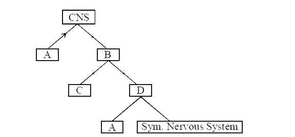
Draw a diagram of transverse section of the spinal cord showing reflect are and label the following parts : (i) Afferent nerve fibres, (ii) Motor nerve fibre, and (iii) Grey matter.
Step-by-Step Solution:
1. Draw the Outline of the Spinal Cord:
- Start by sketching a vertical rectangle to represent the transverse section of the spinal cord. This will serve as the outer boundary of your diagram.
2. Draw the Grey Matter:
- Inside the rectangle, draw a butterfly or 'H' shape to represent the grey matter. The central part of the 'H' will be the central canal, and the two wings will represent the dorsal (posterior) and ventral (anterior) horns of the grey matter.
3. Label the Grey Matter:
- Write "Grey Matter" next to the butterfly shape to indicate its location in the spinal cord.
4. Draw the Afferent Nerve Fibres:
- On the dorsal side (the upper part of the 'H'), draw lines coming from the outside towards the grey matter. These lines represent the afferent nerve fibres (sensory neurons) that carry signals from sensory receptors to the spinal cord.
5. Label the Afferent Nerve Fibres:
- Write "Afferent Nerve Fibres" next to the lines you drew to indicate their function.
6. Draw the Motor Nerve Fibres:
- On the ventral side (the lower part of the 'H'), draw lines extending from the grey matter to the outside, representing the motor nerve fibres that carry signals from the spinal cord to muscles or glands.
7. Label the Motor Nerve Fibres:
- Write "Motor Nerve Fibres" next to the lines you drew to indicate their function.
8. Final Touches:
- Make sure all parts are clearly labeled and the diagram is neat. You can also add arrows to indicate the direction of signal transmission for clarity.
- Start by sketching a vertical rectangle to represent the transverse section of the spinal cord. This will serve as the outer boundary of your diagram.
- Inside the rectangle, draw a butterfly or 'H' shape to represent the grey matter. The central part of the 'H' will be the central canal, and the two wings will represent the dorsal (posterior) and ventral (anterior) horns of the grey matter.
- Write "Grey Matter" next to the butterfly shape to indicate its location in the spinal cord.
- On the dorsal side (the upper part of the 'H'), draw lines coming from the outside towards the grey matter. These lines represent the afferent nerve fibres (sensory neurons) that carry signals from sensory receptors to the spinal cord.
- Write "Afferent Nerve Fibres" next to the lines you drew to indicate their function.
- On the ventral side (the lower part of the 'H'), draw lines extending from the grey matter to the outside, representing the motor nerve fibres that carry signals from the spinal cord to muscles or glands.
- Write "Motor Nerve Fibres" next to the lines you drew to indicate their function.
- Make sure all parts are clearly labeled and the diagram is neat. You can also add arrows to indicate the direction of signal transmission for clarity.
Topper's Solved these Questions
- BOOK - PRADEEPCHAPTER - NEURAL CONTROL AND CORRDINATIONEXERCISE - Additional Question (Long Answer Question)29videos
NEURAL CONTROL AND CORRDINATION
- BOOK - PRADEEPCHAPTER - NEURAL CONTROL AND CORRDINATIONEXERCISE - Analytical Question With Answer20videos
NEURAL CONTROL AND CORRDINATION
- BOOK - PRADEEPCHAPTER - NEURAL CONTROL AND CORRDINATIONEXERCISE - Additional Question (Very Short Answer Question)48videos
NEURAL CONTROL AND CORRDINATION
- BOOK - PRADEEPCHAPTER - MORPHOLOGY OF FLOWERING PLANTSEXERCISE - Assertion-Reason Type Questions16videos
MORPHOLOGY OF FLOWERING PLANTS
- BOOK - PRADEEPCHAPTER - PLANT GROWTH AND DEVELOPMENTEXERCISE - Assertion -Reason type question13videos
PLANT GROWTH AND DEVELOPMENT
Similar Questions
Draw a diagram of human excretory system and label the following :
(i) Urinary bladder
(ii) Left kidney
(iii) Left ureterView Solution(a) Draw a diagram of the structure of a human ovum surrounded by corona radiata. Label the following parts :
(i) Ovum, (ii) Plasma Membrane, (iii) Zona Pellucida
(b) State the function of Zona Pellucida.View SolutionDraw a diagram showing germination of pollen on stigma of a flower and mark on it the following organs/ parts :
(i) Pollen grain
(ii) Pollen tube
(iii) Stigma
(iv) Female germ cellView SolutionDraw a sectional view of human female reproductive system and label the following parts:
(i) Where the development of egg occurs.
(ii) Where fertilisation takes place.
(iii) Where fertilised eggs get implanted.
(b) Describe the changes the uterus undergoes :
(i) To receive the zygote.
(ii) If zygote is not formed.View SolutionWhich of the following answer shows the correct arrangement of nerve fibre
 View Solution
Draw diagram of embryo sac and label the following:
(i) Egg cell (ii) Polar Nuclei (iii) SynergidsView SolutionDraw diagram of a germinating seed and label the following parts of it:
(i) Part that gives rise to shoot system
(ii) Part that gives rise to root system.
(iii) Part that contains stored food.View Solution(a) Draw a diagram of human alimentary canal and label the following parts:
(i) largest gland.
(ii) gland that secretes digestive enzymes and hormone.
(iii) Part where HCl is produced.
(iv) part where digested food is absorbed.
(b) What are villi? Explain their function in the digestive system.View Solution(a) Draw a diagram showing germination of pollen on stigma of a flower and mark on it the following organs/parts:
(i) Pollen Grain
(ii) Pollen tube
(iii) Stigma
(iv) Female germ cell
(b) State the significance of pollen tube.
(c ) Name the parts of flower that develop after fertilisation into
(i) Seed (ii) FruitView SolutionDraw a diagram of internal structure of human heart and label the following:
(i) Mitral valve (ti) Eustachius valve (iii) Chordae tendinae (iv) Pulmonary arch.View Solution

