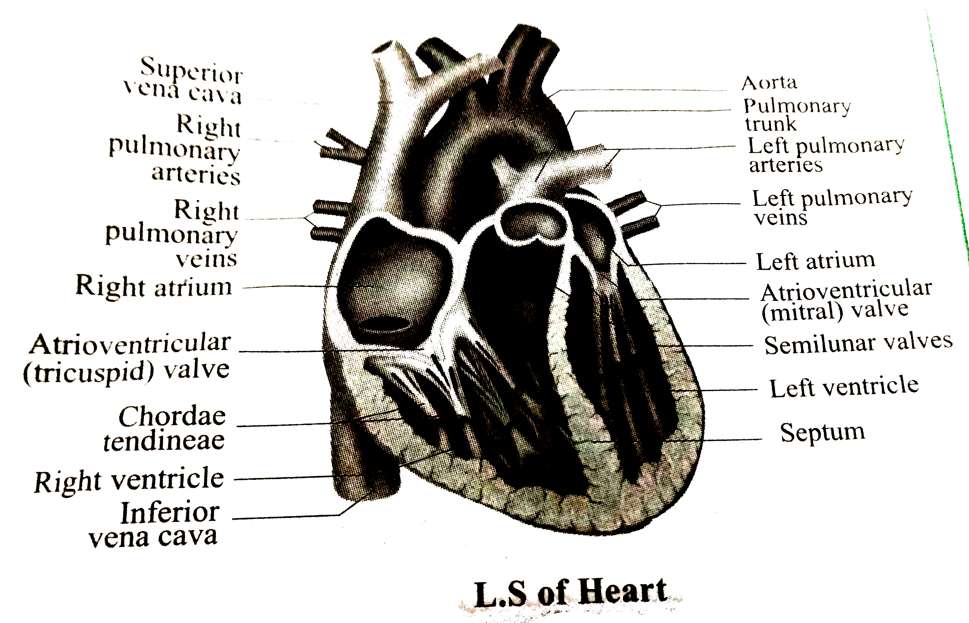
Explain the structure of human heart.
Text Solution
Structure of human heart (L.S)
1. The structure of the heart was described by Raymond de viessens, in 1706.
2. Human heart is made of special type of muscle called the cardiac muscle.
3. It is situated in the thoracic cavity and its apex portion is slightly tilted towards left. It weighs about 300g in an adult.
4. Heart is divided into four chambers, upper two small auricles or atrium and lower two large ventricles. The walls of the ventricles are thicker than the auricles due to the presence of papillary muscles.

5. The heart wall is made up of three layers, the outer epicardium, middle myocardium and inner endocardium. The space present between the membranes is called pericardial space and is filled with pericardial fluid.
6. The two auricles are separated by inter auricular septum and the two ventricles are separated by inter ventricular septum The separation of chambers avoids mixing of oxygenated and deoxygenated blood.
7. The auricle communicates with the ventricle through an opening called auriculo ventricular aperture which is guarded by the auriculo ventricular valves.
6. The opening between the right atrium and the right ventricle is guarded by the tricuspid valve (three flaps or cusps), whereas a bicuspid (two flaps or cusps) or mitral valve guards the opening between the left atriuin and left ventricle.
6. The valves of the heart allows the blood to flow only in one direction, i.e., from the atria to the ventricles and from the ventricles to the pulmonary artery or the aorta. These valves prevent backward flow of blood.
8. The valves of the heart allows the blood to flow only in one direction, i.e., from the atria to the ventricles and from the ventricles to the pulmonary artery or the aorta. These valves prevent backward flow of blood.
9. The myocardium of the ventricle is thrown into irregular muscular ridges called trabeculae cornese which are modified into chordae tendinae. The opening and closing of the semilunar valves are achieved by the chordae tendinae. The chordae tendinae are attached to the lower end of the heart by papillary muscles.
10. Heart receives deoxygenated blood from various parts of the body through the inferior venacava and superior venacava which open into the right auricle. Oxygenated blood from lungs is drained into the left auricle through four pulmonary veins.
|
Similar Questions
With the hepl of a diagram, explain the structure of human eye.
View SolutionWith the help of labelled diagram explain the structure of human respiratory system.
View SolutionDescribe the structure of human heart.
View SolutionDescribe the structure of human heart.
View SolutionDescribe internal structure of a human heart.
View SolutionDescribe internal structure of a human heart.
View SolutionDraw the internal structure of human heart.
View SolutionDraw a diagram to show the internal structure of human heart. Label only one heart chamber, any two heart valves and any three other structures.
View SolutionWith the help of well labelled diagram describe the internal structure of human heart .
View SolutionWith the help of a labelled diagram describe the internal structure of human heart.
View Solution
