You have prepared a stained temporary mount of human cheek cells. Examine it under low and high power of microscope and then write your observations and conclusions.
Step-by-Step Text Solution
1. Preparation of Temporary Mount:
- A stained temporary mount of human cheek cells was prepared for microscopic examination.
2. Observation Under Low Power (40x):
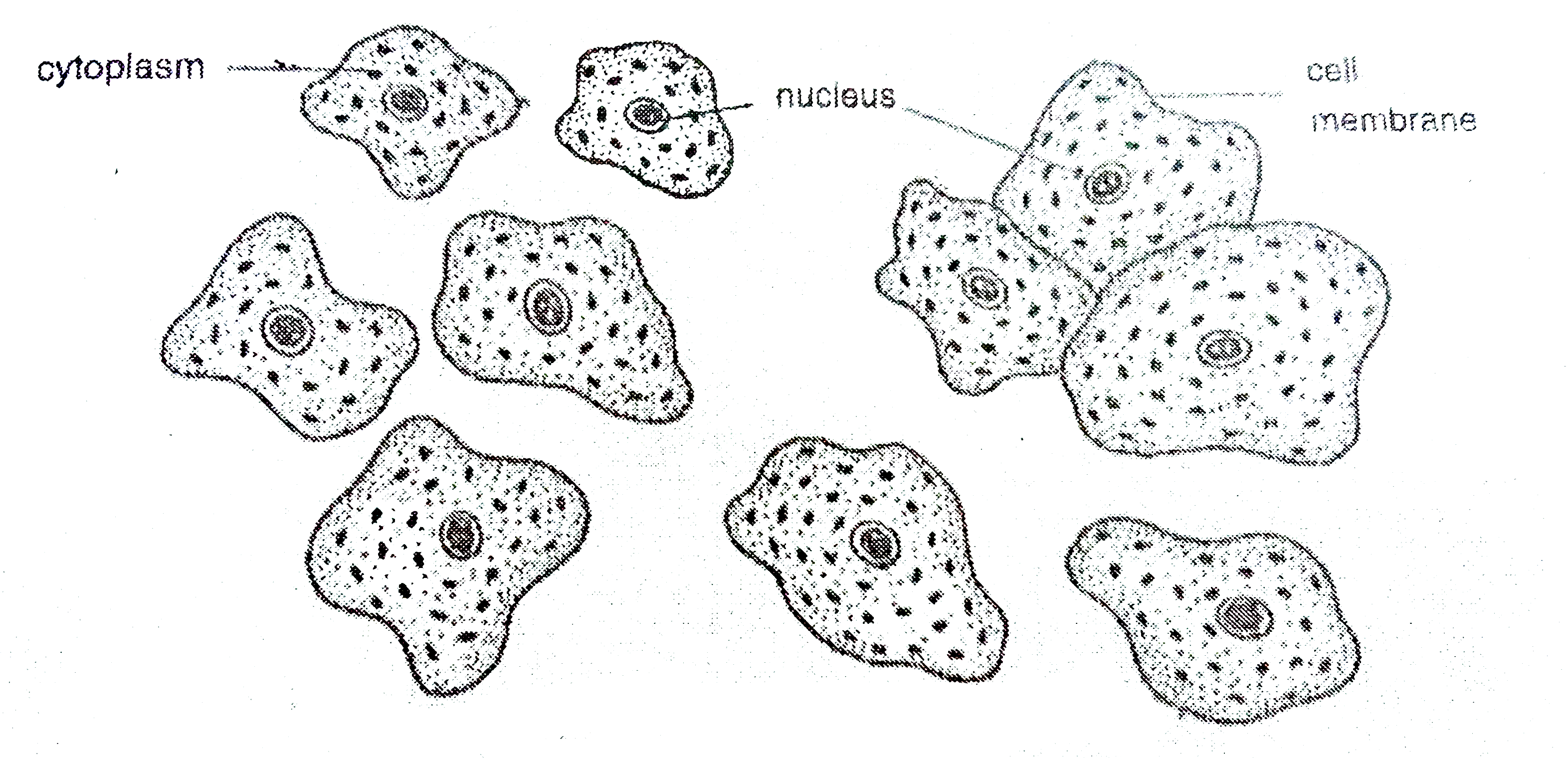
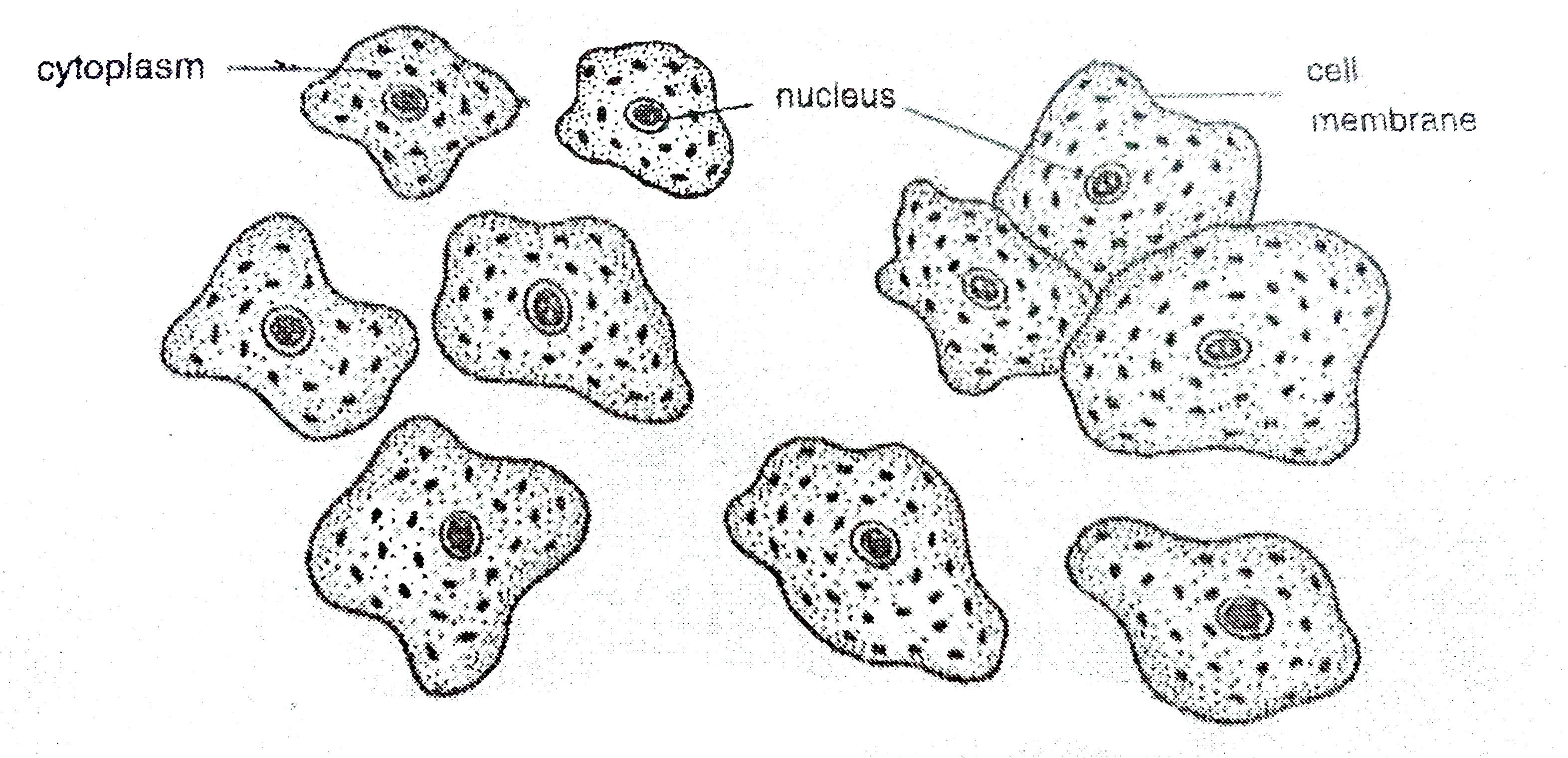
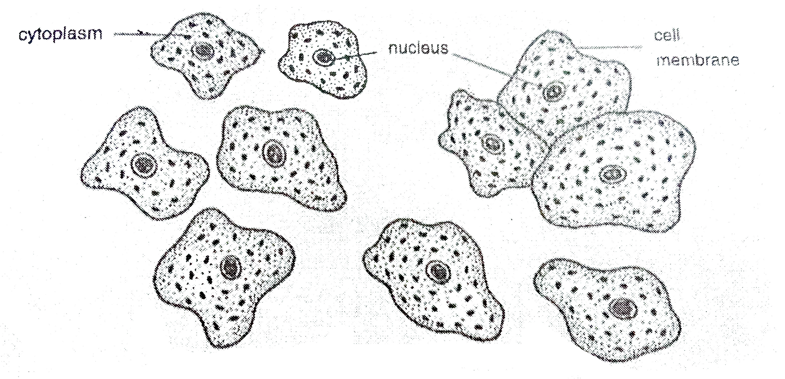
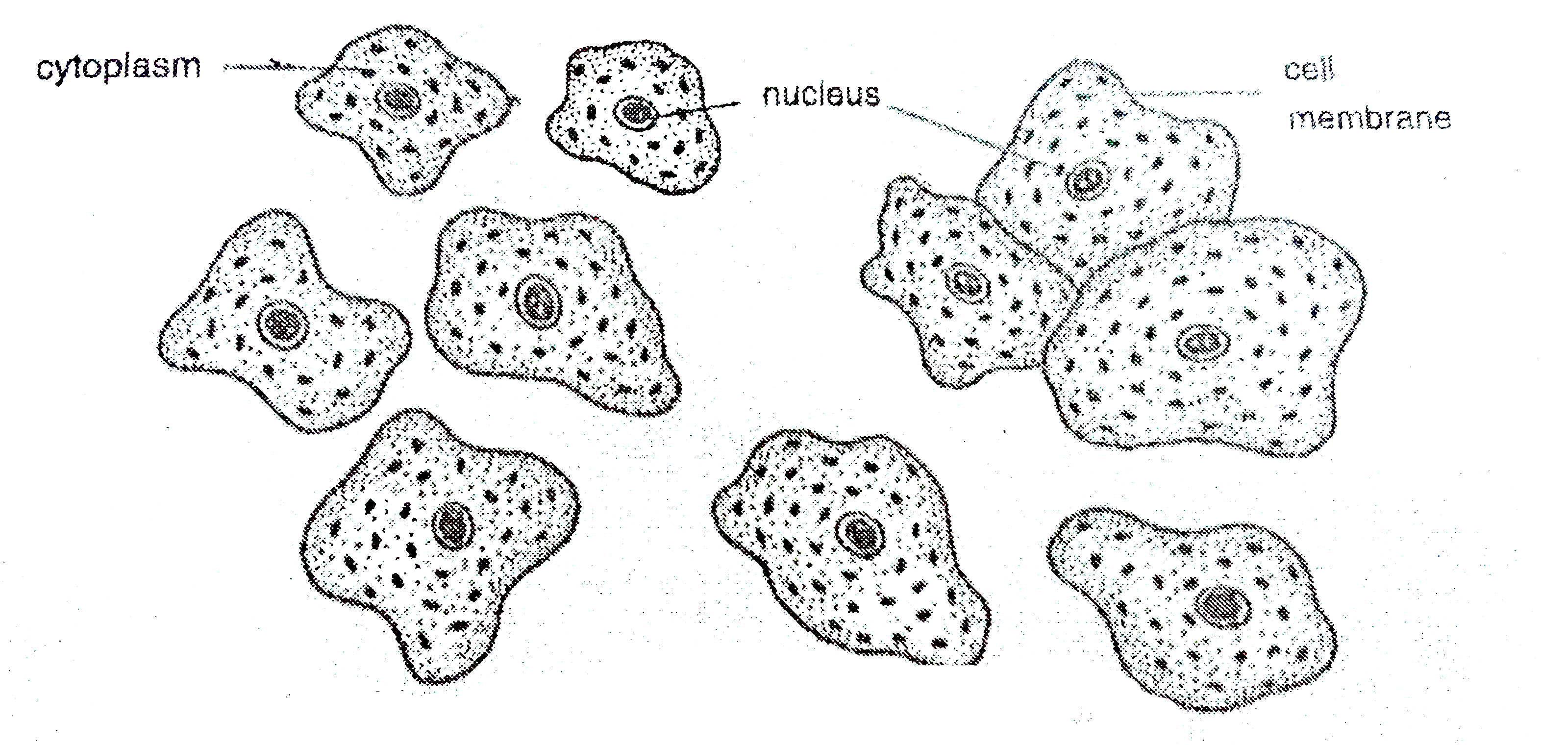
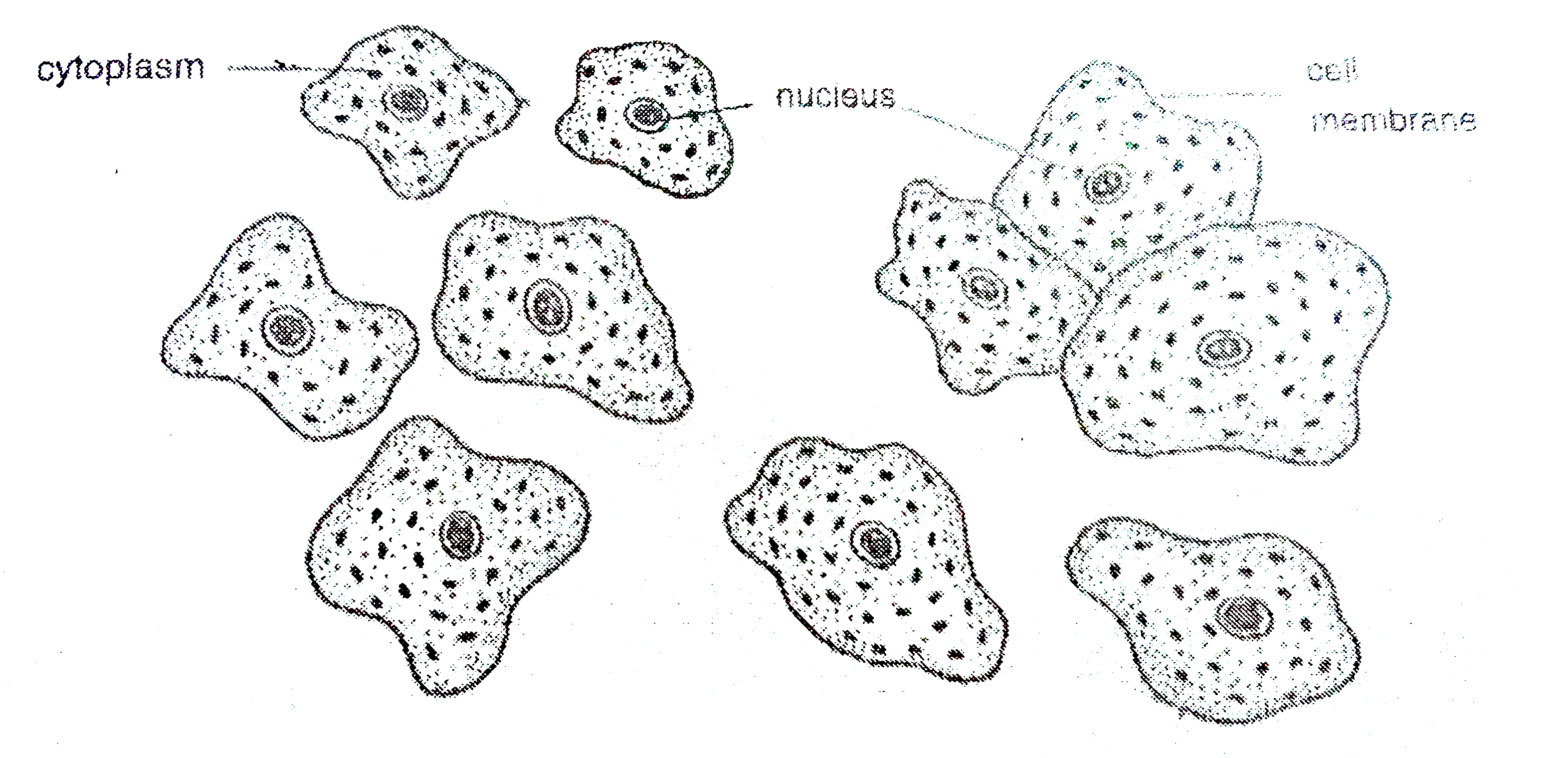
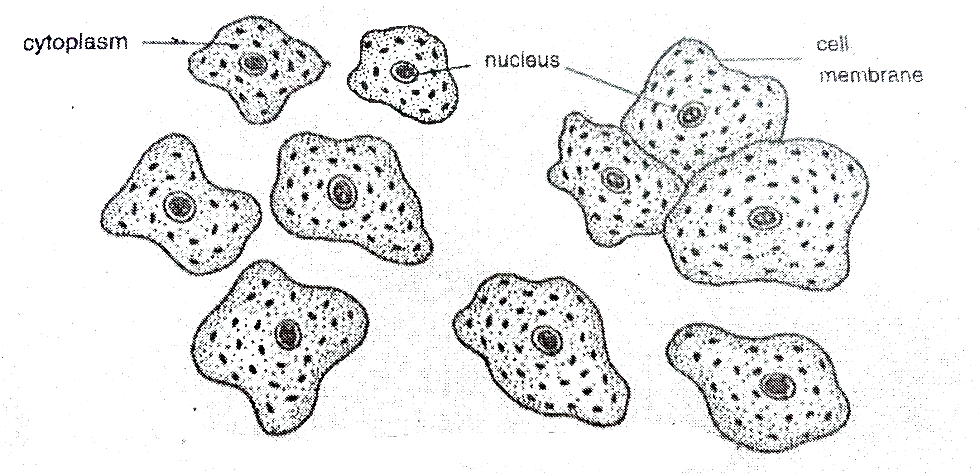
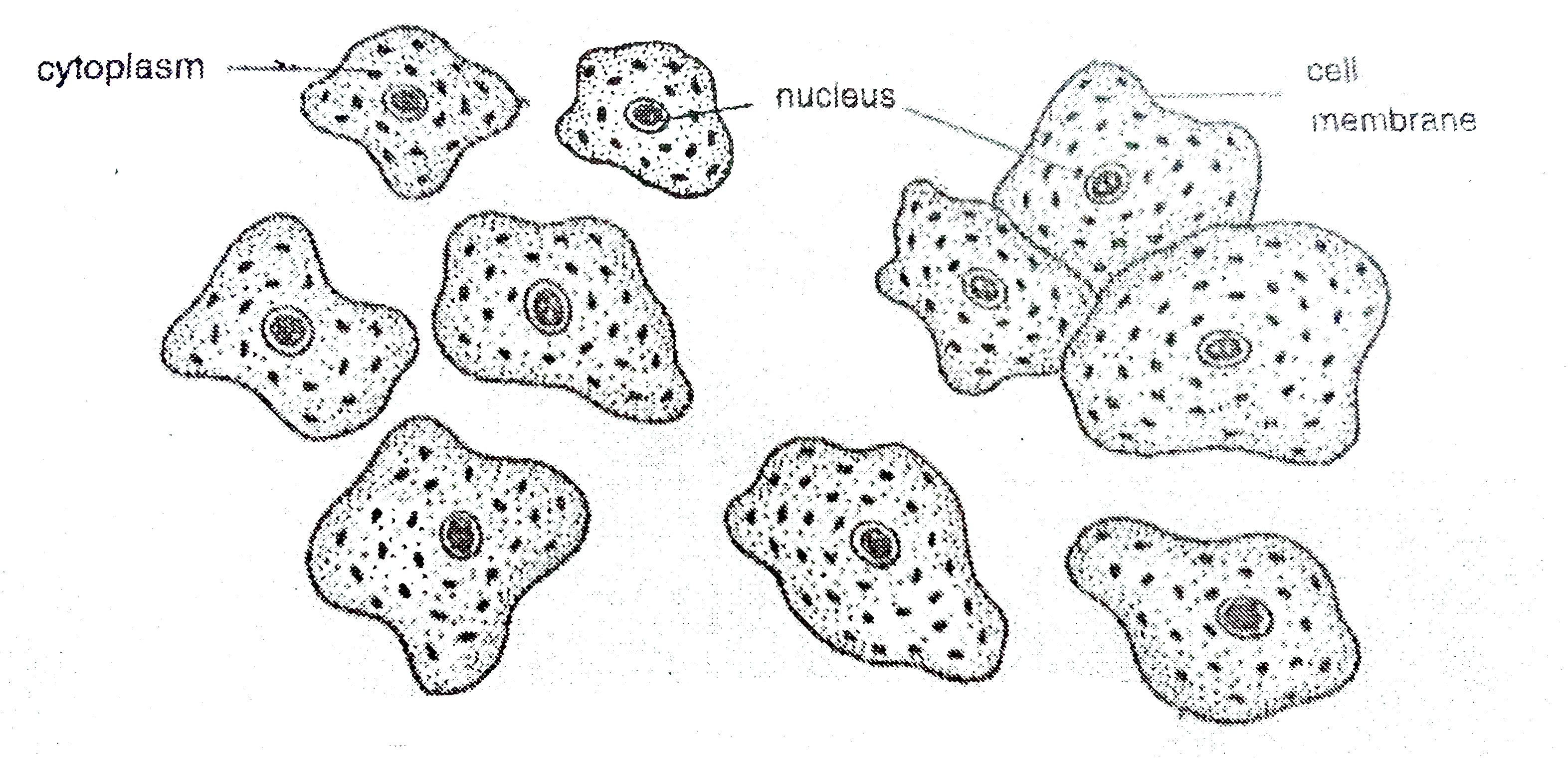
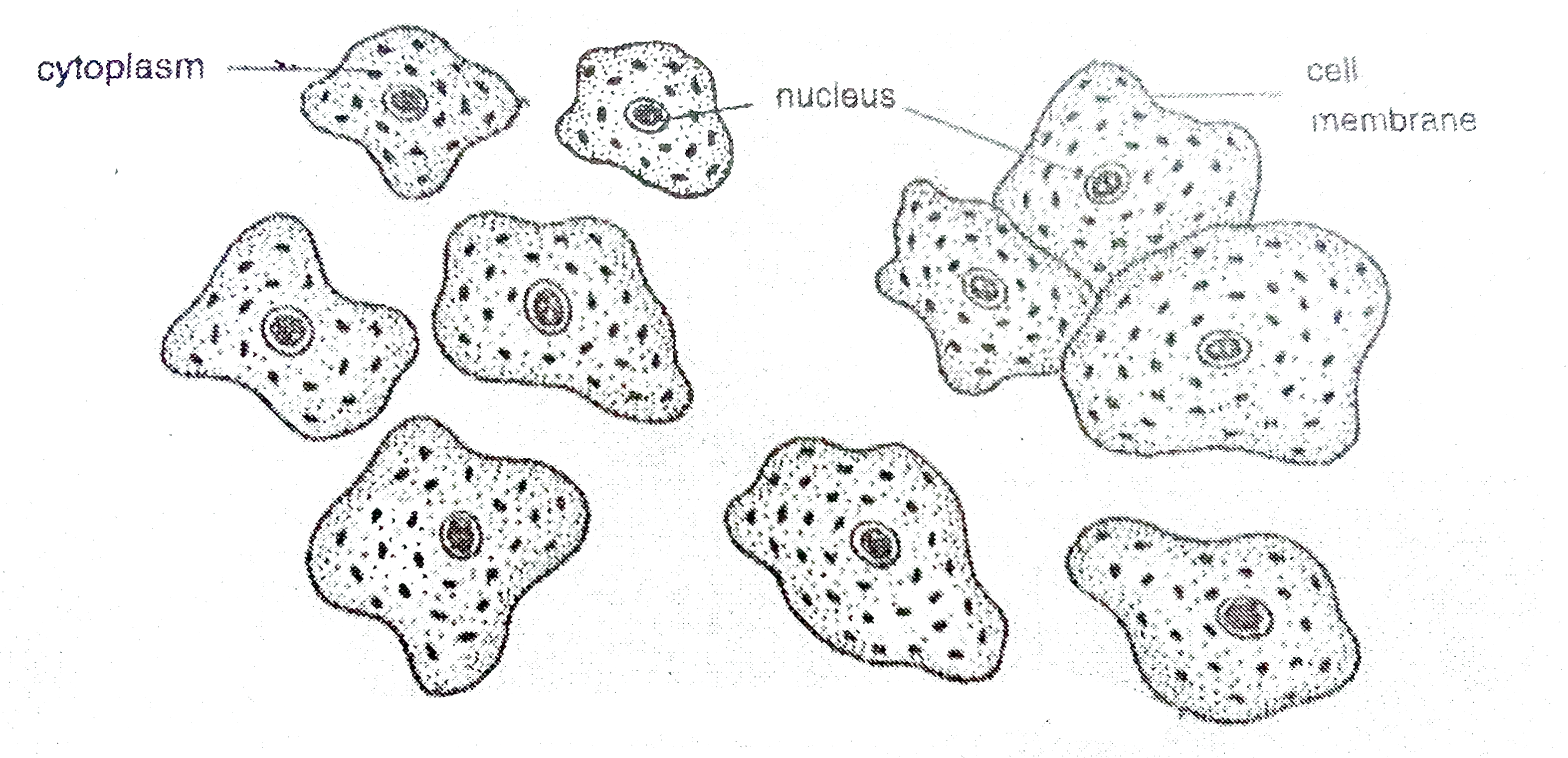
- When viewed under low power (40x magnification), a large number of irregularly shaped cells were observed. These cells appeared flat and were grouped together.
3. Observation Under High Power (1000x):
- Upon switching to high power (1000x magnification), the details of individual cells became clearer. Each cell displayed:
- A distinct cell membrane.
- A prominent nucleus, which appeared as a dot-like structure in the center of the cell.
- Cytoplasm surrounding the nucleus.
- Additionally, oral bacteria were observed, which are smaller than the cheek cells. This indicates the presence of microorganisms that can inhabit the mouth.
4. Detailed Observations:
- The cheek cells were irregularly shaped and flat, lacking a defined shape due to the absence of a cell wall (typical of animal cells).
- The nucleus was clearly visible in the center of each cell.
- The cell membrane was present, providing structure and support.
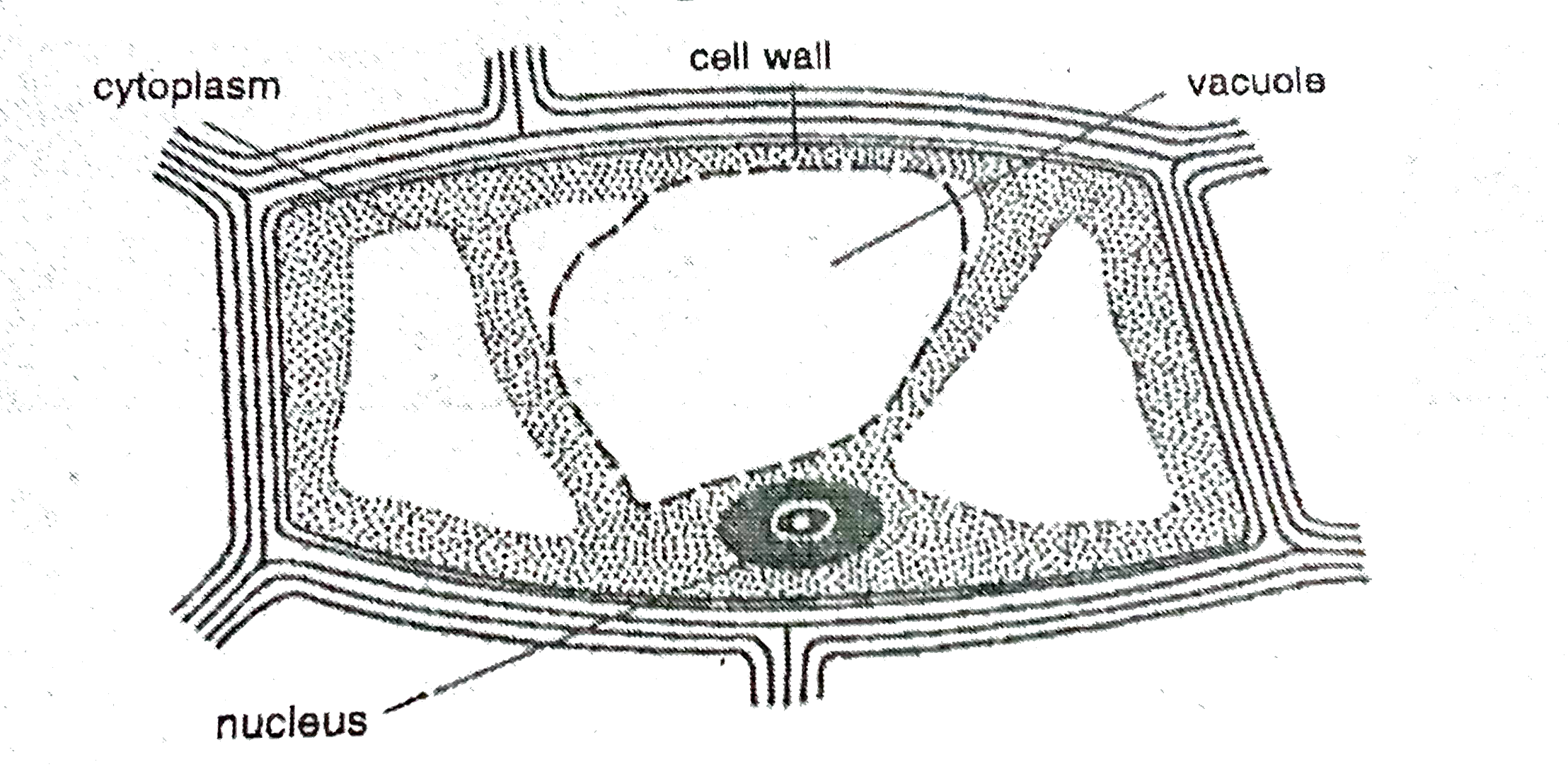
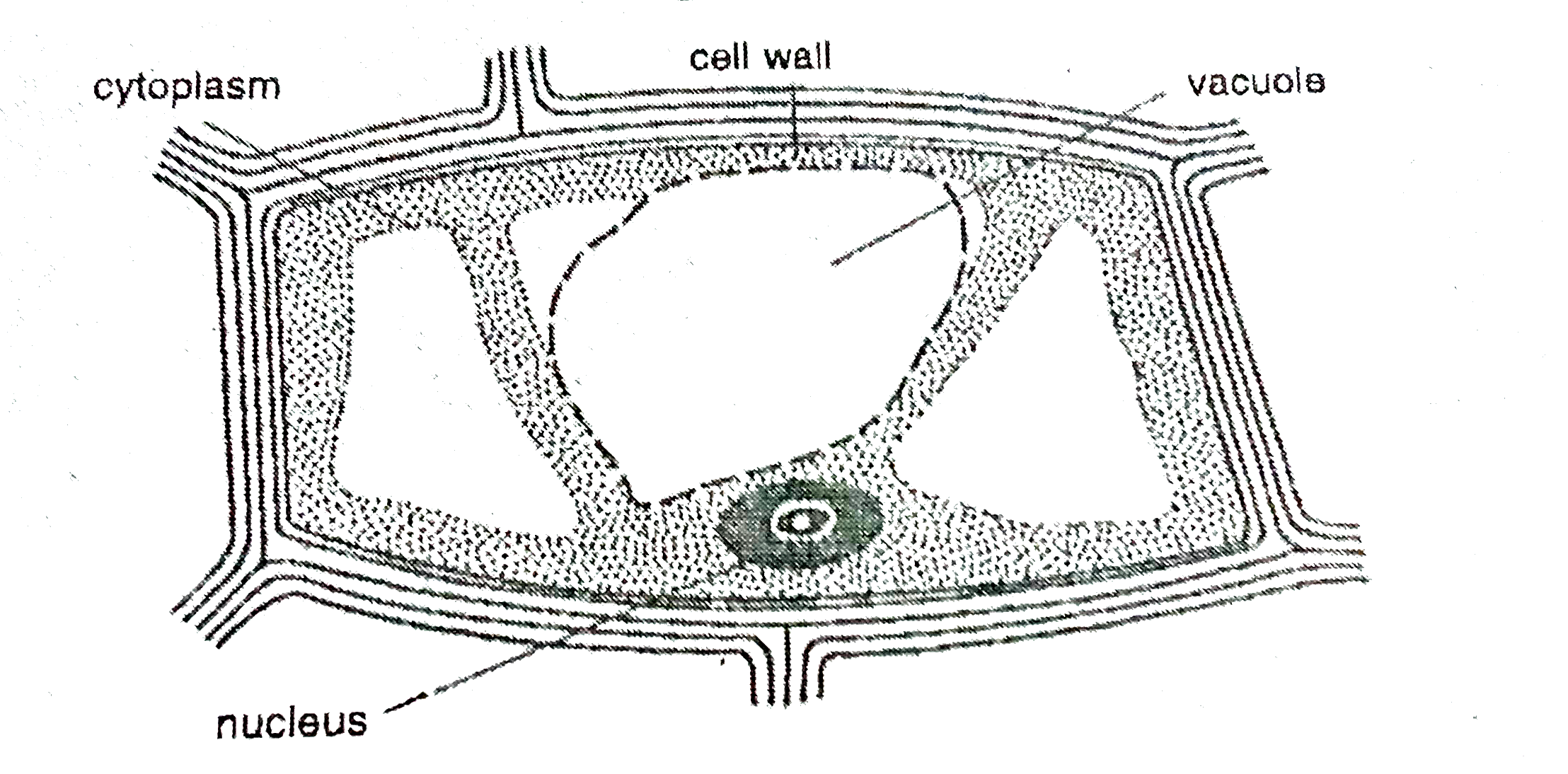
- No vacuoles were observed in the cheek cells.
5. Conclusions:
- Based on the observations, it was concluded that the cells observed are animal cells. This conclusion is supported by the presence of a prominent nucleus, the absence of a cell wall, and the lack of vacuoles.
- A stained temporary mount of human cheek cells was prepared for microscopic examination.
- When viewed under low power (40x magnification), a large number of irregularly shaped cells were observed. These cells appeared flat and were grouped together.
- Upon switching to high power (1000x magnification), the details of individual cells became clearer. Each cell displayed:
- A distinct cell membrane.
- A prominent nucleus, which appeared as a dot-like structure in the center of the cell.
- Cytoplasm surrounding the nucleus.
- Additionally, oral bacteria were observed, which are smaller than the cheek cells. This indicates the presence of microorganisms that can inhabit the mouth.
- The cheek cells were irregularly shaped and flat, lacking a defined shape due to the absence of a cell wall (typical of animal cells).
- The nucleus was clearly visible in the center of each cell.
- The cell membrane was present, providing structure and support.
- No vacuoles were observed in the cheek cells.
- Based on the observations, it was concluded that the cells observed are animal cells. This conclusion is supported by the presence of a prominent nucleus, the absence of a cell wall, and the lack of vacuoles.
Topper's Solved these Questions
- THE FUNDAMENTAL UNIT OF LIFE BOOK - PRADEEPCHAPTER - THE FUNDAMENTAL UNIT OF LIFE EXERCISE - HIGHER ORDER THINKING SKILLS
6 Videos
- THE FUNDAMENTAL UNIT OF LIFE BOOK - PRADEEPCHAPTER - THE FUNDAMENTAL UNIT OF LIFE EXERCISE - ANALYTICAL
11 Videos
- THE FUNDAMENTAL UNIT OF LIFE BOOK - PRADEEPCHAPTER - THE FUNDAMENTAL UNIT OF LIFE EXERCISE - D. LONG ANSWER QUESTIONS
14 Videos
- NATURAL RESOURCES : OUR ENVIRONMENTBOOK - PRADEEPCHAPTER - NATURAL RESOURCES : OUR ENVIRONMENTEXERCISE - QUESTIONS WITH ANSWERS
20 Videos
- TISSUESBOOK - PRADEEPCHAPTER - TISSUESEXERCISE - Analytical Questions with Answers
10 Videos
Similar Questions
Class teacher asked you to perform an experiment to prepare stained temporary mount of human cheek cells. He asked you to answer the following questions :
(i) Name the stains you would use for staining scrap of inside of your cheek.
(ii) Where would you see coloured spherical or oval dot-like structures in each cheek cell ? Name this structure.
View SolutionTo prepare stained temporary mounts of (a) onion peel, and (b) human cheek cells and to record observations and draw their labelled diagram.
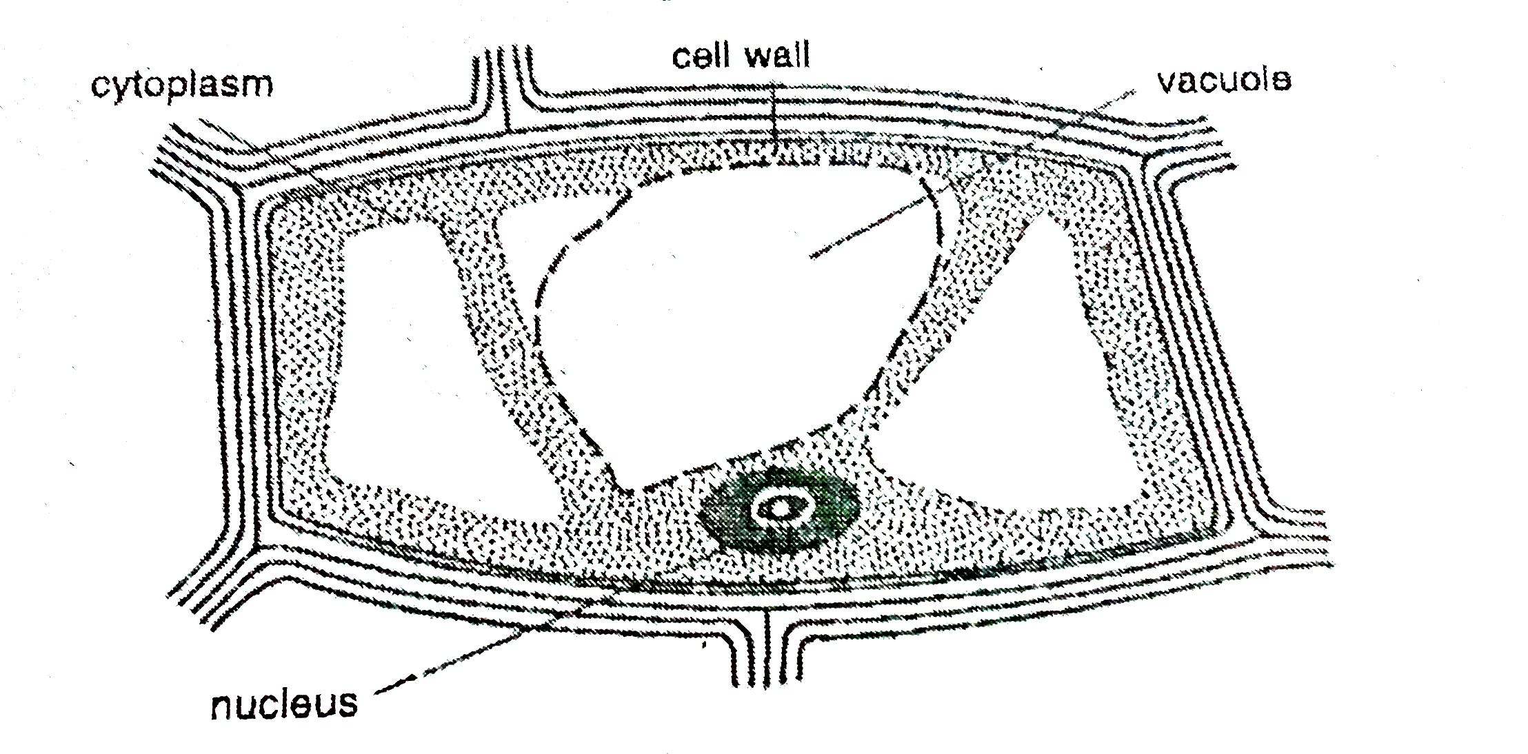
Human cheek cells are commonly stained with
View Solution
Knowledge Check
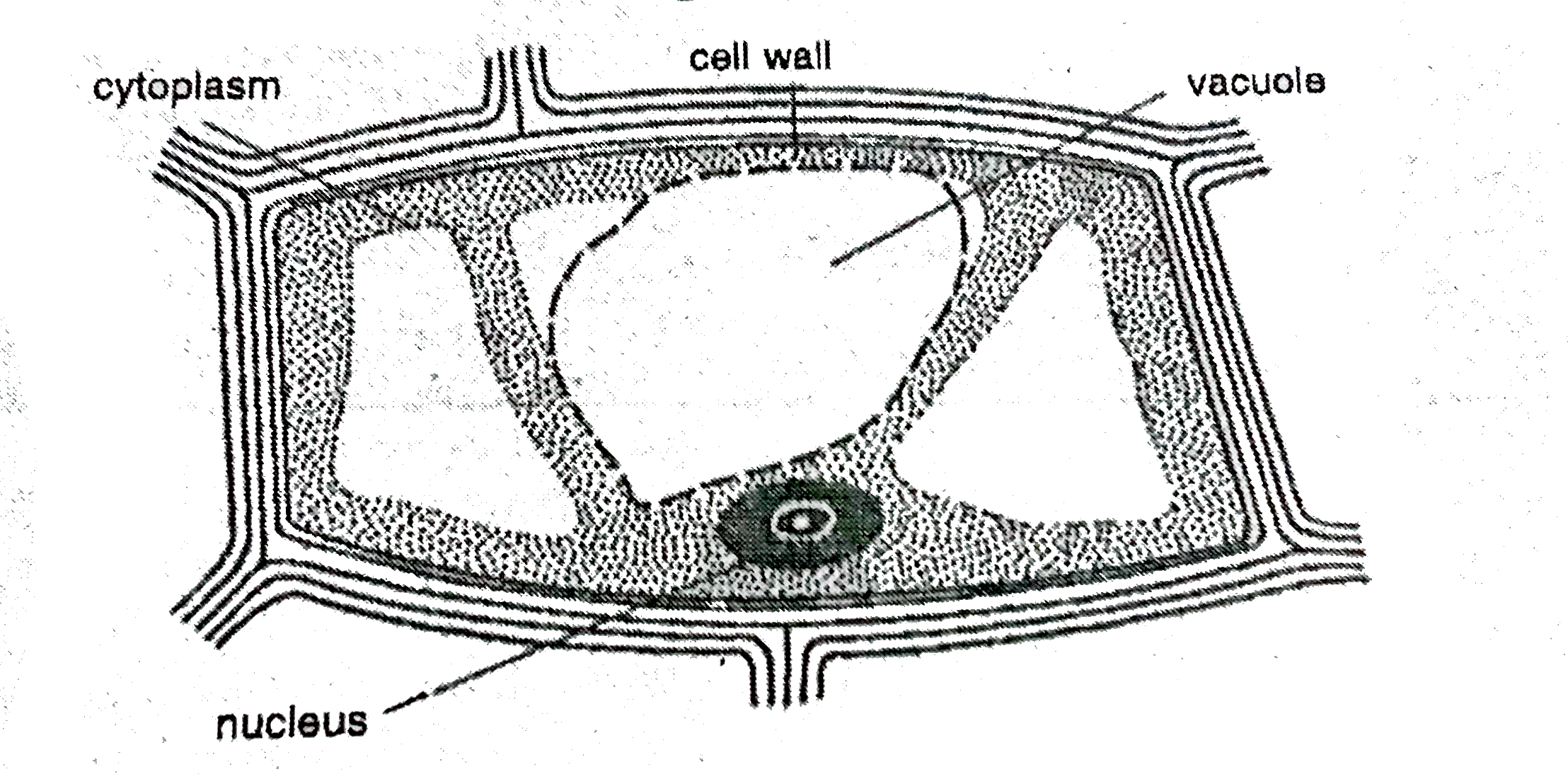
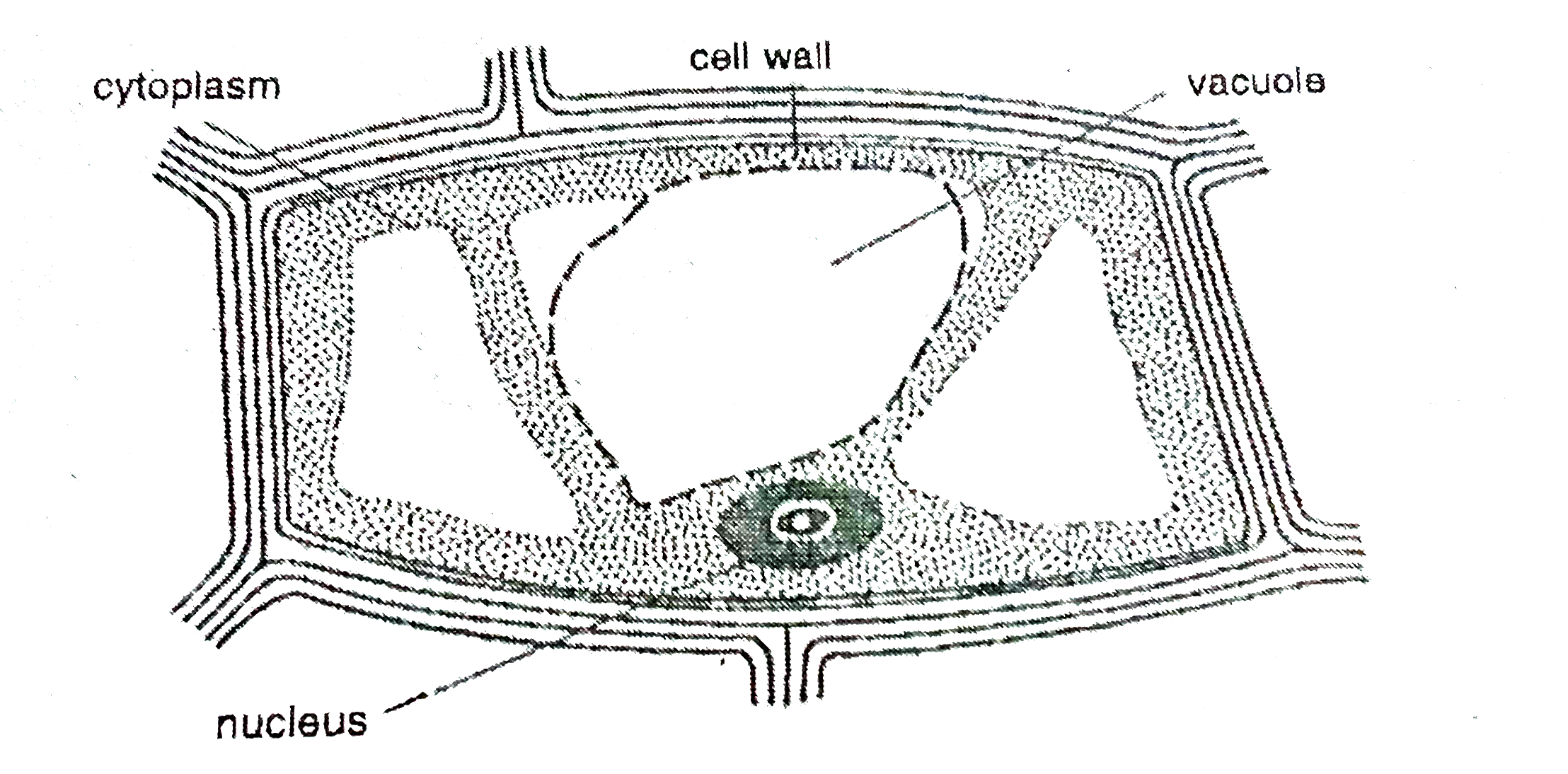
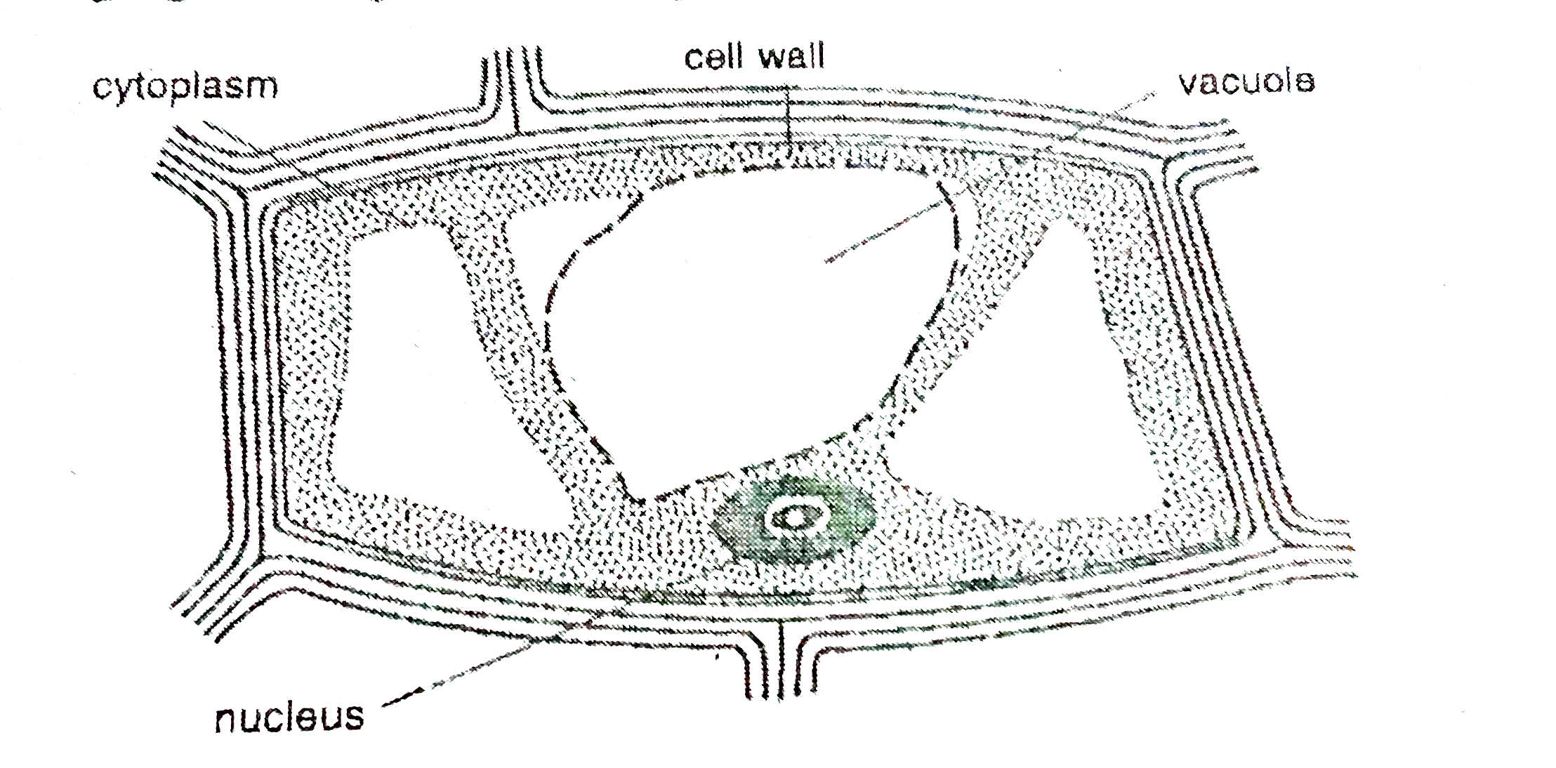
To prepare stained temporary mounts of (a) onion peel, and (b) human cheek cells and to record observations and draw their labelled diagram.
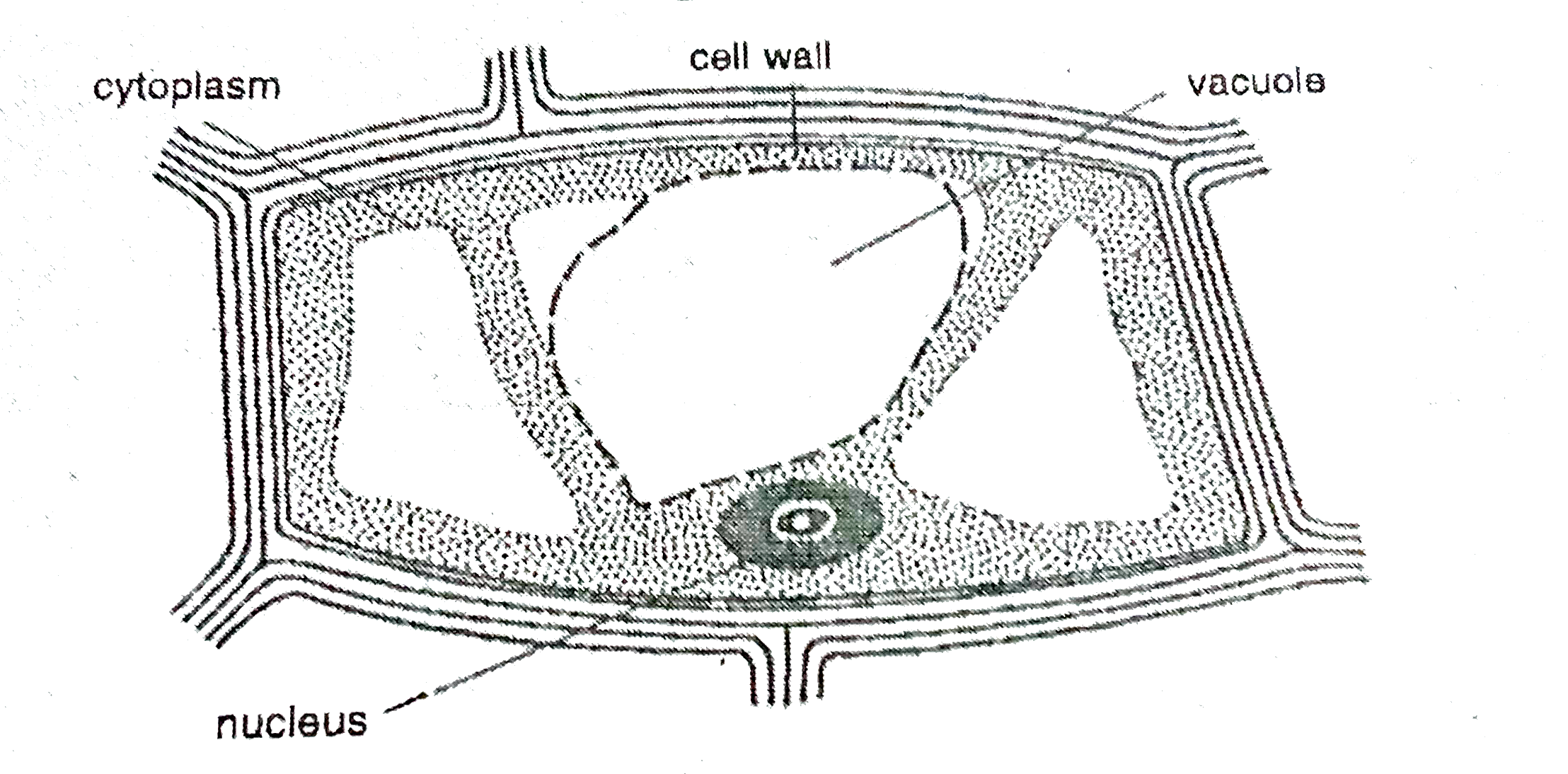
In plant cells, nucleus is generally placedAin the centreBon one side of the cellCattached to plasma membraneDon one corner of the cellTo prepare stained temporary mounts of (a) onion peel, and (b) human cheek cells and to record observations and draw their labelled diagram.
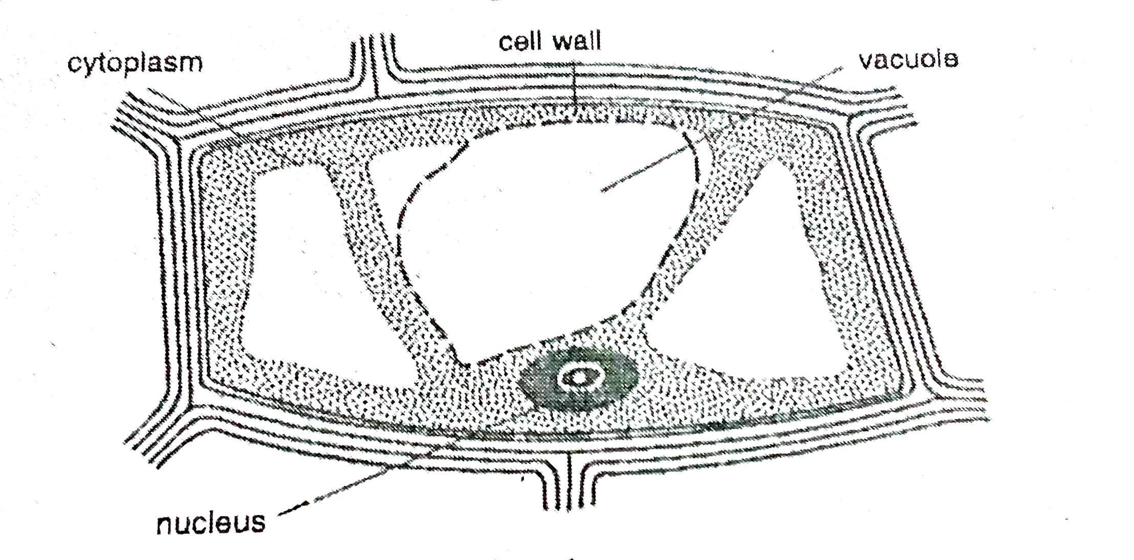
In plant cells, nucleus is generallyAcylindricalBroundedCdiscoidalDellipticalTo prepare stained temporary mounts of (a) onion peel, and (b) human cheek cells and to record observations and draw their labelled diagram.
In juman cheek cells, nucleus is generally locatedAnear the plasma membraneBon one sideCin the centreDon border
To prepare stained temporary mounts of (a) onion peel, and (b) human cheek cells and to record observations and draw their labelled diagram.
Cell wall in plant cells is made of
View SolutionTo prepare stained temporary mounts of (a) onion peel, and (b) human cheek cells and to record observations and draw their labelled diagram.
Definite shape of cell is seen in case of
View SolutionTo prepare stained temporary mounts of (a) onion peel, and (b) human cheek cells and to record observations and draw their labelled diagram.
The outer most covering of a plant cell is
View SolutionTo prepare stained temporary mounts of (a) onion peel, and (b) human cheek cells and to record observations and draw their labelled diagram.
In plant cells, cell to cell contact is maintained through
View SolutionA well stained leaf peel mount when observed under the high power of a microscope shows nuclei in:
View Solution
