T.S. of penis of man shows
1 corpora cavernosa and a pair of corpus spongiosum
1 pair corpora cavermosa and single corpus spongiosum
2 corpora cavernosa and 2 corpus spongiosum
All of the above
The correct Answer is:B
Step-by-Step Solution:
1. Understanding the Structure: The transverse section (T.S.) of the penis reveals different types of tissues and structures. The primary tissues of interest are the corpora cavernosa and the corpus spongiosum.
2. Identifying the Tissues:
- Corpora Cavernosa: There are two cylindrical structures known as corpora cavernosa. These tissues are responsible for the erectile function of the penis. They fill with blood during sexual arousal, leading to an erection.
- Corpus Spongiosum: This is a single structure that surrounds the urethra. It is crucial for maintaining the patency of the urethra during erection and also contains blood vessels that contribute to the erectile process.
3. Location of the Tissues: The corpora cavernosa are located on the dorsal side of the penis, while the corpus spongiosum is located ventrally, surrounding the urethra.
4. Conclusion: In summary, the T.S. of the penis shows one pair of corpora cavernosa and one corpus spongiosum. Therefore, the correct identification of the structures in the T.S. of the penis is:
- 1 pair of corpora cavernosa
- 1 corpus spongiosum
Final Answer:
The transverse section of the penis shows one pair of corpora cavernosa and one corpus spongiosum.
---
- Corpora Cavernosa: There are two cylindrical structures known as corpora cavernosa. These tissues are responsible for the erectile function of the penis. They fill with blood during sexual arousal, leading to an erection.
- Corpus Spongiosum: This is a single structure that surrounds the urethra. It is crucial for maintaining the patency of the urethra during erection and also contains blood vessels that contribute to the erectile process.
- 1 pair of corpora cavernosa
- 1 corpus spongiosum
The transverse section of the penis shows one pair of corpora cavernosa and one corpus spongiosum.
Similar Questions
Knowledge Check
- Question 1 - Select One
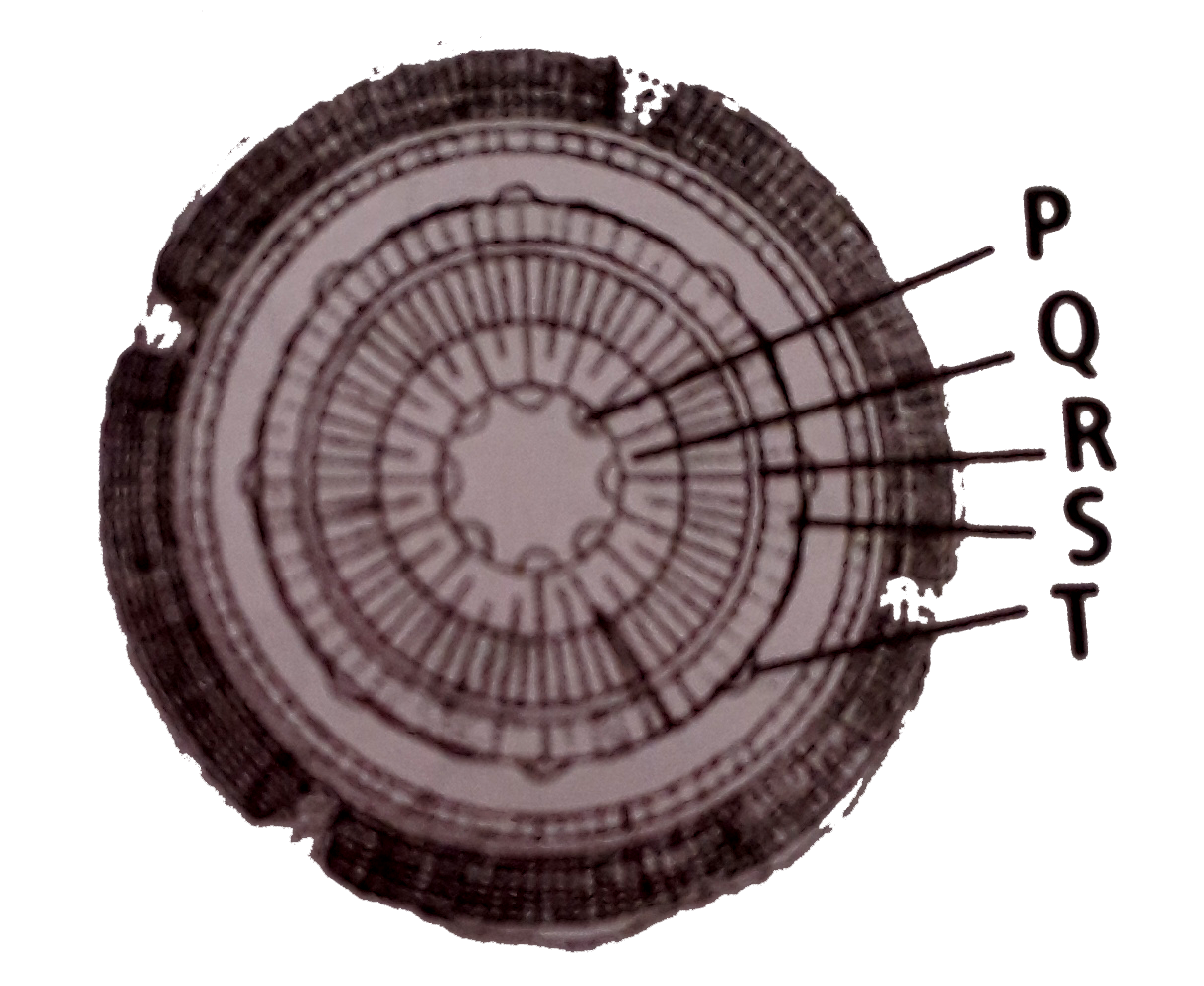
A.T.S. of dicot stem shows
Avascular bundles arranged in a ringBscattered vascular bundles.Cclosed vascular bundlesDradial vascular bundles. - Question 2 - Select One
The penis of a male is:
Aprimary sex organBaccessory sex organCsecondary sex organDexternl sex organ - Question 3 - Select One
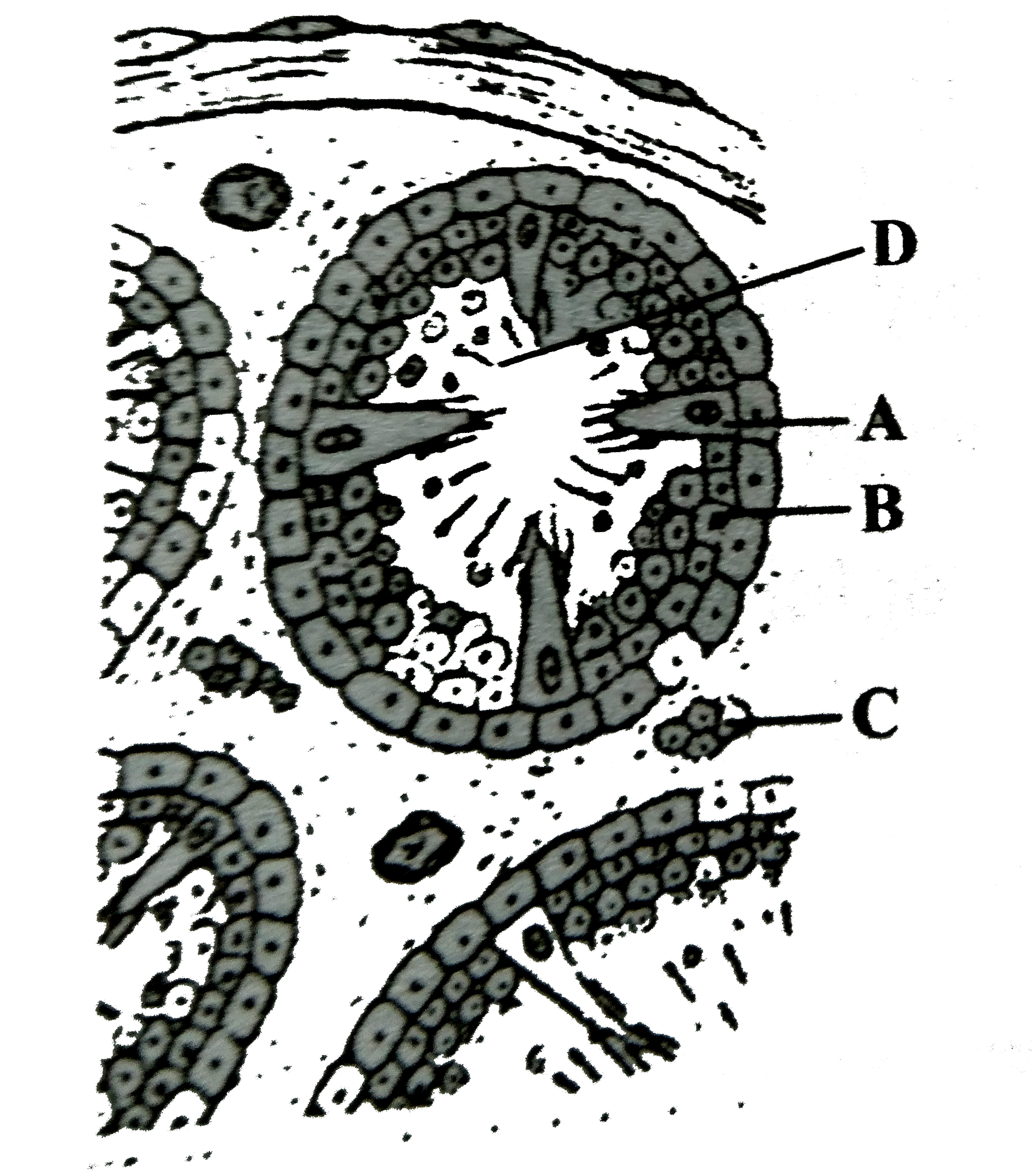
The given diagram refers to T.S. of testis showing sectional view of a few seminiferous tubules. Identify the parts labelled A-D and select the correct option.
 AA-Sertoli cell, B-Spermatozoa,
C-Interstitial cell, D-spermsBA-Sertoli cell, B-Secondary spermatocyte,
C-Interstitial cell, D-SpermsCA-Interstitial cell, B-Spermatogonia,
C-Sertoli cells, D-SpermsDA-Sertoli cells, B-Spermatogonia,
C-Interstitial cells, D-Sperms


