Ventricular contraction in command of :−
S.A. Node
A.V. Node
Purkinje fibres
Papillary muscles
The correct Answer is:1
Step-by-Step Solution:
1. Understanding the Question: The question asks about which structure is responsible for commanding ventricular contraction in the heart.
2. Identifying the Structures: The options given are:
- SC node (Sinoatrial node)
- AV node (Atrioventricular node)
- Purkinje fibers
- Papillary muscles
3. Role of the SC Node: The SC node, also known as the pacemaker of the heart, initiates the electrical impulse that triggers the heartbeat. It primarily controls the rhythm of the heart but does not directly command ventricular contraction.
4. Role of the AV Node: The AV node acts as a relay station between the atria and ventricles. It delays the electrical signal before it passes to the ventricles, allowing them to fill with blood. However, it does not directly command the contraction of the ventricles.
5. Role of Purkinje Fibers: The Purkinje fibers are specialized conductive fibers that spread the electrical impulse throughout the ventricles. They ensure that the ventricles contract in a coordinated manner, effectively commanding the contraction of the ventricular myocardium.
6. Role of Papillary Muscles: Papillary muscles are attached to the heart valves and help in their functioning during ventricular contraction. However, they do not command the contraction itself.
7. Conclusion: Based on the roles of each structure, the Purkinje fibers are the ones that command the contraction of the ventricles.
Final Answer: Ventricular contraction is commanded by Purkinje fibers.
---
- SC node (Sinoatrial node)
- AV node (Atrioventricular node)
- Purkinje fibers
- Papillary muscles
Similar Questions
Knowledge Check
- Question 1 - Select One
Ventricular contraction in command of :−
ASA nodeBAV nodeCPurkinje fibersDPapillary muscles - Question 2 - Select One
The strength of ventricular contraction increases when SAN is stimulated by
AVagus nerveBParasympathetic nerveCSympathetic nerveDAll of these - Question 3 - Select One
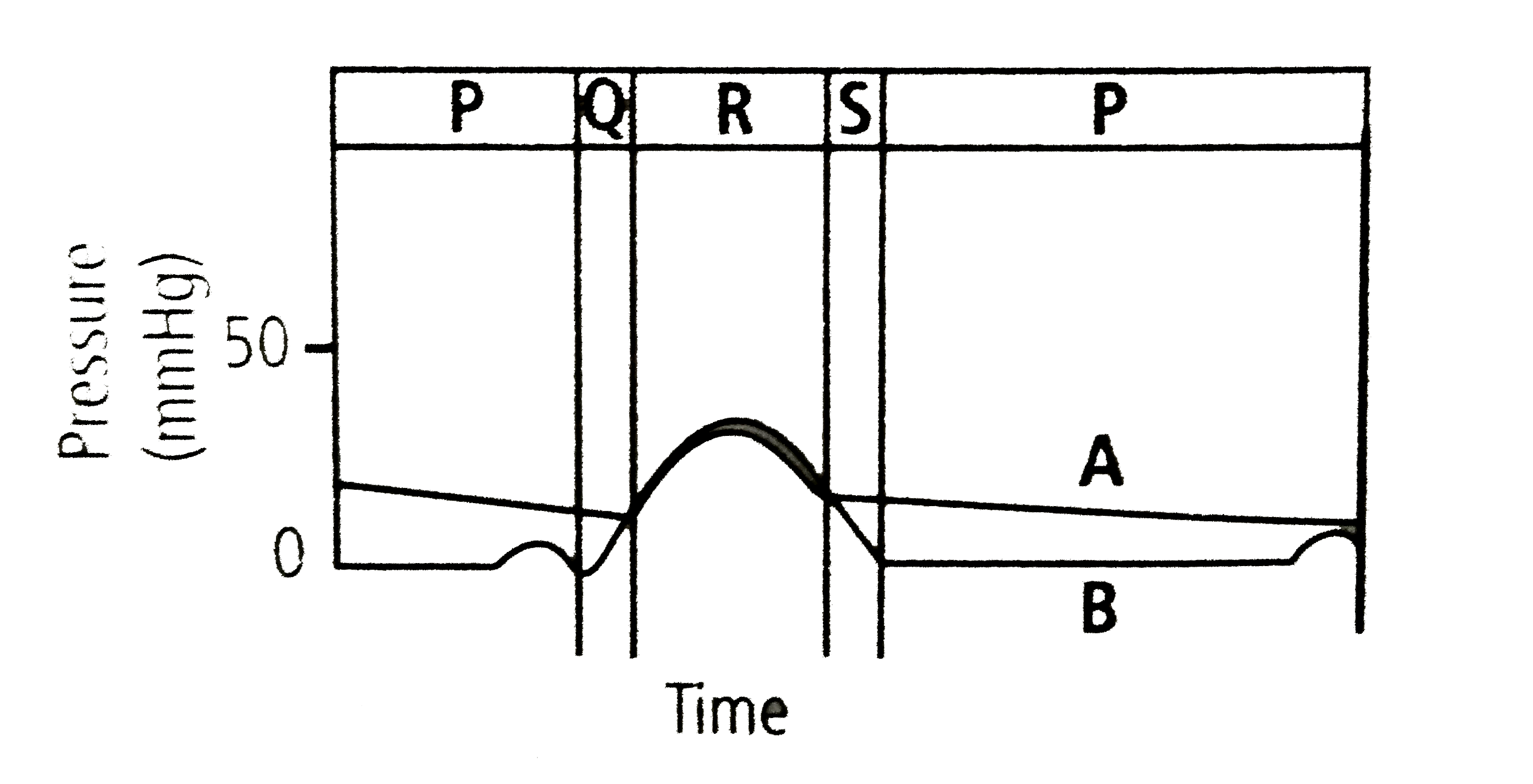
Refer to the given figure in which A refers to pulmonary artery pressure and B refers to right ventricular pressure. Identify P,Q,R and S in the figure and match with the list (i-iv) given below
(i) Isovolumetric ventricular contraction
(ii) Ventricular ejection
(iii) Isovolumetric ventricular relaxation
(iv) Ventricular filling
 AP-(iv),Q-(iii),R-(ii),S-(i)BP-(ii),Q-(iii),R-(i),S-(iv)CP-(iv),Q-(i),R-(ii),S-(iii)DP-(i),Q-(ii),R-(iii),S-(iv)
Assertion : Neutral signals through sympathetic nerves can increase the strength of ventricular contraction
Reason : Parasympathetic neutral signals synergistically act with sympathetic neutral signal to increase the cardiac output.View SolutionWhich phase of the ECG indicates ventricular excitation and contraction?
View SolutionVentricular depolarization is represented by __________.
View SolutionSelect the correct passive form of the given sentence.
Give the command.View SolutionVentricular systole is stimulated by
View Solution

