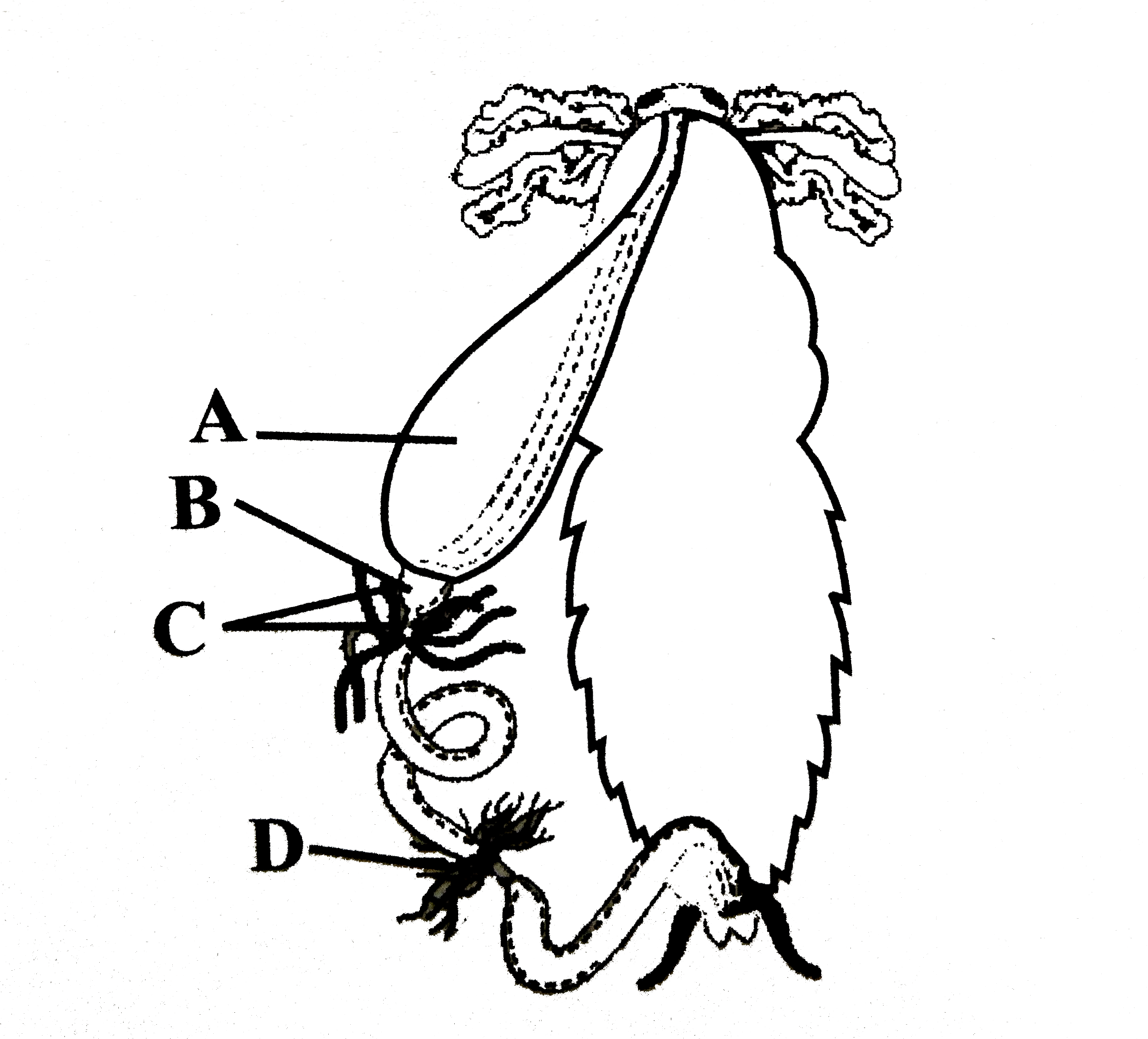
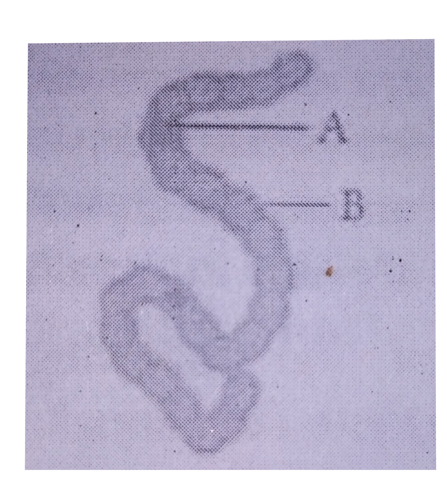
The given figure shows alimentary cancal of cockroach. Identify the parts labelled as A to D and select the correct option
A
Stomach, ileum, hepatic caecae, malpighian tubules
B
Gizzard, crop, hepatic caecae, malpighian tubules
C
Crop, gizzard, malpighian tubules, hepatic caecae
D
Crop, gizzard, hepatic caecae, malpighian tubules
Video Solution
Text Solution
The correct Answer is:D
Crop, gizzard, hepatic caecae, malpighian tubules
|
Topper's Solved these Questions
- STRUCTURAL ORGANISATION IN ANIMALS (CONTD . . .) (ANIMAL MORPHOLOGY)BOOK - AAKASH INSTITUTECHAPTER - STRUCTURAL ORGANISATION IN ANIMALS (CONTD . . .) (ANIMAL MORPHOLOGY)EXERCISE - Assignment (Section-C)
28 Videos
- STRUCTURAL ORGANISATION IN ANIMALS (CONTD . . .) (ANIMAL MORPHOLOGY)BOOK - AAKASH INSTITUTECHAPTER - STRUCTURAL ORGANISATION IN ANIMALS (CONTD . . .) (ANIMAL MORPHOLOGY)EXERCISE - Assignment (Section-D)
10 Videos
- STRUCTURAL ORGANISATION IN ANIMALS (CONTD . . .) (ANIMAL MORPHOLOGY)BOOK - AAKASH INSTITUTECHAPTER - STRUCTURAL ORGANISATION IN ANIMALS (CONTD . . .) (ANIMAL MORPHOLOGY)EXERCISE - Assignment (Section-A)
40 Videos
- STRUCTURAL ORGANISATION IN ANIMALS ( ANIMAL TISSUES ) BOOK - AAKASH INSTITUTECHAPTER - STRUCTURAL ORGANISATION IN ANIMALS ( ANIMAL TISSUES ) EXERCISE - Assignment (Section-D)
10 Videos
- TEST 1BOOK - AAKASH INSTITUTECHAPTER - TEST 1EXERCISE - Examplle
18 Videos
Similar Questions
Knowledge Check
The given figure shows alimentary canal of cockroach. Identify the parts labelled as A to D and select the correct option.
 AStomach, ileum, hepatic caecae, malpighian tubulesBGizzard, crop, hepatic caecae, malpighian tubulesCCrop, gizzard, malpighian tubules, hepatic caecaeDCrop, gizzard, hepatic caecae, malpighian tubules
Identify the parts labelled a, b, c and d and select the correct option
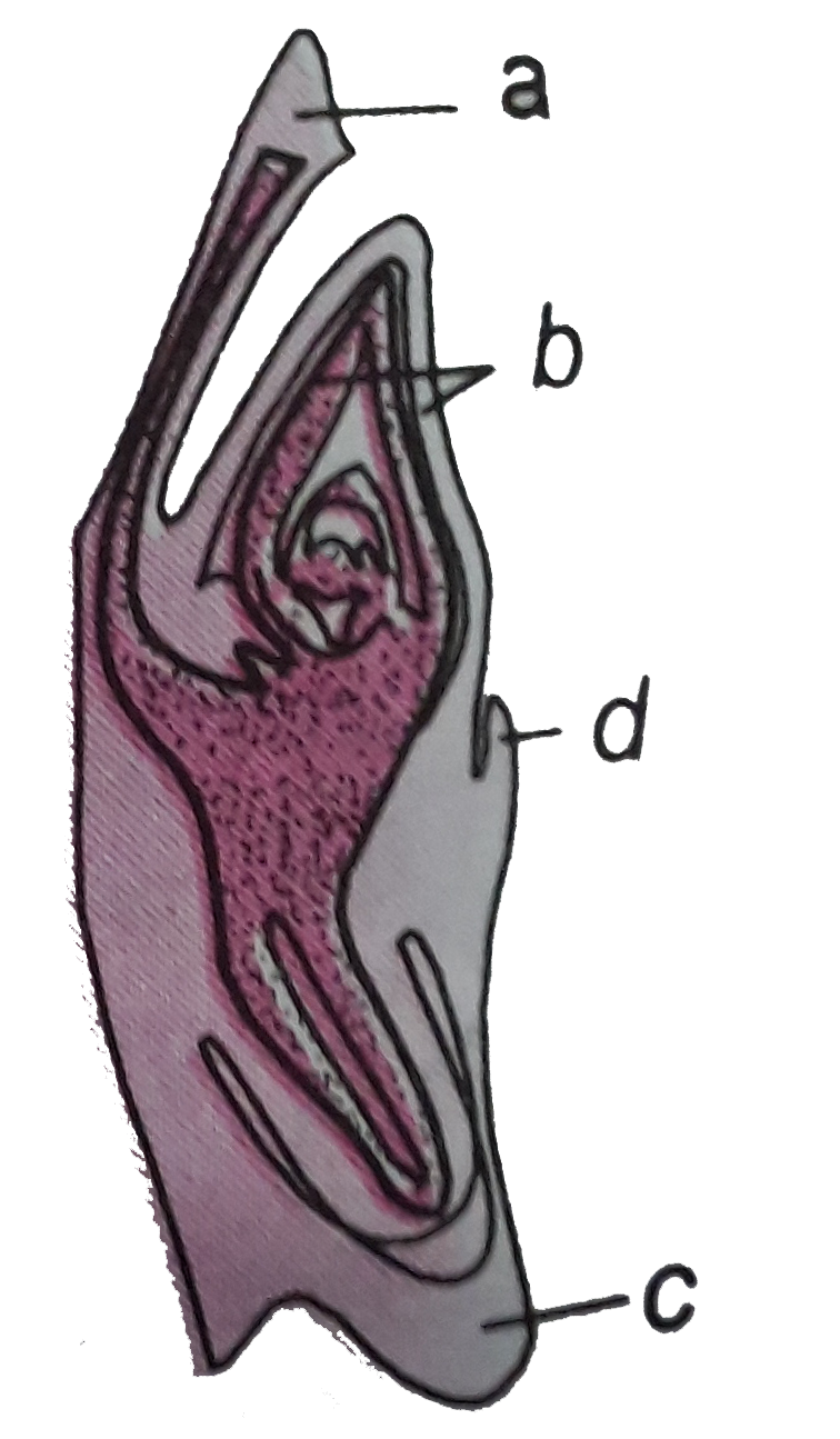 .Aa-scutellum, b-epiblast, c-coleoptile, d-coleorhizaBb-coleorhiza, c-coleoptile, d-epiblastCa-scutellum, b-coleoptile, c-coleorhiza, d-epiblastDa-epiblast, b-coleoptile, c-coleorhiza, d-scutellum.
The given figure shows lateral view of the human brain.Identify the parts labelled as A to E and select the correct option.
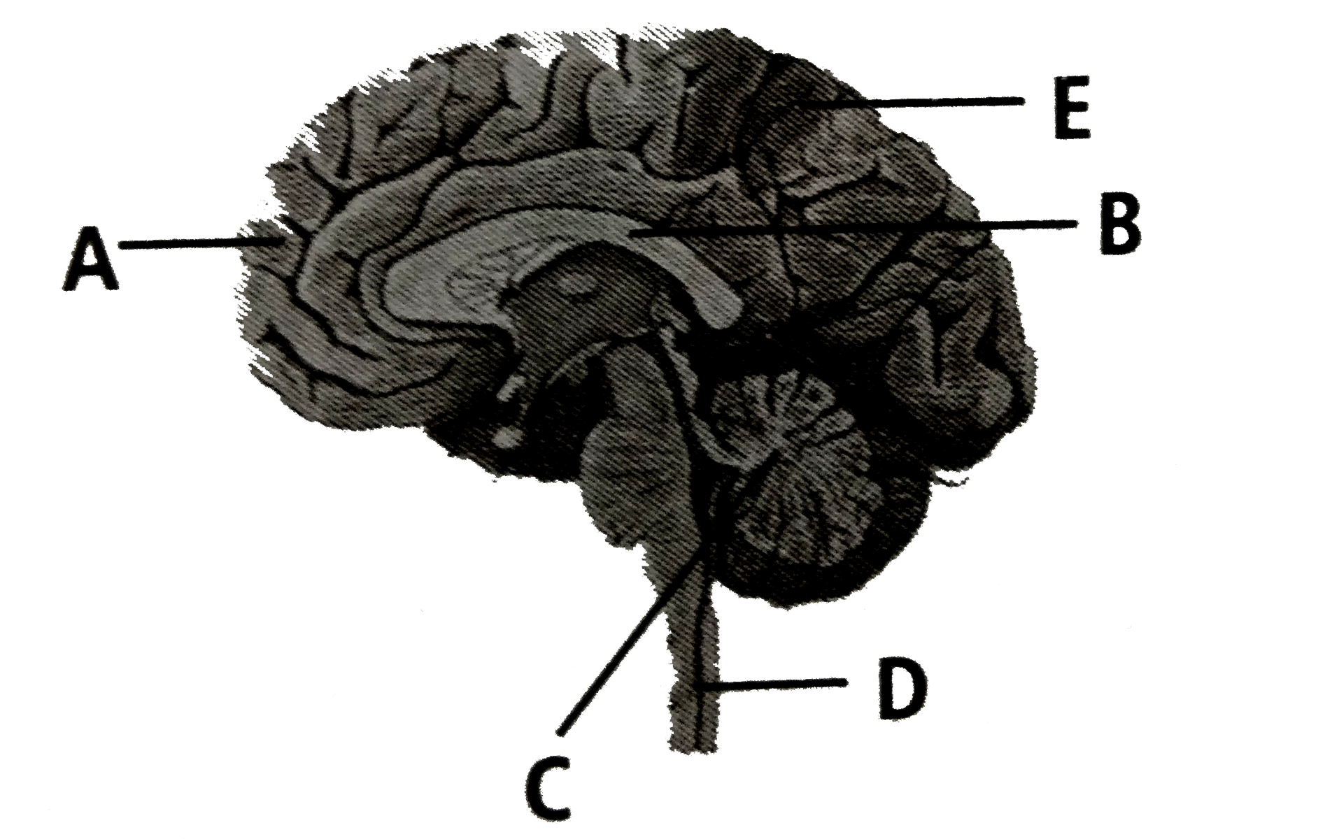 AABCDETemporal lobeCorpus callosumCerebrumMedulla oblongataFrontal lobeBABCDEFrontal lobeThalamusCerebrumMedulla oblongataOccipital lobeCABCDETemporal lobePonsCerebrumMedulla oblongataFrontal lobeDABCDEFrontal lobeCorpus callosumCerebellumMedulla oblongataParietal lobe
Given below is the figure of a sarcomere. Identify the parts labelled as A to D and select the correct option.
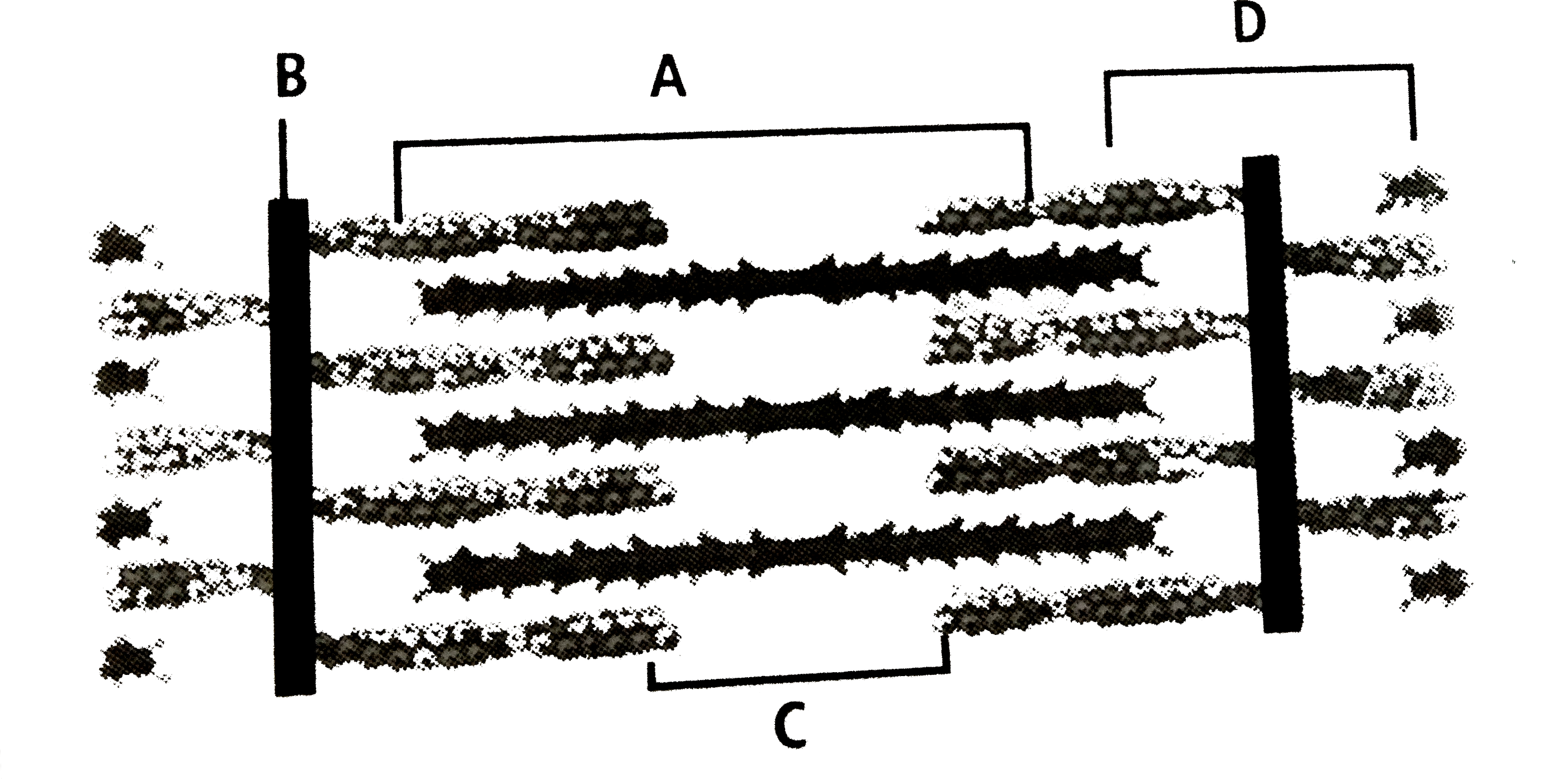
View SolutionIdentify the stage shows in the figure and select the correct option

View SolutionThe figure given here is of rib cage. Identify the parts labelled as A, B and C and select the correct option .
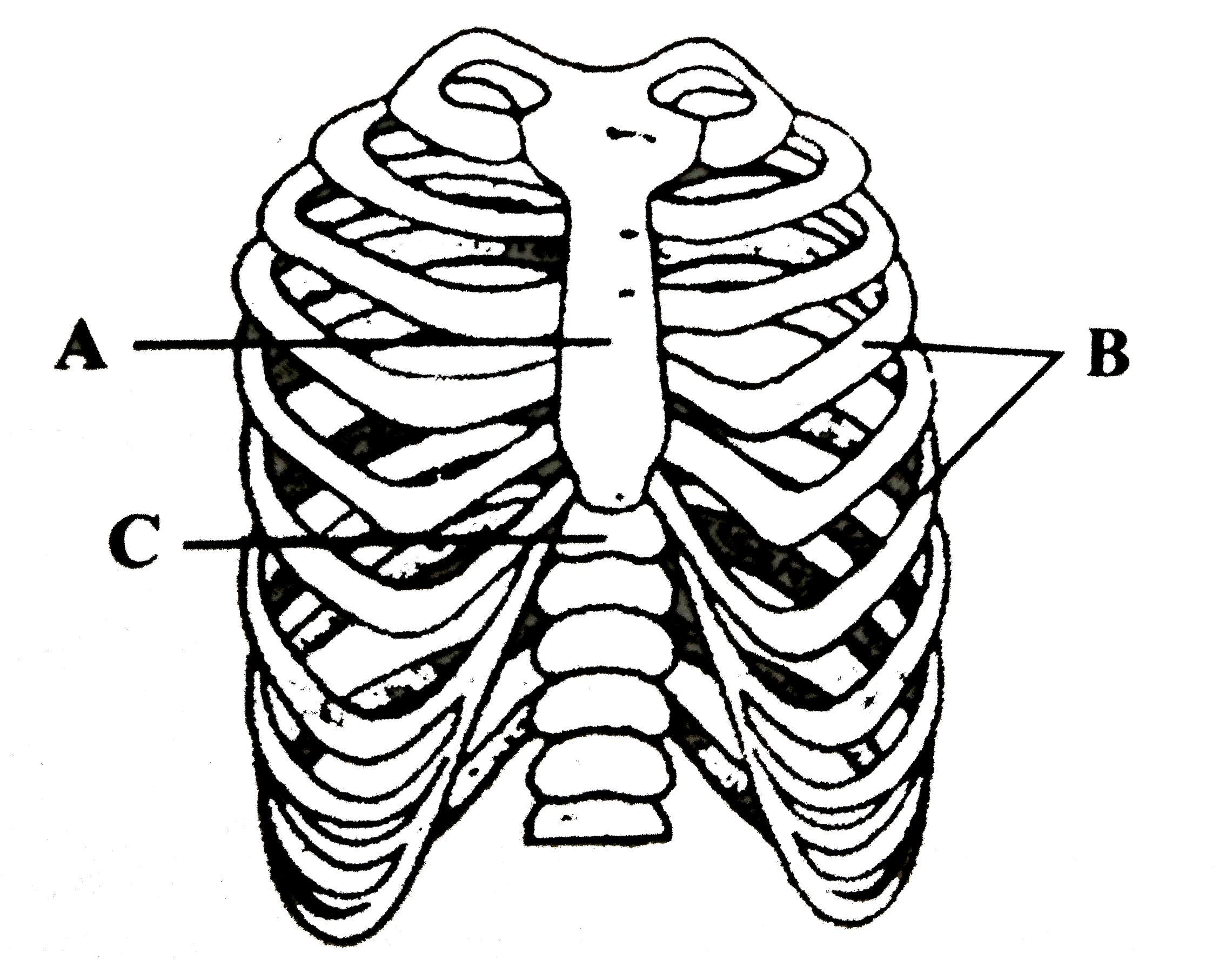
View SolutionIdentify the labeled part in the given figure and select the correct option.
View SolutionThe given figure shows an actin (thin) filament. Identify the labelled parts A, B, and C and select the correct option.

View Solution

