Starting with the zygote, draw the diagrams of the different stages of embryo development in a dicot.
Step-by-Step Solution for the Development of a Dicot Embryo
1. Zygote Formation:
- The process begins with the formation of the zygote, which is a diploid cell formed by the fusion of male and female gametes.
- Diagram: Draw a single circle representing the zygote, labeled "Zygote (2n)".
Hint: Remember that the zygote is the first cell of the new organism, containing genetic material from both parents.
2. First Division:
- The zygote undergoes its first mitotic division, resulting in two cells: the terminal cell and the basal cell.
- Diagram: Draw two circles, one above the other, labeled "Terminal Cell" and "Basal Cell".
Hint: The terminal cell will eventually develop into the embryo, while the basal cell will form the suspensor.
3. Further Division:
- The terminal cell continues to divide, leading to the formation of more cells.
- Diagram: Illustrate the terminal cell dividing into two or more cells, while the basal cell remains as is.
Hint: Focus on the growth of the terminal cell, as it is crucial for embryo development.
4. Formation of Suspensor and Hypophysis:
- The basal cell develops into the suspensor, and the hypophysis (future root) begins to form.
- Diagram: Show the basal cell elongating into a suspensor and the hypophysis appearing as a small structure at the end.
Hint: The suspensor provides nutrients to the developing embryo.
5. Development of Cotyledons:
- The embryo begins to develop cotyledons, which are the first leaves of the plant.
- Diagram: Illustrate the cotyledons starting to form on either side of the developing embryo.
Hint: Remember that dicots have two cotyledons, which is a key characteristic.
6. Heart Stage:
- The embryo reaches the heart stage, where the cotyledons are more pronounced and the overall shape resembles a heart.
- Diagram: Draw the embryo with two prominent cotyledons and a central structure representing the radicle.
Hint: The heart shape is a significant developmental milestone in dicot embryos.
7. Maturation of the Embryo:
- The embryo continues to grow, with the suspensor becoming shorter and the cotyledons enlarging.
- Diagram: Illustrate a more developed embryo with shorter suspensor and larger cotyledons.
Hint: The shortening of the suspensor indicates that the embryo is nearing maturity.
8. Final Structure:
- The final structure of the dicot embryo includes the radicle, plumule, and two cotyledons.
- Diagram: Draw the fully developed embryo with the radicle at the bottom, the plumule at the top, and two cotyledons on either side.
Hint: Ensure to label all parts clearly: radicle, plumule, and cotyledons.
Summary of Diagrams:
1. Zygote (2n)
2. Terminal and Basal Cells
3. Further Division of Terminal Cell
4. Suspensor and Hypophysis Formation
5. Development of Cotyledons
6. Heart Stage
7. Maturation of the Embryo
8. Final Structure of the Dicot Embryo
- The process begins with the formation of the zygote, which is a diploid cell formed by the fusion of male and female gametes.
- Diagram: Draw a single circle representing the zygote, labeled "Zygote (2n)".
- The zygote undergoes its first mitotic division, resulting in two cells: the terminal cell and the basal cell.
- Diagram: Draw two circles, one above the other, labeled "Terminal Cell" and "Basal Cell".
- The terminal cell continues to divide, leading to the formation of more cells.
- Diagram: Illustrate the terminal cell dividing into two or more cells, while the basal cell remains as is.
- The basal cell develops into the suspensor, and the hypophysis (future root) begins to form.
- Diagram: Show the basal cell elongating into a suspensor and the hypophysis appearing as a small structure at the end.
- The embryo begins to develop cotyledons, which are the first leaves of the plant.
- Diagram: Illustrate the cotyledons starting to form on either side of the developing embryo.
- The embryo reaches the heart stage, where the cotyledons are more pronounced and the overall shape resembles a heart.
- Diagram: Draw the embryo with two prominent cotyledons and a central structure representing the radicle.
- The embryo continues to grow, with the suspensor becoming shorter and the cotyledons enlarging.
- Diagram: Illustrate a more developed embryo with shorter suspensor and larger cotyledons.
- The final structure of the dicot embryo includes the radicle, plumule, and two cotyledons.
- Diagram: Draw the fully developed embryo with the radicle at the bottom, the plumule at the top, and two cotyledons on either side.
1. Zygote (2n)
2. Terminal and Basal Cells
3. Further Division of Terminal Cell
4. Suspensor and Hypophysis Formation
5. Development of Cotyledons
6. Heart Stage
7. Maturation of the Embryo
8. Final Structure of the Dicot Embryo
Topper's Solved these Questions
- BOOK - NCERT EXEMPLAR ENGLISHCHAPTER - SEXUAL REPRODUCTION IN FIOWERING PIANTSEXERCISE - Short Answer Type Questions10videos
SEXUAL REPRODUCTION IN FIOWERING PIANTS
- BOOK - NCERT EXEMPLAR ENGLISHCHAPTER - REPRODUCTIVE HEALTH EXERCISE - LONG ANSWER TYPE QUESTIONS5videos
REPRODUCTIVE HEALTH
- BOOK - NCERT EXEMPLAR ENGLISHCHAPTER - STRATEGIES FOR ENHANCMENT IN FOOD PRODUCTIONEXERCISE - Long Answer type Questions8videos
STRATEGIES FOR ENHANCMENT IN FOOD PRODUCTION
Similar Questions
Knowledge Check
- Question 1 - Select One
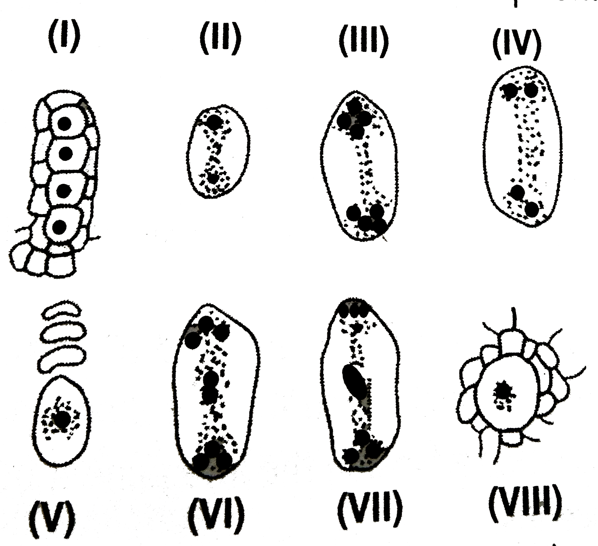
In angiosperms, female gametophte is called as embryo sac. Given figures represent different stages of embryo sac development in angiosperms. Arrange them in correct order and select the correct option.
 A(V)→(I)→(IV)→(II)→(III)→(VII)→(VI)→(VIII)B(VIII)→(V)→(II)→(IV)→(III)→(VII)→(VI)→(I)C(I)→(II)→(IV)→(V)→(VIII)→(III)→(VII)→(VI)D(VIII)→(I)→(V)→(II)→(IV)→(III)→(VI)→(VII)
Identify the correct sequence for different stages of embryo development in dicots .
View SolutionEmbryo development is present in
View SolutionIn Selaginella the embryo develops into
View SolutionHuman embryo completes development in
View SolutionThe correct difference between the embryonic development of monocot and dicot plants is
View Solution(a) Draw a schematic labelled diagram of a fertilised embryo sac of an Angiosperm.
(b) Describe the stage in embryo development in a dicot plant.View Solution(a) Draw a schematic labelled diagram of a fertilised embryo sac of an Angiosperm.
(b) Describe the stage in embryo development in a dicot plant.View Solution

