Nucleic acid exhibit secondary structure. Describe through Watson-Crick model.
To describe the secondary structure of nucleic acids through the Watson-Crick model, we can break down the explanation into several key steps:
1. Definition of Nucleic Acids:
- Nucleic acids are biomolecules essential for life, primarily DNA (deoxyribonucleic acid) and RNA (ribonucleic acid). DNA consists of four nitrogenous bases: adenine (A), guanine (G), thymine (T), and cytosine (C). RNA contains uracil (U) instead of thymine.
Hint: Remember that nucleic acids are made up of nucleotides, which consist of a sugar, a phosphate group, and a nitrogenous base.
2. Secondary Structure:
- The secondary structure of nucleic acids refers to the specific three-dimensional shapes that they can adopt. In the case of DNA, this is primarily a double helical structure.
Hint: Think about how the arrangement of bases and the sugar-phosphate backbone contribute to the overall shape of the molecule.
3. Watson-Crick Model:
- The Watson-Crick model describes the structure of DNA as a double helix. This model was proposed by James Watson and Francis Crick. The two strands of DNA run in opposite directions, which is referred to as being "antiparallel." One strand runs from the 5' end to the 3' end, while the other runs from the 3' end to the 5' end.
Hint: Visualize the double helix as a twisted ladder, where the sides are the sugar-phosphate backbones and the rungs are the base pairs.
4. Backbone Structure:
- The backbone of the DNA strands is composed of alternating sugar (deoxyribose) and phosphate groups. This structure provides stability and support for the nitrogenous bases that extend inward.
Hint: Consider how the sugar and phosphate groups are linked together to form the backbone.
5. Base Pairing:
- The nitrogenous bases pair specifically: adenine pairs with thymine (A-T) via two hydrogen bonds, and guanine pairs with cytosine (G-C) via three hydrogen bonds. This specific pairing is crucial for the stability of the DNA structure.
Hint: Remember the base pairing rules (A-T and G-C) as they are fundamental to DNA structure and function.
6. Helical Structure:
- The DNA double helix has a diameter of approximately 2 nanometers, and the bases are spaced about 0.34 nanometers apart. The helical structure is stabilized by hydrogen bonds between the paired bases and by base stacking interactions.
Hint: Think about how the helical twist of the DNA contributes to its compactness and stability.
7. Hydrogen Bonds:
- Hydrogen bonds play a critical role in maintaining the structure of the DNA double helix. The hydrogen bonds form between the nitrogenous bases, helping to hold the two strands together.
Hint: Consider how the strength of hydrogen bonds contributes to the overall stability of the DNA molecule.
Conclusion:
The secondary structure of nucleic acids, particularly DNA, is elegantly described by the Watson-Crick model, which illustrates the double helical formation, the antiparallel nature of the strands, the sugar-phosphate backbone, and the specific base pairing that underpins the stability and functionality of the molecule.
- Nucleic acids are biomolecules essential for life, primarily DNA (deoxyribonucleic acid) and RNA (ribonucleic acid). DNA consists of four nitrogenous bases: adenine (A), guanine (G), thymine (T), and cytosine (C). RNA contains uracil (U) instead of thymine.
- The secondary structure of nucleic acids refers to the specific three-dimensional shapes that they can adopt. In the case of DNA, this is primarily a double helical structure.
- The Watson-Crick model describes the structure of DNA as a double helix. This model was proposed by James Watson and Francis Crick. The two strands of DNA run in opposite directions, which is referred to as being "antiparallel." One strand runs from the 5' end to the 3' end, while the other runs from the 3' end to the 5' end.
- The backbone of the DNA strands is composed of alternating sugar (deoxyribose) and phosphate groups. This structure provides stability and support for the nitrogenous bases that extend inward.
- The nitrogenous bases pair specifically: adenine pairs with thymine (A-T) via two hydrogen bonds, and guanine pairs with cytosine (G-C) via three hydrogen bonds. This specific pairing is crucial for the stability of the DNA structure.
- The DNA double helix has a diameter of approximately 2 nanometers, and the bases are spaced about 0.34 nanometers apart. The helical structure is stabilized by hydrogen bonds between the paired bases and by base stacking interactions.
- Hydrogen bonds play a critical role in maintaining the structure of the DNA double helix. The hydrogen bonds form between the nitrogenous bases, helping to hold the two strands together.
The secondary structure of nucleic acids, particularly DNA, is elegantly described by the Watson-Crick model, which illustrates the double helical formation, the antiparallel nature of the strands, the sugar-phosphate backbone, and the specific base pairing that underpins the stability and functionality of the molecule.
Topper's Solved these Questions
- BOOK - NCERT EXEMPLAR ENGLISHCHAPTER - BIOMOLECULES EXERCISE - Short Answer Type Questions5videos
BIOMOLECULES
- BOOK - NCERT EXEMPLAR ENGLISHCHAPTER - BIOLOGICAL CLASSIFICATIONEXERCISE - Long answer5videos
BIOLOGICAL CLASSIFICATION
- BOOK - NCERT EXEMPLAR ENGLISHCHAPTER - Body Fluids and Circulation EXERCISE - LONG ANSWER TYPE Q5videos
Body Fluids and Circulation
Similar Questions
Knowledge Check
- Question 1 - Select One
The chemical formula of deoxyribonucleic acid
(DNA) is now well established. The molecule is a
very long chain, the backbone of which consists of a
regular alternation of sugar and phosphate groups.
5 To each sugar is attached a nitrogenous base, which
can be of four different types. Two of the possible
bases—adenine and guanine—are purines, and the
other two—thymine and cytosine—are pyrimidines.
So far as is known, the sequence of bases along the
10 chain is irregular. The monomer unit, consisting of
phosphate, sugar and base, is known as a nucleotide.
The first feature of our structure which is of
biological interest is that it consists not of one chain,
but of two. These two chains are both coiled around
15 a common fiber axis. It has often been assumed that
since there was only one chain in the chemical
formula there would only be one in the structural
unit. However, the density, taken with the X-ray
evidence, suggests very strongly that there are two.
20. The other biologically important feature is the
manner in which the two chains are held together.
This is done by hydrogen bonds between the bases.
The bases are joined together in pairs, a single base
from one chain being hydrogen-bonded to a single
25 base from the other. The important point is that only
certain pairs of bases will fit into the structure.
One member of a pair must be a purine and the other
a pyrimidine in order to bridge between the two
chains. If a pair consisted of two purines, for
30 example, there would not be room for it.
We believe that the bases will be present almost
entirely in their most probable forms. If this is true,
the conditions for forming hydrogen bonds are more
restrictive, and the only pairs of bases possible are:
35 adenine with thymine, and guanine with cytosine.
Adenine, for example, can occur on either chain, but
when it does, its partner on the other chain must
always be thymine.
The phosphate-sugar backbone of our model is
40 completely regular, but any sequence of the pairs of
bases can fit into the structure. It follows that in a
long molecule many different permutations are
possible, and it therefore seems likely that the precise
sequence of bases is the code which carries the
45 genetical information. If the actual order of the bases
on one of the pair of chains were given, one could
write down the exact order of the bases on the other
one, because of the specific pairing. Thus one chain
is, as it were, the complement of the other, and it is
50 this feature which suggests how the deoxyribonucleic
acid molecule might duplicate itself.
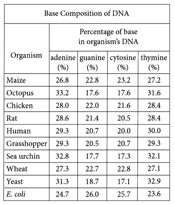
The table shows, for various organisms, the percentage of
each of the four types of nitrogenous bases in that
organism’s DNA.

Adapted from Manju Bansal, “DNA Structure: Revisiting the
Watson-Crick Double Helix.” ©2003 by Current Science Association,
Bangalore
31. Based on the table, is the percentage of adenine in each organism’s DNA the same or does it vary, and which statement made by the authors is most consistent with that data?AThe same, “Two of . . . pyrimidines” (lines 6-8)BThe same, “The important . . . structure” (lines 25-26)CIt varies, “Adenine . . . thymine” (lines 36-38)DIt varies, “It follows . . . information” (lines 41-45) - Question 2 - Select One
The chemical formula of deoxyribonucleic acid
(DNA) is now well established. The molecule is a
very long chain, the backbone of which consists of a
regular alternation of sugar and phosphate groups.
5 To each sugar is attached a nitrogenous base, which
can be of four different types. Two of the possible
bases—adenine and guanine—are purines, and the
other two—thymine and cytosine—are pyrimidines.
So far as is known, the sequence of bases along the
10 chain is irregular. The monomer unit, consisting of
phosphate, sugar and base, is known as a nucleotide.
The first feature of our structure which is of
biological interest is that it consists not of one chain,
but of two. These two chains are both coiled around
15 a common fiber axis. It has often been assumed that
since there was only one chain in the chemical
formula there would only be one in the structural
unit. However, the density, taken with the X-ray
evidence, suggests very strongly that there are two.
20. The other biologically important feature is the
manner in which the two chains are held together.
This is done by hydrogen bonds between the bases.
The bases are joined together in pairs, a single base
from one chain being hydrogen-bonded to a single
25 base from the other. The important point is that only
certain pairs of bases will fit into the structure.
One member of a pair must be a purine and the other
a pyrimidine in order to bridge between the two
chains. If a pair consisted of two purines, for
30 example, there would not be room for it.
We believe that the bases will be present almost
entirely in their most probable forms. If this is true,
the conditions for forming hydrogen bonds are more
restrictive, and the only pairs of bases possible are:
35 adenine with thymine, and guanine with cytosine.
Adenine, for example, can occur on either chain, but
when it does, its partner on the other chain must
always be thymine.
The phosphate-sugar backbone of our model is
40 completely regular, but any sequence of the pairs of
bases can fit into the structure. It follows that in a
long molecule many different permutations are
possible, and it therefore seems likely that the precise
sequence of bases is the code which carries the
45 genetical information. If the actual order of the bases
on one of the pair of chains were given, one could
write down the exact order of the bases on the other
one, because of the specific pairing. Thus one chain
is, as it were, the complement of the other, and it is
50 this feature which suggests how the deoxyribonucleic
acid molecule might duplicate itself.
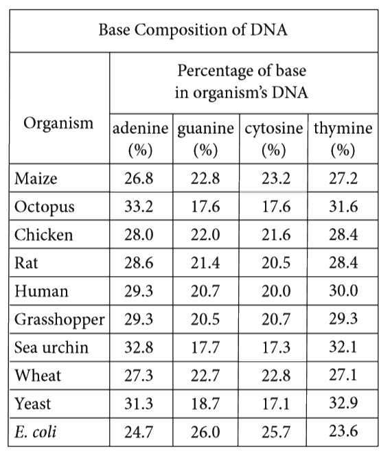
The table shows, for various organisms, the percentage of
each of the four types of nitrogenous bases in that
organism’s DNA.

Adapted from Manju Bansal, “DNA Structure: Revisiting the
Watson-Crick Double Helix.” ©2003 by Current Science Association,
Bangalore
25. The authors’ main purpose of including the information about X-ray evidence and density is toAestablish that DNA is the molecule that carries the genetic information.Bpresent an alternate hypothesis about the composition of a nucleotide.Cprovide support for the authors’ claim about the number of chains in a molecule of DNA.Dconfirm the relationship between the density of DNA and the known chemical formula of DNA.
Define nucleic acid. Describe the structure of DNA model proposed by Watson and Crick.
View SolutionClover leaf model of holley for tRNA is a 2D model giving secondary structure. A 3D model showing tertiary structure was
View SolutionAssertion : All proteinaceous enzymes have a three dimensional structure
Reason : The secondary structure of protein is according to amino
acids present inside the polypeptideView SolutionWhile proposing double helical structure for DNA, Watson and Crick proposed scheme for
View SolutionWhat are the functions of nucleic acids ? What are the structures of sugar molecules present in DNA and RNA.
View SolutionIn α− helix secondary structure, hydrogen bonds lie between oxygen of carboxylic group of first amino acid to the -NH group of the
View Solution

