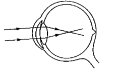
A person cannot see distinctly the book placed nearer than 60 cm from his eyes.
(i) Name the defect of vision the person is suffering from.
(ii) List two possible causes of this defect.
(iii) Draw a ray diagram to show this vision defect.
(iv) How can this defect be corrected ?
(v) Draw a ray diagram to show the correction of this defect.
Step-by-Step Solution:
1. Decrease in the size of the eyeball: In hypermetropia, the eyeball may be shorter than normal, which causes light rays to focus behind the retina instead of directly on it.
2. Increase in the focal length of the eye lens: The eye lens may have a longer focal length than normal, which also results in the image being focused behind the retina.
(iii) Draw a ray diagram to show this vision defect.
To illustrate hypermetropia, we can draw a ray diagram:
1. Draw a horizontal line to represent the retina.
2. Draw a convex lens to represent the eye lens.
3. Place an object (like a book) to the left of the lens.
4. Draw rays coming from the object: one ray going straight through the center of the lens and another ray hitting the lens at an angle.
5. Show that these rays converge to a point behind the retina instead of on it.
(iv) How can this defect be corrected?
Hypermetropia can be corrected by using a convex lens (converging lens). The convex lens helps to converge the light rays before they enter the eye, allowing the image to be focused directly on the retina.
(v) Draw a ray diagram to show the correction of this defect.
To illustrate the correction of hypermetropia using a convex lens:
1. Draw the same horizontal line for the retina.
2. Place the convex lens in front of the eye.
3. Draw the same object (like a book) to the left of the lens.
4. Draw rays coming from the object: one ray going straight through the center of the convex lens and another ray hitting the lens at an angle.
5. Show that these rays converge at a point on the retina, indicating that the image is now correctly focused.
2. Increase in the focal length of the eye lens: The eye lens may have a longer focal length than normal, which also results in the image being focused behind the retina.
2. Draw a convex lens to represent the eye lens.
3. Place an object (like a book) to the left of the lens.
4. Draw rays coming from the object: one ray going straight through the center of the lens and another ray hitting the lens at an angle.
5. Show that these rays converge to a point behind the retina instead of on it.
2. Place the convex lens in front of the eye.
3. Draw the same object (like a book) to the left of the lens.
4. Draw rays coming from the object: one ray going straight through the center of the convex lens and another ray hitting the lens at an angle.
5. Show that these rays converge at a point on the retina, indicating that the image is now correctly focused.
Similar Questions
A person is unable to see objects distinctly placed within 50 cm from his eyes.
(a) Name the defect of vision the person is suffering from and list its two possible causes.
(b) Draw ray diagram to show the defect in the above case.
(c) Mention the type of lens used by him for the correction of the defect and calculate its power. Assume that the near point for the normal eye is 25 cm.
(d) Draw a labelled diagram for the correction of the defect in the above case.
View Solution(a) A student is unable to see clearly the words written on the black board placed at a distance of approximately 3 m from him. Name the defect of vision the boy is suffering from. State the possible causes of this defect and explain the method of correcting it.
View SolutionA person cannot see distinctly object kept beyond 2 m. This defect can be corrected by using a lens of power
View SolutionNooper needs a lens of power −4.5D for correction of her vision.
(a) What kind of defect in vision is she suffering from ?
(b) What is the focal length and nature of corrective lens ?
( c) Draw ray diagrams showing the defected eye and correction for the defect.
(d) What are the causes of this defect ?
View SolutionGiven below is a diagram depicting a defect of the human eye, study the same and then answer the questions that follow:

(i) Name the defect shown in the diagram.
(ii) What are the two possible reasons that cause this defect?
(iii) Name the type of lens used to correct this defect.
(iv) With the help of a diagram show how the defect shown above is rectified using a suitable lens.
View SolutionA student sitting at the back of the classroom cannot read clearly the letters written on the blackward. What advice will a doctor give to her ? Draw ray diagram for the correction of this defect.
View SolutionA student wants to project the image of a candle flame on a screen 60cm in front of a mirror by keeping the flame at a distance of 15 cm from its pole.
(i) Write the type of mirror he should use.
(ii) Find the liner magnification of the image produced.
(iii) What is the distance between the object and its image ?
(iv) Draw a ray diagram to show the image formation in this case.
View Solution(a) A student suffering from myopia is not able to see distinctly the objects placed beyond 5m. Two possible reasons due to which this defect of vision may have arisen. With the help of ray diagrams, explain :
(i) Why the student is unable to see distinctly the objects placed beyond 5m from his eyes.
(ii) The type of corrective lens used to restore proper vision and how this defect is corrected by the use of this lens.
(b) If in this case, numerical value of focal length of corrective lens is 5m, find the power of the lens as per the new cartesion sign convention.
View SolutionA person may suffer from both myopia and hypermetropia defects. (a) What is this condition called? (b) When does it happen? (c) Name the type of lens often required by the persons suffering from this defect. Draw labelled diagram of such lenses.
View Solution(i) Neeta needs a lens of power - 4.5D for correction of her vision.
(a) What kind of defect in vision is she suffering from ?
(b) What is the focal length and nature of corrective lens ?
(c) Explain with the help of a diagram how this defect can be corrected ?
(ii) State the functions of each of the following parts of human eye.
(a) Cornea (b) Pupil (c) Iris (d) Retina
View Solution
