Discuss the microscopic structure of skeletal muscle tissue.
Text Solution
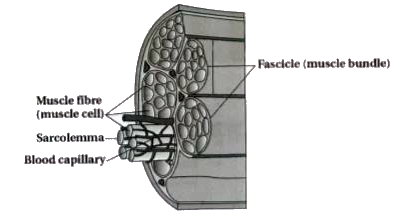
Each organised skeletal muscle in our body is made of number of muscles bundles or fascircles held together by a common collagenous connective tissue layer called fascia. Each muscle bundle contains a number of muscle fibres.

Diagrammatic cross sectional view of a muscle showing muscle bundles and muscle fibres
Each muscle fibre is a lined by the plasma membrance called sarcollema enclosing sarcoplasm.
Muscle fibres is syncitium as the sarcoplasm contains many nuclei.
The endoplasmic reticulum of the muscle fibres (sarcoplasmic reticulum) is the store house of calcium ions.
A characteristics feature of the muscle fibre is the presence of a large number of parallelly arranged filaments in the sarcoplasm calle myofilaments or myofibrils.
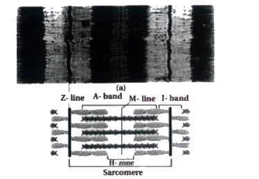
Each myofibril has alternate dark and light bands on it. It is due to the distribution pattern of the two important proteins - Actin and Myosin.
The light bands contain actin is called I-band (Isotropic band).
dark band called .A. (Anisotropic band contains myosin protein).
Both the proteins are arranged as rod-like structures, parallel to each other and to the longitudinal axis of the myofibrils.
Actin filaments are thinner as compared to the myosin filaments.
In the centre of each .I. band is an elastic fibre called .Z. line which bisects it. The thin filaments are firmly attached to the .Z. line.
The thick filaments in the .A. band are also held together in the middle of this band by a thin fibrous membrane called .M. line.
The .A. and .I. bands are arranged alternately throughout the length of the myofibrils.
The portion of the myofibril between two successive .Z. lines is considered as the functional unit of contraction and is called a sarcomere.
In a resting state, the edges of thin filaments on either side of the thick filaments partially overlap the free ends of the thick filaments leaving the central part of the thick filaments. This central part of thick filament, not overlapped by thin filaments is called .H. zone.

|
Topper's Solved these Questions
- LOCOMOTION AND MOVEMENT BOOK - KUMAR PRAKASHANCHAPTER - LOCOMOTION AND MOVEMENT EXERCISE - SECTION B (Difference /Scientific Reasons)
7 Videos
- LOCOMOTION AND MOVEMENT BOOK - KUMAR PRAKASHANCHAPTER - LOCOMOTION AND MOVEMENT EXERCISE - SECTION -C (Defination /Explanation -terms /Location -Function /Full Name )
17 Videos
- DIGESTION AND ABSORPTIONBOOK - KUMAR PRAKASHANCHAPTER - DIGESTION AND ABSORPTIONEXERCISE - Question from Module (Question Paper)
11 Videos
- MINERAL NUTRITIONBOOK - KUMAR PRAKASHANCHAPTER - MINERAL NUTRITIONEXERCISE - QUESTIONS FOR MODULE (IMPORTANT MCQ FOR NEET)
4 Videos
Similar Questions
Knowledge Check
A skeletal muscle can show
Aisotonic contractionBisometric contractionCtwitch contractionDAll of the aboveMyoglobin of skeletal muscles
Ais present in sarcoplasmBstores carbon dioxideCstores oxygenDBoth (a) and (c)Summarizing the structural and functional properties of skeletal muscle tissues, it can be considered :
Anonstriated involuntary muscleBnonstriated voluntary muscleCstriated voluntary muscleDstriated involuntary muscle
Summarizing the structural and functional properties of skeletal muscle tissues, it can be considered :
View SolutionDescribe the structure of a vertebrate skeletal muscle. Explain the sliding filament model of muscle contraction.
View SolutionA common collagenous connective tissue layer which hold the number of muscle bundles of skeletal muscle is called :
View SolutionHow smooth muscle is more like cardiac muscle than skeletal muscle, with respect to both its structure and function?
View SolutionA common collagenous connective tissue layer which hold the number of muscle bundles of skeletal muscle is called :
View Solution
