if the DNA of a virus is labelled with 32P and the protein of the virus is labelled with .32p and the protein of the virus is labelled with ^(35)S, after transduction which molecule (s) would be present inside the bacterial cells ?
.^(32)P only
.^(35 )S only
Both (a) and (b)
None of these
The correct Answer is:a
To solve the question, we need to analyze the information given about the labeling of the virus's DNA and protein and understand the process of transduction.
1. Understanding the Labels:
- The DNA of the virus is labeled with 32P (a radioactive isotope of phosphorus).
- The protein of the virus is labeled with 35S (a radioactive isotope of sulfur).
2. Transduction Process:
- Transduction is a process where a virus (bacteriophage) transfers genetic material from one bacterium to another.
- In this case, the virus infects bacterial cells and can introduce its genetic material (DNA) into the bacterial cell.
3. Identifying Genetic Material:
- Historically, experiments (like those by Hershey and Chase) have shown that DNA, not protein, is the genetic material that gets transferred during viral infection.
- The radioactive labeling indicates which components are being transferred. Since the DNA is labeled with 32P, it will be incorporated into the bacterial cells.
4. Result of Infection:
- After the transduction, the bacterial cells will contain the viral DNA labeled with 32P.
- The protein labeled with 35S does not enter the bacterial cells and remains outside, as proteins do not serve as genetic material.
5. Conclusion:
- Therefore, after transduction, the only molecule that would be present inside the bacterial cells is the DNA labeled with 32P.
Final Answer:
The molecule present inside the bacterial cells after transduction would be 32P (DNA).
- The DNA of the virus is labeled with 32P (a radioactive isotope of phosphorus).
- The protein of the virus is labeled with 35S (a radioactive isotope of sulfur).
- Transduction is a process where a virus (bacteriophage) transfers genetic material from one bacterium to another.
- In this case, the virus infects bacterial cells and can introduce its genetic material (DNA) into the bacterial cell.
- Historically, experiments (like those by Hershey and Chase) have shown that DNA, not protein, is the genetic material that gets transferred during viral infection.
- The radioactive labeling indicates which components are being transferred. Since the DNA is labeled with 32P, it will be incorporated into the bacterial cells.
- After the transduction, the bacterial cells will contain the viral DNA labeled with 32P.
- The protein labeled with 35S does not enter the bacterial cells and remains outside, as proteins do not serve as genetic material.
- Therefore, after transduction, the only molecule that would be present inside the bacterial cells is the DNA labeled with 32P.
The molecule present inside the bacterial cells after transduction would be 32P (DNA).
Topper's Solved these Questions
- BOOK - NCERT FINGERTPS ENGLISHCHAPTER - MOLECULAR BASIS OF INHERITANCEEXERCISE - HOTS8videos
MOLECULAR BASIS OF INHERITANCE
- BOOK - NCERT FINGERTPS ENGLISHCHAPTER - MOLECULAR BASIS OF INHERITANCEEXERCISE - NCERT28videos
MOLECULAR BASIS OF INHERITANCE
- BOOK - NCERT FINGERTPS ENGLISHCHAPTER - MICROBES IN HUMAN WELFAREEXERCISE - Microbes In Human Welfare138videos
MICROBES IN HUMAN WELFARE
- BOOK - NCERT FINGERTPS ENGLISHCHAPTER - ORGANISMS AND POPULATIONEXERCISE - Organisms And Population175videos
ORGANISMS AND POPULATION
Similar Questions
Knowledge Check
- Question 1 - Select One
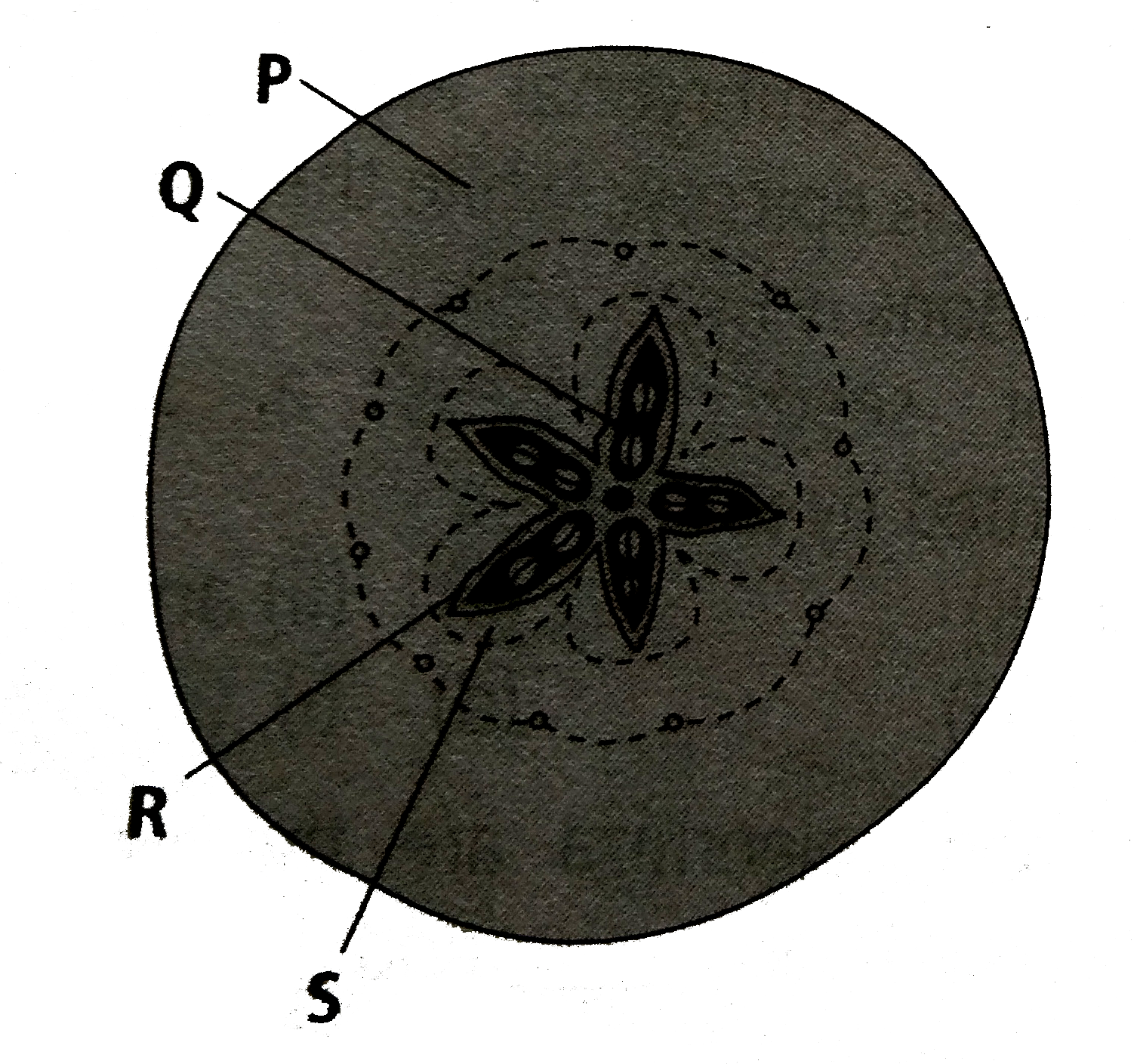
Refer to the given figure and identify the parts labelled P,Q,R and S.
 APQRSSeedThalamusMesocarpEndocarpBPQRSThalamusSeedEndocarpMesocarpCPQRSSeedThalamusEndocarpMesocarpDPQRSPericapSeedMesocarpEndocarp
 - Question 2 - Select One
A scientist was studying the production of a protein that was released by an animal cell into a culture medium, she found that the protein only appeared in the culture medium after the added a few drops of a hormone to the cell. Before adding the hormone, she labelled the protein inside the cell with a fluorescent dye and looked at the cell under the lgiht microscope. The dye was seen in flattened sheets and tube. like structures throughout the cell, and in stacks of flattened sac-like structures. after adding the hormone, the dye was aslo seen as small dots clustered against the cell membrane, Which statement most likely explains these observations?
AThe hormone stimulates protein synthesis in the cell vacuole, the protein is then passed to the Golgi apparatus, and eventually passess through the cell membrane by passive diffusion.BThe hormone triggers the synthesis of the protein in the endoplasmic reticulum and it is then secreted outside of the cell via channel proteins in the cell membraneCThe protein is made in the endoplasmic reticulum, is passed to the Golgi appartus and is secreted through hormone-stimulated exocytosis.DThe protein is made in the Golgi apparatus, is passed to the endoplasmic recticulum and is secreted through hormone-stimulated pinocytosis.
Protein which is secreted by virus infected cell and form cytokine barrier is :-
View SolutionWhich of the following statements are false regarding 'Hershey and Chase' experiment, proving DNA is genetic material ?
(i) They made use of rats.
(ii) Radioactivity was seen in supernatant for the medium that was infected by phase radiolabelled with .32P.
(iii) Used radioisotope to label sulphur with .35S in sulphur- containing amino acids.
(iv) Radioactive phages were allowed to attach to E. coli bacteria.View SolutionIn the long or modern form of the periodic table, the element in the periodic table have been divided into four blocks, s−p−d-and f-. Each period begins with the filling of new energy shell. Two series of f-block elements are placed at the bottom of the periodic table.
If the elements were discovered in future in which the electrons would be present in g-orbitals. Then g-orbital starts, and ends up with what atomic number?View SolutionMark four points on a sheet of paper. Label them P,Q. R and S.
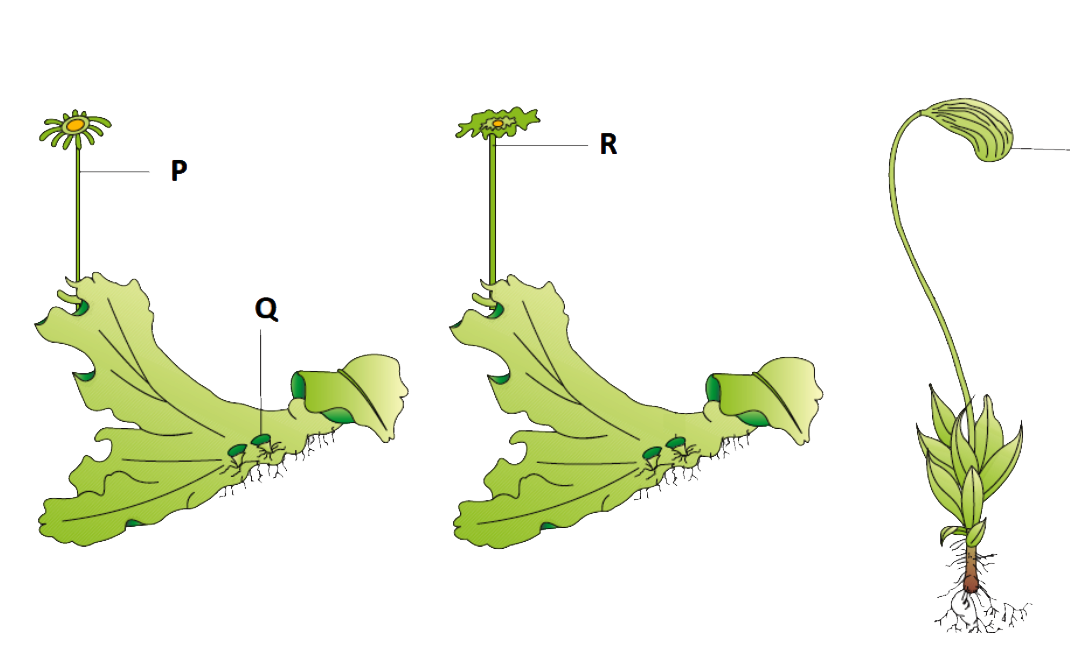
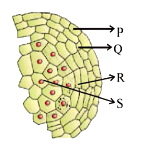
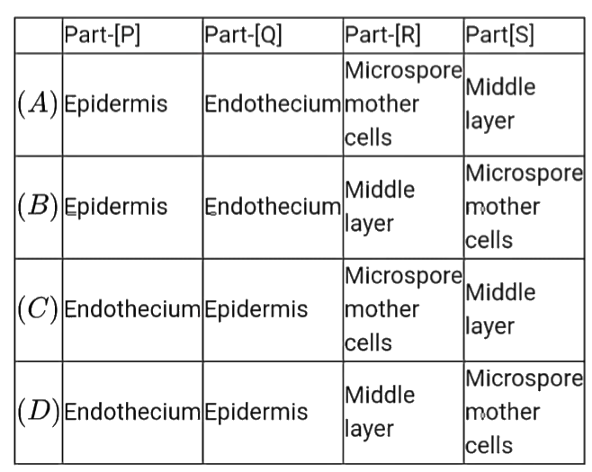
View SolutionObserve the following figures and identify the structures of parts labelled as P, Q, R and S.
 View Solution
In the first biological applicativon of bucky ball , chemists at the University of Califorminaat San F Francisco and Santa Barbra made a discovery in 1993 that could help I designing drugs to rtreat AIDS. The human immunodeficienrcy virus (HIV) that causes AIES reproduces by syntheising a long protein chain , which is cut into smaller segments by an enzymecalled HiV-proteases . ONe way to stop AIDS, then might be to inacivate the enzyme. when the chemists reacted a water-soluble derivative of bucky ball with HIV-protease , they found that itbinds to the portion of th enzyme that would ordinarily clareve the repreouctive protein , preventing the HIV virus from reproducing . Consequently the virus could no bnolgre infect the human cells they had frown in the laboratory . The buycky ball compound itself is not a suitable drug for use against AIDS because of potential side effects and delivery difficaultes , but it does provide a model for the development of such drugs .
Bucky ball is the allotrope of :View Solution


