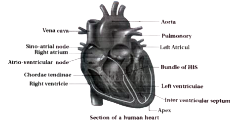
Draw a labelled diagram showing interal structure of heart .

The entire heart is made of cardiac muscles. The walls of ventricles are much thinker tan that of the atria.
A specialised cardiac musculature called the nodal tissue is also distributed in the heart. A patch of this tissue is present in the right upper cornor if the right atrium called he sino-artical node (SAN).
Another mass of this tissue is seen in lower left coron of the right atrium close to the atrio ventricular septum called the atrio ventricular node (AVN).
A bundle of nodal fibres atrio through the atrio ventricular septa to emerge on the top of the inter ventricular septum and immediately divides into a right and left bundle.
These branches give rise to minute fibres throughout the ventricular musculature of the respective sides and are called purkinje fibres.
These fibres along with right and left bundle are known as bundle of HIS.
The nodal musculature has the ability to generate action potentials without any external stimuli. ie. it is autoexcitable.
However the number of action potentials that could br generated in a minute vary at different parts of nodal system. The SAN can generate the maximum number of action potentials. ie. - 70 -75 −1min and is responsible for intitiating and maintaining the rhythmic contractile activity of the heart. Therefore it is called the pacemaker.
Our heart normally beats 70-75 times in a minute (average 72 beats −1min.
Topper's Solved these Questions
- BODY FLUIDS AND CIRCULATIONBOOK - KUMAR PRAKASHANCHAPTER - BODY FLUIDS AND CIRCULATIONEXERCISE - SECTION - B (DIFFERENCE / SCIENTIFIC REASONS)
8 Videos
- BODY FLUIDS AND CIRCULATIONBOOK - KUMAR PRAKASHANCHAPTER - BODY FLUIDS AND CIRCULATIONEXERCISE - SECTION - C (DEFINITION / EXPLANATION - TERMS / LOCATION - FUNCTION.)
7 Videos
- BIOMOLECULES BOOK - KUMAR PRAKASHANCHAPTER - BIOMOLECULES EXERCISE - QUESTION PAPER (DESCRIBE IN DETAIL):
1 Videos
- BREATHING AND EXCHANGE OF GASES BOOK - KUMAR PRAKASHANCHAPTER - BREATHING AND EXCHANGE OF GASES EXERCISE - Questions from Module (Question Paper)
11 Videos
Similar Questions
(a)Draw a labelled diagram of internal structure of human heart.
(b) What are chordae tendinae? Give their function.
View SolutionDraw a labelled diagram to show the structure of earth.
View SolutionDraw labelled diagram of internal structure of human heart.
Label right atrium, mitral valve, left ventricle and pulmonary semilunar valve.
Write a function of Eustachian and tricuspid valve found in human heart.
View SolutionDraw a neat and labelled diagram showing the structure of an atom.
View SolutionDraw a well labelled diagram of internal structure of human heart.
View SolutionAn element has a face-centred cubic (fcc) structure with a cell edge of a. The distance between the centres of two nearest tetrahedral voids in the lattice is:
View SolutionDraw a labelled diagram of the internal structure of human ovary.
View SolutionAn element has a face-centred cubic (fcc) structure with a cell edge of a. The distance between the centres of two nearest tetrahedral voids in the lattice is:
View SolutionDraw a diagram to show the internal structure of human heart. Label only one heart chamber, any two heart valves and any three other structures.
View SolutionDraw a neat labelled diagram to show the structure of the human eye.
View Solution
