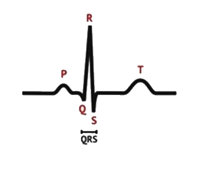
The given figure is the ECG of a normal human. Which one of its components is correctly interpreted below ?
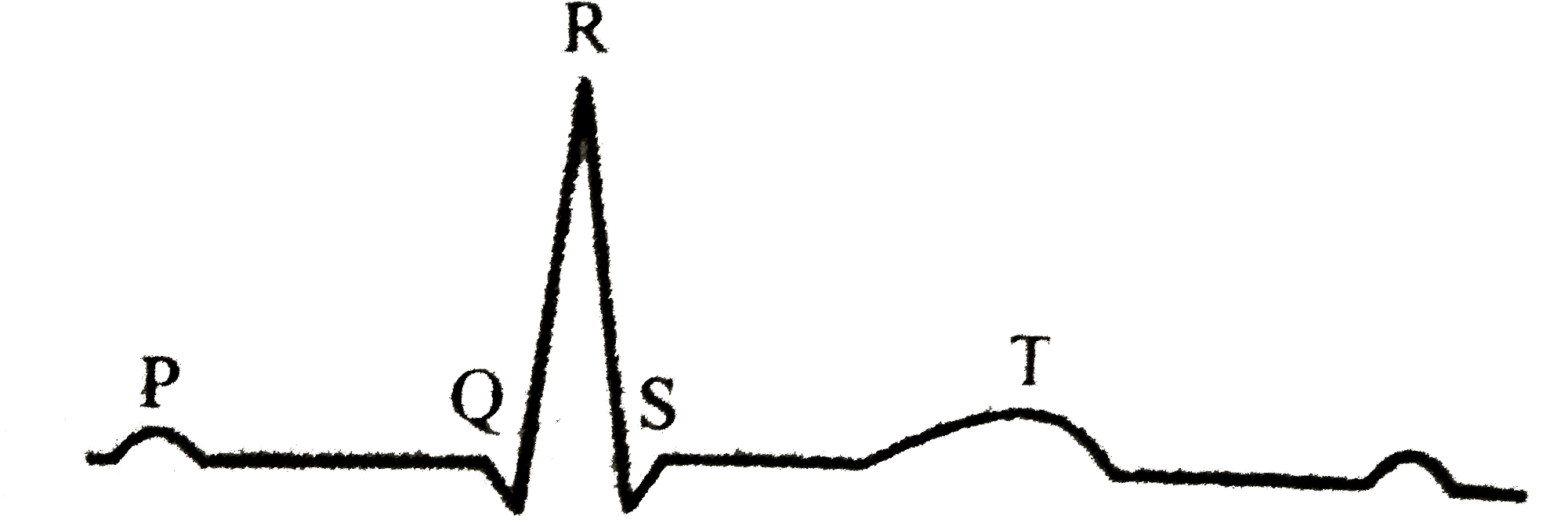
Complex QRS- one complete pulse
Peak T-Initiation of total cardiac contraction
Peak P and Peak R together -systolic and diastolic blood pressures
Peak P -Initiation of left atrial contraction only
The correct Answer is:D
Step by step video & image solution for The given figure is the ECG of a normal human. Which one of its components is correctly interpreted below ? by Biology experts to help you in doubts & scoring excellent marks in Class 12 exams.
Updated on:21/07/2023Topper's Solved these Questions
- BIOTECHNOLOGY : PRINCIPLES AND PROCESSESBook:TRUEMEN BIOLOGY ENGLISHChapter:BIOTECHNOLOGY : PRINCIPLES AND PROCESSESExercise:MULTIPLE CHOICE QUESTIONS
Explore 124 Videos
- BREATHING AND EXCHANGE OF GASES Book:TRUEMEN BIOLOGY ENGLISHChapter:BREATHING AND EXCHANGE OF GASES Exercise:MULTIPLE CHOICE QUESTIONS
Explore 199 Videos
Similar Questions
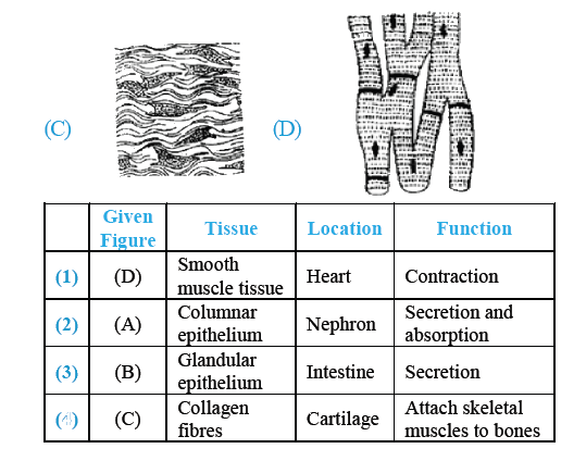
The four figures (A, B, C and D) given below represent four different types of animal tissues. Which one of these is correctly identified in the given options along with its correct location and function ?

View SolutionThe four sketches (A, B, C and D) given below, represent four different types of animal tissues. Which one of these is correctly identified in the options given, along with its correct location and function ?

View Solution
Knowledge Check
Which of the following reactions are correctly interpreted?
A B
B C
C D
D
Which one of the following is correct for a normal human?
ApH of urine is around 8BOn an average, 75-80 mg of urea is excreted via urine per day.CPresence of ketone bodies in urine is an indicator of diabetes mellitusDRelaxation of smooth muscles of bladder and simultaneous contraction of urethral sphincter causes release of urine.ECG depicts the depolarisation and repolarisation process during the cardiac cycle. In the ECG of a normal healthy individual one of the following waves is not represented.
ADepolarisation of atriaBRepolarisation of atriaCDepolarisation of ventriclesDRepolarisation of ventricles
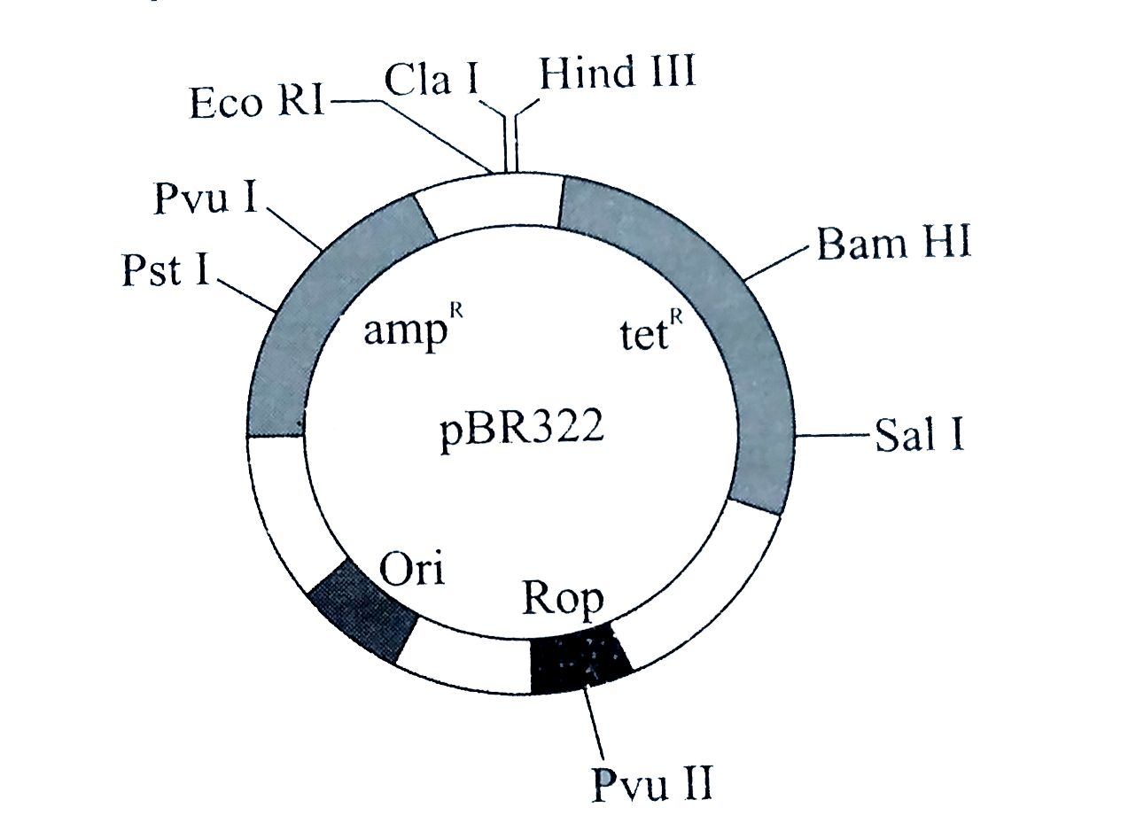
In the diagram of pBR 322, which identifies components correctly ?
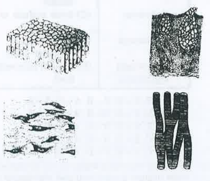
View SolutionThe four sketches A, B,C and D given below
represent four different types of animal tissues .
which one of these is correctly identified
in the options given along with its correct location and function ?
View SolutionGiven below is the ECG of a normal human . Identify the right match of wave and its associated electrical activity.
View SolutionECG depicts the depolarisation and repolarisation process during the cardiac cycle. In the ECG of a normal healthy individual one of the following waves is not represented.
View SolutionECG depicts the depolarisation and repolarisation process during the cardiac cycle. In the ECG of a normal healthy individual one of the following waves is not represented.
View Solution