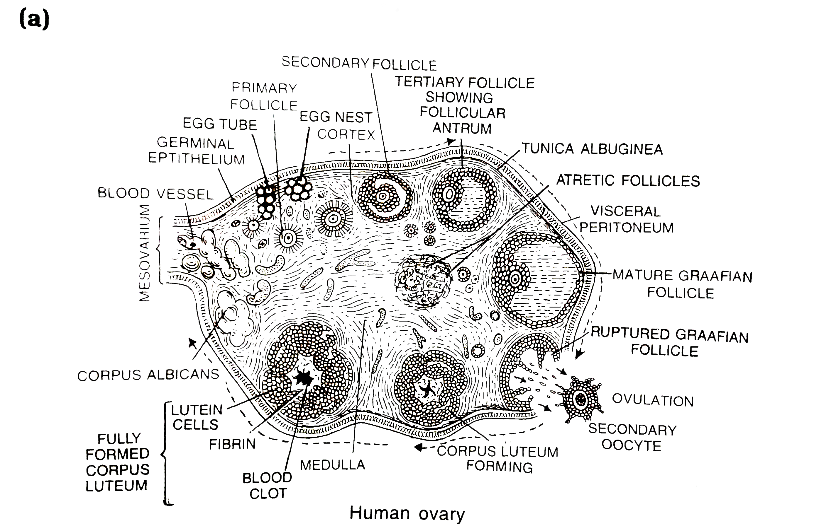
(a) Draw a labelled diagram of sectional view of human ovary showing different stages of oogenesis.
(b) Where is morula formed in humans? Explain the process of its development from the zygote.

(b) Morula is formed in Fallopian tube. The zygote divides mitotically in successive steps. In the first division two blastomeres are formed during second division four and during third division eight blastomeres are formed. The eight to sixteen cell stage embryo is called morula
Topper's Solved these Questions
- REPRODUCTIONBOOK - TRUEMEN BIOLOGY ENGLISHCHAPTER - REPRODUCTIONEXERCISE - SECTION -C
22 Videos
- Protista BOOK - TRUEMEN BIOLOGY ENGLISHCHAPTER - Protista EXERCISE - ASSERTION AND REASON
6 Videos
- Reproduction In OrganismsBOOK - TRUEMEN BIOLOGY ENGLISHCHAPTER - Reproduction In OrganismsEXERCISE - MULTIPLE CHOICE QUESTION
131 Videos
Similar Questions
Draw a diagramatic sectional view of human ovary showin stages of oogenesis along with corpus luteum
Where is morula formed in humans? Explain the process of its development from zygote
View SolutionThe ovary is half inferior in flowers of
View SolutionDraw a labelled diagram of cross section of human spinal cord.
View SolutionDraw a labelled diagram of the internal structure of human ovary.
View SolutionAn element has a face-centred cubic (fcc) structure with a cell edge of a. The distance between the centres of two nearest tetrahedral voids in the lattice is:
View SolutionDraw a neat labelled diagram of a transverse section of human ovary.
View SolutionAn element has a face-centred cubic (fcc) structure with a cell edge of a. The distance between the centres of two nearest tetrahedral voids in the lattice is:
View SolutionDraw a labelled diagram of the sagittal section of the human kidney.
View SolutionDraw a labelled sectional view of seminiferous tubule of a human male.
View SolutionDraw a labelled diagram of a portion of sectional view of retina showing its basic structure (no description is required)
View Solution