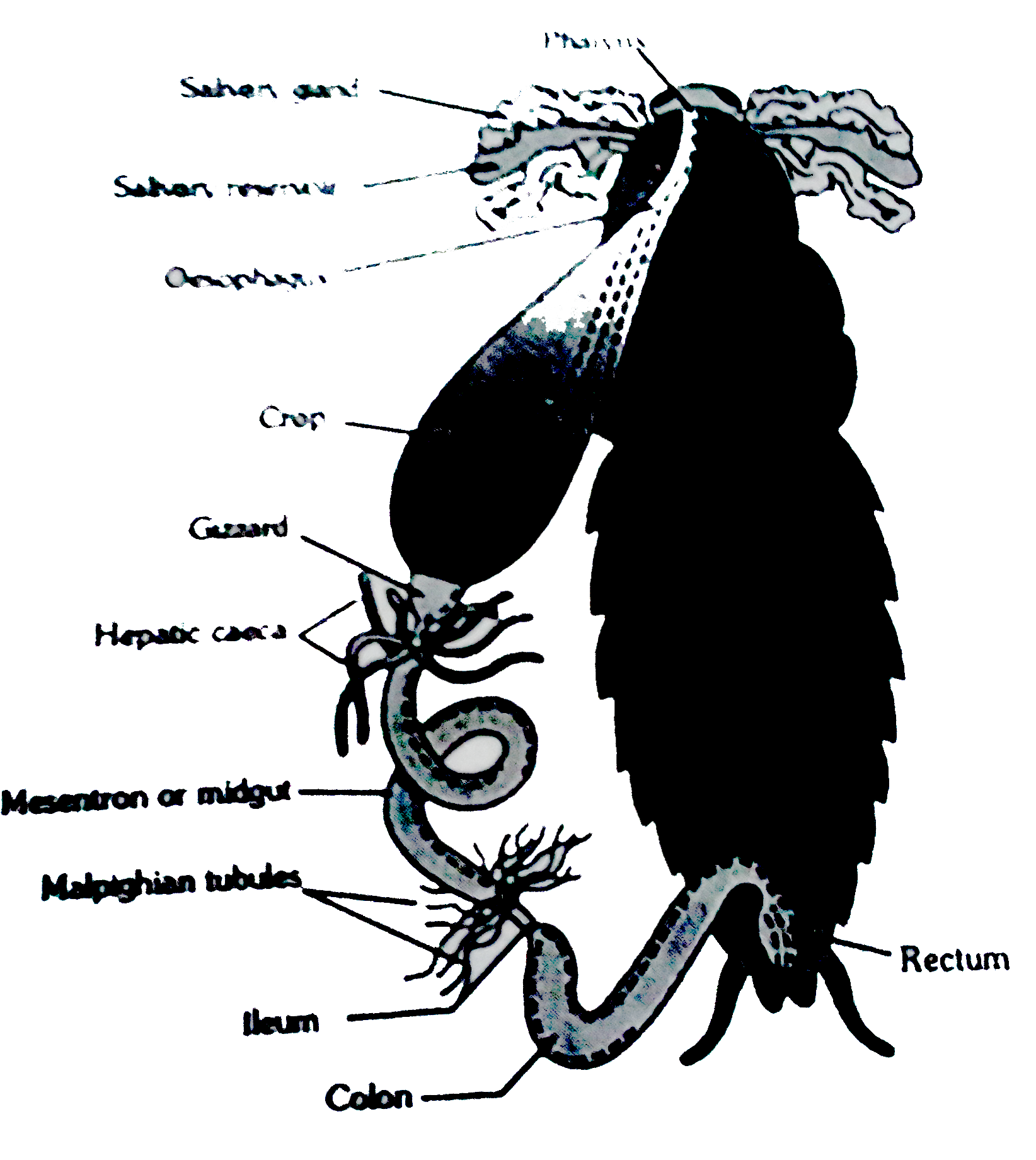
Draw a labelled diagram of alimentary canal of a cockroach
Step-by-Step Solution to Draw a Labeled Diagram of the Alimentary Canal of a Cockroach
1. Start with the Outline: Begin by drawing the basic outline of the cockroach's body. This will help you position the alimentary canal accurately within the body.
2. Draw the Foregut:
- Mouth: At the front of the cockroach, draw a small opening to represent the mouth.
- Pharynx: From the mouth, extend a short tubular structure upwards to represent the pharynx.
- Esophagus: Draw a narrow tube leading down from the pharynx to represent the esophagus.
- Crop: Below the esophagus, draw a sack-like structure to represent the crop, which stores food.
- Gizzard: Following the crop, draw a muscular structure to represent the gizzard, which grinds the food.
3. Draw the Midgut:
- After the gizzard, extend a wider tube to represent the midgut, which is the main site for digestion.
4. Add the Malpighian Tubules:
- At the junction of the midgut and hindgut, draw several yellow, thin tubular structures to represent the Malpighian tubules, which are involved in excretion.
5. Draw the Hindgut:
- Extend the midgut into a broader tube to represent the hindgut.
- Rectum: At the end of the hindgut, draw a short tube leading to the anus to represent the rectum.
6. Label the Parts: Clearly label each part of the alimentary canal:
- Mouth
- Pharynx
- Esophagus
- Crop
- Gizzard
- Midgut
- Malpighian Tubules
- Hindgut
- Rectum
- Anus
7. Final Touches: Ensure all parts are clearly labeled and the diagram is neat. You may want to use different colors for different parts for better visualization.
- Mouth: At the front of the cockroach, draw a small opening to represent the mouth.
- Pharynx: From the mouth, extend a short tubular structure upwards to represent the pharynx.
- Esophagus: Draw a narrow tube leading down from the pharynx to represent the esophagus.
- Crop: Below the esophagus, draw a sack-like structure to represent the crop, which stores food.
- Gizzard: Following the crop, draw a muscular structure to represent the gizzard, which grinds the food.
- After the gizzard, extend a wider tube to represent the midgut, which is the main site for digestion.
- At the junction of the midgut and hindgut, draw several yellow, thin tubular structures to represent the Malpighian tubules, which are involved in excretion.
- Extend the midgut into a broader tube to represent the hindgut.
- Rectum: At the end of the hindgut, draw a short tube leading to the anus to represent the rectum.
- Mouth
- Pharynx
- Esophagus
- Crop
- Gizzard
- Midgut
- Malpighian Tubules
- Hindgut
- Rectum
- Anus
Topper's Solved these Questions
- STRUCTURAL ORGANISATION IN ANIMALS BOOK - ICSECHAPTER - STRUCTURAL ORGANISATION IN ANIMALS EXERCISE - Ncert Exercises (Differentiate Between )
28 Videos
- STRUCTURAL ORGANISATION IN ANIMALS BOOK - ICSECHAPTER - STRUCTURAL ORGANISATION IN ANIMALS EXERCISE - Test Your Progress (HOTS)
8 Videos
- SELF ASSESSMENT PAPER-3BOOK - ICSECHAPTER - SELF ASSESSMENT PAPER-3EXERCISE - PART II (SECTION C)
4 Videos
- STRUCTURE ORGANISATION IN ANIMALS -ANIMAL TISSUESBOOK - ICSECHAPTER - STRUCTURE ORGANISATION IN ANIMALS -ANIMAL TISSUESEXERCISE - REVIEW QUESTIONS (LONG ANSWER TYPE QUESTIONS)
12 Videos
Similar Questions
Draw a labelled diagram of a neuron
View SolutionDraw a labeled diagram of sperm.
View SolutionDraw a labeled diagram of sperm.
View SolutionDraw a labelled diagram of synapse.
View SolutionDraw a labelled diagram of a neuron.
View SolutionDraw a labelled diagram of a nephron.
View SolutionDraw a labelled diagram of human alimentary canal and its associated glands
View SolutionGiven below the diagram of alimentary canal of cockroach which type of structure responsible for storing of food.

View SolutionDraw a labelled diagram of a nerve cell.
View SolutionDraw a labelled diagram of human ovum.
View Solution
