Which one of the following forms the pigmented layer in eye ?
(a) choroid
(b) retina
(c) sclera
(d) fovea
choroid
retina
sclera
fovea
To determine which structure forms the pigmented layer in the eye, let's analyze the options provided:
1. Choroid: The choroid is a layer of blood vessels and connective tissue located between the retina and the sclera. It contains a lot of pigment, which helps to absorb excess light and prevent scattering within the eye. This layer is crucial for providing nutrients to the retina and is indeed the pigmented layer of the eye.
2. Retina: The retina is the innermost layer of the eye that contains photoreceptor cells (rods and cones) responsible for detecting light. While it does have a pigmented layer at its back (the retinal pigment epithelium), the retina itself is not primarily described as a pigmented layer.
3. Sclera: The sclera is the white outer layer of the eyeball. It provides structure and protection but does not have pigmentation like the choroid.
4. Fovea: The fovea is a small pit in the retina that contains a high concentration of cones and is responsible for sharp central vision. It does not form a pigmented layer.
Based on this analysis, the correct answer is:
(a) Choroid
Topper's Solved these Questions
- NEURAL CONTROL AND COORDINATIONBOOK - ICSECHAPTER - NEURAL CONTROL AND COORDINATIONEXERCISE - (Review question) Fill in the blanks
10 Videos
- NEURAL CONTROL AND COORDINATIONBOOK - ICSECHAPTER - NEURAL CONTROL AND COORDINATIONEXERCISE - (Review question) Matching
1 Videos
- NEURAL CONTROL AND CO-ORDINATION BOOK - ICSECHAPTER - NEURAL CONTROL AND CO-ORDINATION EXERCISE - NCERT EXERCISES WITH ANSWER (DIFFERENTIATE BETWEEN)
11 Videos
- PHOTOMORPHOGENESIS IN PLANTSBOOK - ICSECHAPTER - PHOTOMORPHOGENESIS IN PLANTSEXERCISE - REVIEW QUESTIONS (LONG ANSWER TYPE QUESTIONS)
6 Videos
Similar Questions
Knowledge Check
Which one of the following consists of a thin layer of endothelium only?
AarteriolesBvenulesCcapillariesDvalves
Which one of the following pollutants causes irritation in eye?
View SolutionWhich of the following embryonic layer formed gametes :
View SolutionWhich one of the following is the smallest odd whole number? (a) 0 (b) 1 (c) 3 (d) 5
View SolutionWhich one of the following is a prime number?
(a) 161
(b) 221
(c) 373
(d) 437
View SolutionIn which of the following form is glucose stored in liver
(a) Starch
(b) Glycogen
(c) Cellulose
(d) Sucrose
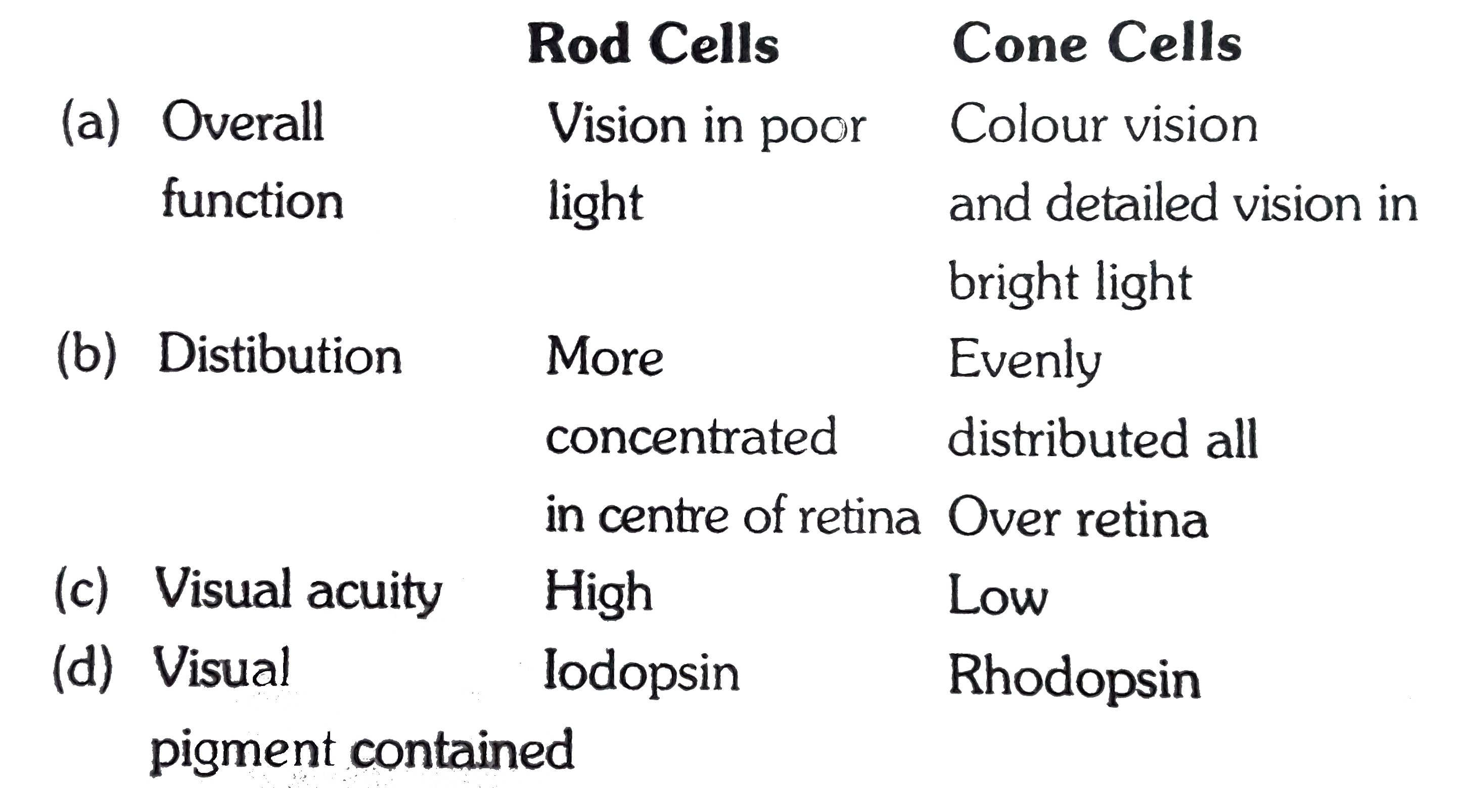
View SolutionWhich one of the following is the correct difference between Rod Cells and cone cells of our retina

View SolutionWhich one of the following is the correct difference between Rod Cells and cone cells of our retina

View Solution
